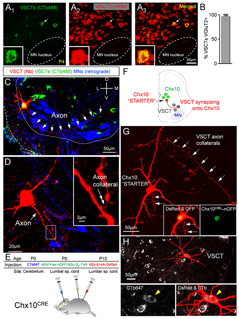
Figure 6. VSCTs are glutamatergic, possess spinal axon collaterals and make synapses with Chx10 spinal neurons.

(A1-3) VSCTs (CTb-488 from cerebellum, green) and VGluT2 mRNA (red) and merged image. Inset: VSCT (arrow in A1-3) expressing VGluT2. (B) Percentage of VSCTs expressing VGluT2 (N=3). (C) A P4 L2 VSCT filled with Neurobiotin (red) after intracellular recording colocalizing with CTb-488 (cerebellum at P0). MNs were backfilled with a dextran dye from the VR (blue). White arrows mark VSCT axon. (D) Higher mag of VSCT [in (C)], showing its main axon (white arrow) and an ipsilaterally projecting axon collateral (inset, double arrow; magnified from the dotted box). (E) AAV2/1-Flex-nGFP-N2c-Gp-TVA injected in spinal cord at P0 to introduce the rabies G protein to Chx10 neurons (Chx10CRE mice). Concurrently, CTb-647 injected in cerebellum to mark VSCTs. Rabies-N2c-EnvA-dsRed was injected in spinal cord at P10. (F) The “starter” Chx10 neurons are labeled with nuclear GFP and cytoplasmic dsRed. Neurons providing monosynaptic input to “starter” neurons are labeled with dsRed, but not nGFP. VSCTs are identified through CTb647. (G) A Chx10 “starter” neuron identified by co-localization of dsRed and nGFP (insets). Arrows indicate VSCT axon collaterals synapsing onto the Chx10 “starter” neuron. (H) VSCT (yellow arrowhead in insets) labeled by transynaptic transfection of its axon collaterals onto the Chx10 neuron. Data are represented as mean ± SEM. See also Figs. S6 and S7.
