PLOS ONE logoLink to PLOS ONE
. 2022 Feb 17;17(2):e0263873. doi: 10.1371/journal.pone.0263873

Relative changes in brain and kidney biomarkers with Exertional Heat Illness during a cool weather marathon

Michael J Stacey 1,2,3,*, Neil E Hill 4, Iain T Parsons 1,3,5, Jenny Wallace 6, Natalie Taylor 6, Rachael Grimaldi 7, Nishma Shah 8, Anna Marshall 8, Carol House 9, John P O’Hara 3, Stephen J Brett 2,10, David R Woods 1,3
Editor: Samuel Penna Wanner11
PMCID: PMC8853487  PMID: 35176088

Abstract

Background

Medical personnel may find it challenging to distinguish severe Exertional Heat Illness (EHI), with attendant risks of organ-injury and longer-term sequalae, from lesser forms of incapacity associated with strenuous physical exertion. Early evidence for injury at point-of-incapacity could aid the development and application of targeted interventions to improve outcomes. We aimed to investigate whether biomarker surrogates for end-organ damage sampled at point-of-care (POC) could discriminate EHI versus successful marathon performance.

Methods

Eight runners diagnosed as EHI cases upon reception to medical treatment facilities and 30 successful finishers of the same cool weather marathon (ambient temperature 8 rising to 12 ºC) were recruited. Emerging clinical markers associated with injury affecting the brain (neuron specific enolase, NSE; S100 calcium-binding protein B, S100β) and renal system (cystatin C, cysC; kidney-injury molecule-1, KIM-1; neutrophil gelatinase-associated lipocalin, NGAL), plus copeptin as a surrogate for fluid-regulatory stress, were sampled in blood upon marathon collapse/completion, as well as beforehand at rest (successful finishers only).

Results

Versus successful finishers, EHI showed significantly higher NSE (10.33 [6.37, 20.00] vs. 3.17 [2.71, 3.92] ug.L-1, P<0.0001), cysC (1.48 [1.10, 1.67] vs. 1.10 [0.95, 1.21] mg.L-1, P = 0.0092) and copeptin (339.4 [77.0, 943] vs. 18.7 [7.1, 67.9] pmol.L-1, P = 0.0050). Discrimination of EHI by ROC (Area-Under-the-Curve) showed performance that was outstanding for NSE (0.97, P<0.0001) and excellent for copeptin (AUC = 0.83, P = 0.0066).

Conclusions

As novel biomarker candidates for EHI outcomes in cool-weather endurance exercise, early elevations in NSE and copeptin provided sufficient discrimination to suggest utility at point-of-incapacity. Further investigation is warranted in patients exposed to greater thermal insult, followed up over a more extended period.

Introduction

As global temperatures rise, elite athletes, sports participants, recreational runners and hikers, and a range of occupational groups share an increasing risk of EHI (Exertional Heat Illness). A spectrum of EHI [13] severity is described in the literature, ranging from transient reductions in consciousness and lesser elevations in core temperature with mild EHI to the pronounced hyperthermia, central nervous system (CNS) dysfunction and multi-organ failure observed in exertional heat stroke (EHS) [4]. In civilian mass participation events, the occurrence of severe EHI may reach 31 case per 10000 competitors in higher-risk races [5]. A lower EHS incidence of 1 in 10,000 finishers has been reported in marathon runners [6], though cases may increase with the severity of climatic conditions [7] and can be even greater in and other athletic disciplines [8].

Larger scale mass participation recreational events may attract clinical and laboratory support familiar with the presentation and management of EHI. Capacity and capability may be more limited, however, for detached emergency responders, ‘pop-up’ medical facilities at smaller events and in certain industrial and agricultural settings. This risks EHI events being under-estimated, under-resourced or under-managed by non-specialists. Indeed, where prior experience is lacking and EHI not recognised or distinguished from benign exertional hyperthermia, cooling may be altogether neglected [4]. This is highly relevant to more remote field settings, including those with a military focus, in which the need for early recognition of end-organ injury—for example acute kidney injury (AKI)—has been highlighted [9]. In such circumstances, prompt decisions regarding escalation and evacuation to higher echelons of medical care may have a definitive impact on outcome both for the individual and the event or mission [4].

On occasion, EHS may manifest in an indolent or subacute fashion, whereby initial clinical progress provides false reassurance [10]. Even with fulminating illness, standard laboratory parameters may not immediately or reliably reflect or predict the risk of organ injury [4, 11]. As such, the safety and appropriateness of releasing from medical care or advising on return-to-exercise can be difficult to judge in a patient affected by EHI, especially in the relatively short-space of time available to emergency providers. This may be particularly important where the affected individual will be unaccompanied upon discharge, or subsequent heat tolerance testing is available and relatively indicated [12]. In such circumstances, an early indication of established or evolving organ injury could help inform whether ongoing clinical supervision is indicated and/or aid assessment of the likelihood of increased risk associated with further bouts of exercise and thermal stress [13]. In this sense, a reproducible physiological index to discriminate EHS with downstream clinical sequelae from lesser kinds of incapacity could help tailor a clinical pathway from collapse to recovery.

We have demonstrated compromise to the GI epithelium with early, substantial and sustained elevations in intestinal fatty acid binding protein (I-FABP) in marathon runners diagnosed with EHI [14]. This raises the question of whether these patients experienced significant injury to other organ systems in the evolution of their illness, which might have been detectable at the initial point of care (POC). As central nervous system (CNS) impairment was defining and renal stress anticipated with the substantial cardiovascular strain observed, we further investigated biomarkers with emerging clinical utility in relation to brain (neuron specific enolase, NSE; S100 calcium-binding protein B, S100β), kidneys (cystatin C, cysC; kidney-injury molecule-1, KIM-1; neutrophil gelatinase-associated lipocalin, NGAL) and fluid-regulatory stress (copeptin, the C-terminal part of pro-arginine vasopressin that is a stable surrogate for arginine vasopressin, AVP).

This selection of biomarkers was informed by previous investigations in humans showing prognostic potential when measured close to the point of injury/debility, as for heat stroke (S100B) [15], traumatic and septic encephalopathy (NSE) [16, 17] and clinically significant AKI of various aetiologies (sCr, NGAL, KIM-1) [18, 19]; as well as by studies showing associations with inflammatory responses specific to acute exercise-heat stress (NGAL) [20] and sub-clinical AKI (copeptin) [21, 22]. Our primary aim was to determine whether these markers could effectively discriminate marathon runners affected by EHI from successful finishers completing the event that same day. Secondary aims were to examine changes in these biomarkers over time and to relate indirect evidence for organ injury to likely precipitants, co-factors or other indicators relevant to the episodes of collapse observed.

Materials and methods

We collected data from two groups of runners–people who volunteered for blood and anthropometric assessment to occur before and after the marathon (henceforth referred to as ‘successful finishers’) and a separate group of people not enrolled as a successful finisher, who collapsed and required medical treatment for EHI during the marathon. No successful finishers collapsed or required medical treatment during the marathon event.

Successful finishers

Potential participants were contacted via electronic mailshot from the Brighton Marathon Race team and provided with information pertaining to associated research projects. Inclusion criteria were age 18–60 years. Volunteers were excluded if they had not read and complied with the Medical Advice for participating in the marathon (http://www.brightonmarathonweekend.co.uk/medical-advice/), had a prior history of heat illness, significant preceding head injury, epilepsy, congenital or acquired kidney disease or were taking drugs known to affect the renal system (including non-steroidal anti-inflammatory drugs).

Anthropometric and physiological measurements and blood tests were taken between 10.00 and 19.00 on the day prior to the event. Unshod standing height and minimally clothed body mass were recorded for each control participant using a stadiometer and scales. Participants were then seated for around 10 min prior to the measurements of resting heart rate (HR), systolic (SBP) and diastolic blood pressure (DBP) all using an integrated patient monitoring device (GE Carescape V100, UK). Venepuncture was performed at the antecubital fossa.

Blood samples (13 ml per draw) for these successful finishers were taken at the following time-points: (1) ‘pre-race’ baseline (B), at race registration the day before the marathon; (2) T0; as close to the time of successful completion of event as feasible (<30 minutes); (3) T24; the following day, as close to 24 hours post-run as feasible. Blood was centrifuged at 1500G for 15 minutes then separated and snap frozen in liquid nitrogen on site for subsequent assay.

Collapsed runners

Runners who collapsed during the marathon were assessed for study enrolment following evacuation to the nearest marathon medical facility, clinical re-assessment and immediate essential medical treatment. The cases in question were triaged, treated and confirmed by clinicians experienced in the management of EHI, who were able to discount non-EHI diagnoses including post-exertional hypotension, primary cardiac disorders and exercise-associated hyponatraemia uncomplicated by hyperthermia. Criteria for recruitment were a clinical diagnosis of EHI, where excess body heat was deemed the primary cause of incapacity and core body temperature measured rectally (Intellivue integrated thermistor, Philips Healthcare, Amsterdam, Netherlands) was ≥38.5°C, in association with CNS impairment (for example, abnormal motor control, loss of responsiveness, amnesia for the episode) occurring spontaneously during or soon after marathon run followed by failure to make a prompt recovery with prostration and initial medical care. Explicitly, runners diagnosed with exertional or post-exertion syncope were excluded from recruitment, as were those re-categorised with an alternative aetiology based upon response to initial treatment. Level of consciousness upon presentation to medical staff was defined according to the widely used Alert-Voice-Pain-Unresponsive (AVPU) scale, which assigns the best casualty response to stimulation in a graded fashion and corresponds with the more detailed Glasgow Coma Scale as: <A> Alert: awake but potentially confused, GCS up to 15; <V> Verbal: responsive to verbal stimulation, GCS ~12; <P> Pain: responsive to painful stimuli, GCS ~8; <U> Unresponsive: unconscious, no response to voice or pain, GCS 3.

Blood samples (13 ml per draw) were taken at the following time-points: (1) T0; as close to the time of collapse as feasible (within 30 minutes), (2) T1; at 1 hour following collapse (as clinical considerations allowed) and (3) T4; at 4 hours following collapse (again as clinical considerations allowed). No EHI cases were available for next-day (T24) sampling despite ethical approval being in place to do so. Samples were analysed at Affinity Biomarker Labs (London, UK). Serum was analysed for sCr, cysC, CK and total protein (TP) on a commercial platform (Siemens Advia 1800, Siemens Healthcare Diagnostics Ltd, Camberley, UK). Serum NSE, KIM-1 and NGAL (R & D Systems Europe, Abingdon, UK) were measured by commercially available immunoassay with intra- and inter-assay variability of <10% and an upper limit of detection of 20ug.L-1, 700 ng.L-1 and 200 ug.L-1 respectively. Serum S100β (EMD Merck-Millipore, St. Louis, USA) was measured by sandwich ELISA with variability <5% and upper limit of detection of 200 ng.L-1. Plasma copeptin was measured with Time-Resolved Amplified Cryptate Emission technology (Thermo Fisher-Brahms, Hennigsdorf, Germany).

Data were assessed for normality and expressed as mean ± SD or median [IQR]. Linear relationships between parametric and non-parametric variables were assessed for significance by Pearson’s or Spearman’s rank tests, respectively. Data were compared by t-test (parametric data) or Mann-Whitney test (non-parametric data) and Receiver Operating Characteristic (ROC) curves were constructed to determine the Area-Under-The-Curve and sensitivity and specificity for the biomarkers of interest in discriminating EHI versus successful finishers.

Significance was set to alpha = 0.05. A formal power calculation was not attempted prospectively, due to the study protocol’s dependence upon a convenience sample of willing volunteers and uncertain availability of EHI cases. However, it was noted that, in a sample of 28 patients affected by brain injury with a traumatic mechanism, power was adequate to discriminate 9 survivors with favourable functional outcomes from 19 patients experiencing death or poor functional outcomes according to serum NSE and S100β measured soon after insult, with peak sensitivity and specificity of 88% and 100% for S100β [23]. Elsewhere, a study of 10 subjects showed significantly greater NGAL with exercise under heat stress where increased inflammation and elevated kidney injury biomarkers were induced versus control conditions in the same subjects [20]. With marathon running, both plasma copeptin and NGAL were shown to be significantly higher in 12 runners with AKI defined biochemically versus 10 experiencing sub-threshold elevation in sCr [21].

Ethics approval

Ethical approval for a study of people running the 2019 Brighton Marathon was obtained from London South East ethics committee (19/LO/0340 247967). At the race registration (on the day before the marathon) volunteers (successful finishers) were formally recruited to take part in the study and provided written informed consent. For EHI cases who initially lacked mental capacity to consent for themselves (AVPU grade V to U, or A with any concern for capacity) we proceeded with presumed consent until they were deemed able to give it retrospectively.

Results

On the day of the marathon, ambient temperature measured at the local meteorological station increased from 8 °C during the event muster (race start time 09:45) to peak at 12 °C with runners still on the course between 14:00 and 15:00. Among runners who collapsed while participating and underwent triage upon reception to medical facilities, eight underwent serial clinical review and were confirmed as EHI cases. These runners (5 male, 3 female) had completed a maximum of 22.4 ± 5.5 miles before collapsing and being evacuated forwards to the nearest on-course medical facilities, stationed at, respectively, 14 and 26.3 mile points. The recruited cohort represented 100% of clinically confirmed EHI cases presenting to marathon medical staff, at the only hospital-standard receiving facilities available on-course.

Individual biochemical results and available clinical observations are displayed in Table 1. In the global pandemic context, platform availability and reagent shortages limited measurements of copeptin to 7 of these 8 cases. S1 File shows comparative further biochemical measurements obtained 1 or 4 hours post-collapse, with NSE seen to decline in 2 out of 3 cases with this more complete data.

Table 1. Clinical and biochemical results for eight EHI cases sampled within 30 minutes of incapacity (T0 sampling point).

Loc. 1. Treatment facility stationed at 14 mile-point on course (NB course design resulted in runners up to 21 miles being received here). Location II. Main medical tent stationed 100 m behind finishing line.

Loc. Case/ Time Distance AVPU Tc °C RR breaths.min-1 HR beats.min-1 BP mmHg NSE ng.L-1 S100b ng.L-1 sCr μmol.L-1 cysC mg.L-1 NGAL ug.L-1 KIM-1 ng.L-1 Copeptin pmol.L-1 CK IU.L-1 TP g.L-1
I. 1 14 miles V 40.1 60 115 101/46 6.37 90.52 154 1.53 185.48 14.04 339.4 179.0 78.2
2 25 miles A 41.5 25 155 117/57 6.37 184.79 155 1.74 >200.0 27.74 U/A 324.6 77.3
3 21 miles A 39.0 50 136 120/52 >20.00 98.97 145 0.94 112.94 52.56 104.0 305.7 74.3
II. 4 >21 miles V 39.1 40 106 138/68 9.9 111.91 103 1.08 98.52 22.40 983.3 310.6 63.8
5 >21 miles P >40.0 U/A U/A U/A 26.78 38.63 124 1.16 136.12 30.00 77.0 729.5 56.3
6 >21 miles V 38.6 28 141 109/65 4.29 232.06 192 1.77 351.2 19.31 942.6 484.9 75.6
7 >21 miles V >38.5 U/A U/A U/A >20.00 98.33 142 1.48 179.44 26.69 5.6 802.7 67.1
8 >21 miles V >38.5 U/A U/A U/A 10.76 <2.7 164 1.59 >200.0 40.61 802.7 603.0 68.7

AVPU, Alert-Voice-Pain-Unresponsive scale; BP–blood pressure; CK–creatine kinase; cysC–cystatin C; HR–heart rate; KIM-1 –Kidney Injury Molecule 1; NGAL–neutrophil gelatinase associated lipocalin; NSE–neuron specific enolase; RR–respiratory rate; sCr–serum creatinine; Tc -first measured core temperature upon admission to medical facility ± 1 hour post-admission Tc; TP–Total protein; U/A–data unavailable (data entry spoiled, however all entered participants known to have Tc measured upon reception to medical facility >38.5 °C).

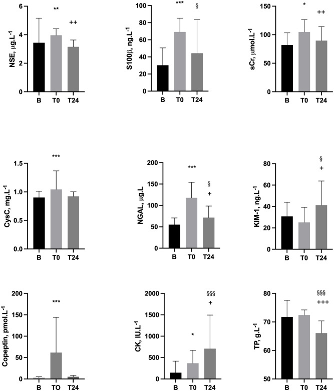
Thirty healthy runners aged 37.7 ± 8.9 years old (16 male, 14 female) successfully completed the marathon distance of 26.2 miles in 4.2 ± 0.8 (range 2.8 to 5.6) hours. Loss of body mass was -2.5 ± 1.6%. S2 File displays physical and biochemical results from B and T0, including laboratory reference ranges in health. For runners who also completed T24 measures (n = 18), sequential changes B-T0-T24 are displayed in Fig 1 and S3 File. For T0 measures, NGAL correlated with T0 sCr (r = 0.63, p = 0.005); S100β correlated with T24 CK (r = 0.66, P = 0.0028); and CK correlated with T24 KIM-1 (r = 0.55, p = 0.018).

Fig 1. Biochemical variables assayed pre-marathon (B), upon completion (T0) and next-day (T24) in 18 successful finishers with complete data across the three measurement points.

Fig 1

T0 vs B ***P<0.0005, **P<0.005, *P<0.05. T24 vs B §§§P<0.0005, §§P<0.005, §<0.05. T24 vs T0 +++P<0.0005, ++P<0.005, +P<0.05. CK–creatine kinase; cysC–cystatin C; KIM-1 –Kidney Injury Molecule 1; NGAL–neutrophil gelatinase associated lipocalin; NSE–neuron specific enolase; sCr–serum creatinine; TP–Total protein.

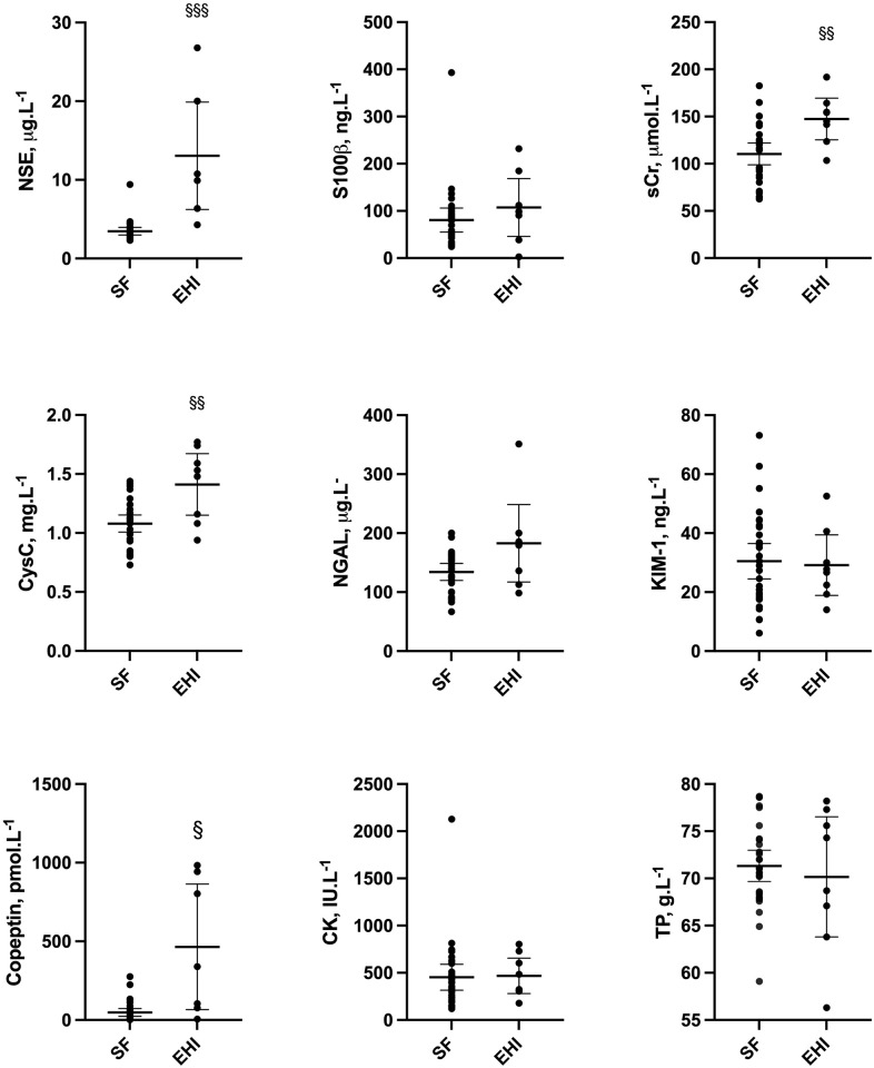
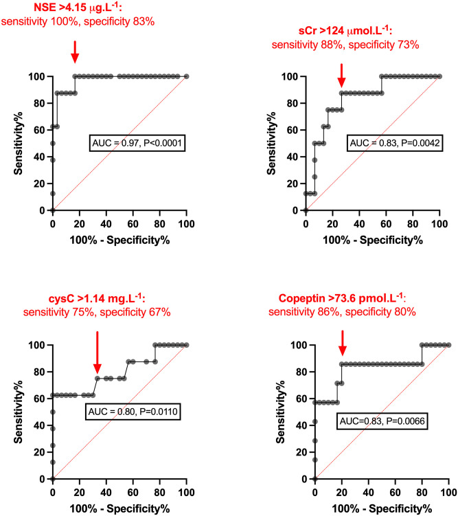
Biochemical profiles for EHI cases versus successful finishers are shown in Fig 2. Analysis by ROC with corresponding AUC, where significant, and performance values (EHI versus successful finishers) is displayed in Fig 3. Versus successful finishers, EHI cases had higher heart rate (131 ± 20 vs 87 ± 14 b.min-1) and lower diastolic blood pressure (58 ± 9 vs 67 ± 8 mmHg, P = 0.0123), but showed no difference (P = 0.4677) in systolic blood pressure.

Fig 2. Biochemical comparison at T0 (marathon completion or withdrawal) for 30 successful finishers (SF) vs 8 Exertional Heat Illness cases (EHI)* §§§P<0.0005, §§P<0.005, §<0.05.

Fig 2

*n = 7 for copeptin. CK–creatine kinase; cysC–cystatin C; KIM-1 –Kidney Injury Molecule 1; NGAL–neutrophil gelatinase associated lipocalin; NSE–neuron specific enolase; sCr–serum creatinine; TP–Total protein.

Fig 3. ROC curves for biochemical parameters showing Area-Under-Curve (AUC) ≥0.7 for 30 successful finishers (SF) vs 8 Exertional Heat Illness cases (EHI),* assayed at T0 (marathon completion or withdrawal).

Fig 3

CysC–cystatin C; NGAL–neutrophil gelatinase associated lipocalin; NSE–neuron specific enolase; sCr–serum creatinine. *n = 7 for copeptin.

Discussion

This study is the first to examine a number of blood biomarkers for human EHI, both in heat-attributed collapse and with successful marathon completion. Our investigation also represents a relatively unique examination of EHI generated against the environmental gradient by metabolic heat production, in runners who faced challenging conditions of relatively cold Spring temperatures and gusting headwinds along the final seafront miles of the marathon course. Despite a tendency for on-course cooling secondary to these conditions, novel variation in NSE, cysC and copeptin was evident between clinically-diagnosed EHI cases and successful finishers. The striking elevations in NSE and copeptin observed in some collapsed runners suggested a substantial degree of neuronal as well as fluid-regulatory and/or cardiovascular stress at point of collapse. This associated with case discrimination by ROC that was outstanding for NSE (AUC>0.9), excellent for sCr and copeptin (>0.8) and robust for cysC (>0.7).

Serum NSE is a neuronal enzyme indicative of neuroinflammation [17], which has been reported to rise early in sepsis-associated encephalopathy (SAE) and show positive associations with elements of systemic inflammation, including IL-6 [18]. Therefore NSE might be expected to increase with the evolution of endotoxaemic encephalopathy concomitant to exertional hyperthermia. This is suggested by the present work and supported by elevated I-FABP in our complementary reporting of the same casualties [14]. While the level of consciousness observed upon initial medical assessment was less depressed in these EHI cases than observed in other positive clinical studies of patients with SAE [18], traumatic brain injury [23] and cardiac arrest [24], this tallies with a discriminant value of NSE in the present investigation that was relatively lower in numerical value. This would also be in keeping with lower grade neuronal injury sustained with evolving EHI, thankfully interruptible with cooling–first passively upon cessation of exercise, then actively under medical supervision–and fortunately associated with eventual recovery to baseline in all cases.

Unlike NSE, S100β was not elevated in EHI cases versus successful finishers. This accords with its lack of discrimination for encephalopathy and lower prognostic potential when measured in the early stages of injury among other cohorts. As a marker of functionally milder brain insults associated with lesser reductions in consciousness at presentation (Glasgow Coma Scale score 13–15, equivalent to ‘A’ or ‘V’ on the AVPU scale), S100β has reported high sensitivity for radiographically-demonstrable traumatic intracranial injury, at a threshold corresponding to the mean and median values observed in EHI cases in the present work (~100ng.L-1) [24]. The specificity of S100β for brain injury is reduced in trauma encompassing extracranial sites, however, perhaps due to release from other tissues including those containing chondrocytes and fat cells [25]. In the present work, correlation with next-day CK suggest that S100β measured close to the point of collapse may have additionally reflected musculoskeletal stress in successful finishers. Being renally excreted with a half-life of 30 to 120 minutes, S100β would be also be expected to show an effect of exercise-associated changes in GFR and AKI in addition to any attributable to any CNS injury.

Risks of dehydration in this study are likely to have been lower than many comparable reports in the literature. Among successful finishers, this was evidenced by loss of body mass that was not excessive for endurance exercise and lower copeptin than has been observed in warmer marathon conditions [21], perhaps due to relatively reduced osmotic/volume stimulus to AVP secretion. In EHI, a range of copeptin values were observed, with 6 out of 7 cases assayed showing levels more than twice the median of successful finishers; several values reached or exceeded those reported in severe sepsis and haemorrhagic shock [26, 27]. This may indicate relatively greater dehydration altogether, with both hypovolaemia and hypertonicity being potent, classical stimuli to the release of AVP/copeptin. Incipient inflammation, cardiovascular stress, reduced renal clearance and AKI or hyperthermia may also have contributed to elevated copeptin levels [22]. In one of two EHI cases sampled 1 hour post-recruitment to the present study, relative doubling of copeptin and KIM-1 suggested renal injury from ongoing relative ischaemia, despite cooling.

Taken together, these findings are supportive of further enquiry into the utility of a number of the biomarkers described in identifying and prognosticating for severe EHI. The requirement for such a tool may be debated, as the majority of EHI patients treated promptly with cooling show a good prognosis following incapacitation [4]. However the perennial challenge of managing heat stress derived from both exercise and the environment, repeated instances of fatal EHS where preventive guidelines were in place (if not wholly followed) and potential difficulties in consistently recognising and differentiating significant EHI from lesser forms of incapacity, especially in cooler conditions, indicate a need for better tools for attendant healthcare professionals. The American College of Sports Medicine acknowledges in its position statement that the clinical changes associated with EHS can be subtle and easy to miss if coaches, medical personnel, and athletes do not maintain a high level of awareness [28].

Athletes deemed to have suffered moderate to severe EHI may be advised to follow-up with a physician after initial recovery and discharge from local medical facilities [12, 28]. Practically speaking, this extends the importance of early, accurate diagnosis into considerations regarding return-to-play/participation. Whether some of the cases reported above represented EHS or lesser forms of EHI may be rather academic in real-time; what may matter more is the ability to detect those at risk of organ-injury early on, to ensure that appropriate management is instituted and followed through to sporting, recreational or occupational recovery. This first requires research tools to help stratify casualties for interventions auxiliary to cooling and determine how initial presentation and treatment relate to longer-term outcomes [9].

One potential limitation of the study is that emergency care providers around the course may have elected to transfer potential EHI cases with more concerning physiology direct to the local permanent hospital Emergency Department, rather than delivering them to the on-course medical facilities from which we recruited. However the triage of marathon casualties is co-ordinated by radio communication with a central hub of medical advice co-located with the main temporary (finish line) medical facility and we were made aware of no other severe cases bypassing this facility, nor did Emergency Department staff at the local hospital report other casualties attending when our team attended in follow up of case 5 at 4 hours post-collapse. Some unidentified EHI cases may have recovered ambulatory capacity and failed to attend for any medical treatment on-course or to present to hospital facilities, though these would almost certainly have represented the milder end of the spectrum of EHI presentations described in the literature, rather than being heat stroke cases.

We acknowledge that in recruiting cases close to the point of individual collapse along the marathon course, the final running distance completed varied within the EHI cohort. A disparity was also present in comparison to successful finishers at T0, who had all completed the full 26.2 mile run. Nevertheless, sampling occurred within a standardised time window (<30 minutes of EHI or marathon completion) and resulted in biomarker values that were largely more pronounced, rather than less, in the EHI group.

Co-factors in the evolution of EHI may also have impacted results in the direction of increased biomarker values, e.g. haemolysis arising with hyperthermia, which may represent an early stage in the development of clotting abnormalities in heatstroke, but can also increase NSE release from erythrocytes [29]. Another limitation is that the exclusion criteria for the successful finisher cohort (listed in the Methods) mean that this group may differ from the collapsed athlete group, who were not subject to these exclusion criteria. Whether collapsed EHI cases run faster, generate more metabolic heat are relatively more or less endurance trained than successful finishers in this context are all relevant questions, reflecting important mechanistic considerations.

However shortfalls in data of this kind need not detract from the practical potential reported in the present work and future studies may be able to recruit, follow and characterise sufficient collapsed cases to be more stringent in this respect. Better demographic characterisation (age, anthropometric characteristics) and improved understanding of the effect of changes in plasma volume on these markers (e.g. Dill and Costill-corrected values [30] for paired data, as in successful finishers) would, respectively, provide reassurance on the generalisability of these initial findings and relevant mechanisms underpinning them. This will require increased resourcing and logistical support to more completely overcome the unfavourable conditions for uniform collection of contextual data in this kind of field setting.

The investigation of a wider panel of biomarkers of relevance to renal and brain injuries may also be considered. For example, glial fibrillary acid (GFAP) has shown prognostic potential in detecting radiographically occult traumatic brain injury at presentation [31]. Although not included in this preliminary investigation, due to lack of data supporting associations with other relevant forms of encephalopathy at the time of writing, GFAP has since shown potential to assist early diagnosis and prognostication for sepsis-associated encephalopathy [32]. This comes with the added possibility of POC measurement [33], which could be highly advantageous in the marathon medical environment, specifically in relation to a diagnosis of heat stroke whereby systemic inflammation shares many parallels with sepsis and as such may similarly benefit from early intervention [4].

In conclusion, both emerging biomarkers in the form of NSE and copeptin, and more established injury surrogates such as sCr, performed well under cold weather conditions in discriminating successful marathon performance from supervening EHI. With significant renal injury identified in 16–25% of individuals affected by heat stroke of both kinds [24], more work is clearly required in this area. Later measurement of both neuro- and renal biomarkers (e.g. Day 1 and Day 3 post-event) could build prognostic value into these insights from the initial hours following collapse and allow further scrutiny for differences versus successful finishers. Extending this enquiry into other thermally and/or physically stressful settings may have potential utility for a range of sporting, recreational and occupational activities, especially where an episode of incapacity needs further substantiation to inform clinical decisions on rest, recuperation and ‘return to play/work’.

Supporting information

S1 File. Clinical and biochemical results for three EHI cases sampled following collapse, within 30 minutes of incapacity (T0) and again 60 minutes (T1) or 4 hours (T4).

Loc. 1. Treatment facility stationed at 14 mile-point on course (NB course design resulted in runners up to 21 miles being received here). Location II. Main medical tent stationed 100 m behind finishing line.

(DOCX)

S2 File. Results for 30 successful finishers, at rested baseline B and upon marathon completion T0.

CK–creatine kinase; cysC–cystatin C; KIM-1 –Kidney Injury Molecule 1; NGAL–neutrophil gelatinase associated lipocalin; NSE–neuron specific enolase; sCr–serum creatinine; sNa–serum sodium; Ur–serum urea; TP–Total protein.

(DOCX)

S3 File. Individual biochemical results for 18 successful finishers (n = 15 for copeptin), at rested baseline B, upon marathon completion T0 and next-day T24.

CK–creatine kinase; cysC–cystatin C; KIM-1 –Kidney Injury Molecule 1; NGAL–neutrophil gelatinase associated lipocalin; NSE–neuron specific enolase; sCr–serum creatinine; sNa–serum sodium; Ur–serum urea; TP–Total protein.

(DOCX)

Acknowledgments

UK Surgeon General’s Department (funding); Dr Amarjit Samra and Yvonne Yau (technical support); Brighton Marathon Medical Research Team, including Dr Rob Galloway and Mrs Carrie Weller (hosting and facilitating); Dr Ed Walter and Dr Oliver Gibson (contribution to data collection).

Data Availability

All relevant data are within the manuscript and its Supporting information files.

Funding Statement

The sources of funding for the study were (a) direct financial support from the Ministry of Defence to cover assay costs, salaries of involved military personnel, their temporary accommodation if required close to the study site and (b) material hosting by the Brighton marathon medical team, who provided medical tentage in which the study was performed and appropriate screens to protect volunteer dignity while being sampled etc. No formal numbered grant award was made, rather the MoD raised and settled purchase orders e.g. with Affinity Biomarkers, the commercial company that assayed the chemistry reported. The funders had no role in study design, data collection and analysis, decision to publish, or preparation of the manuscript.

References

  • 1.Carter R, Cheuvront SN, Williams JO, et al. Epidemiology of hospitalizations and deaths from heat illness in soldiers. Med Sci Sports Exerc. 2005; 37:1338–1344. doi: 10.1249/01.mss.0000174895.19639.ed [DOI] [PubMed] [Google Scholar]
  • 2.Malamud N, Haymaker W, Custer RP. Heat Stroke: Clinico-pathologic study of 125 fatal cases. Mil Surg. 1946; 99: 397–449. [PubMed] [Google Scholar]
  • 3.Shibolet S, Coll R, Gilat T, et al. Heatstroke: Its clinical picture and mechanism in 36 cases. Q J Med. 1967; 36: 525–548. [PubMed] [Google Scholar]
  • 4.Epstein Y, Ranovitch R. Heatstroke. N Engl J Med. 2019; 380: 2449–59. doi: 10.1056/NEJMra1810762 [DOI] [PubMed] [Google Scholar]
  • 5.Hosokawa Y, Adams WM, Belval LN, et al. Exertional heat illness incidence and on-site medical team preparedness in warm weather. Int J Biometeorol. 2018; 62:1147–1153. doi: 10.1007/s00484-018-1517-3 [DOI] [PubMed] [Google Scholar]
  • 6.Roberts WO. Exertional heat stroke in the marathon. Sports Med. 2007; 37:440–3. doi: 10.2165/00007256-200737040-00044 [DOI] [PubMed] [Google Scholar]
  • 7.Roberts WO. Burden of Injury and Illness in the Road Race Medical Tent: A Narrative Review. Breslow RG, Giberson-Chen CC, Clin J Sport Med. 2021; 1;31(6):e499–e505. doi: 10.1097/JSM.0000000000000829 . [DOI] [PubMed] [Google Scholar]
  • 8.Macaluso F, Barone R, Isaacs AW, et al. Heat stroke risk for open-water swimmers during long-distance events. Wilderness Environ Med. 2013;24:362–5. doi: 10.1016/j.wem.2013.04.008 [DOI] [PubMed] [Google Scholar]
  • 9.Wesseling C, Aragón A, González M et al. Kidney function in sugarcane cutters in Nicaragua—a longitudinal study of workers at risk of Mesoamerican nephropathy. Environ Res 2016; 147:125–132. doi: 10.1016/j.envres.2016.02.002 [DOI] [PubMed] [Google Scholar]
  • 10.Dickinson JG. Heat-exercise hyperpyrexia. J R Army Corps. 1989; 135: 27–29. doi: 10.1136/jramc-135-01-08 [DOI] [PubMed] [Google Scholar]
  • 11.Leon LR, Bouchama A. Heat stroke. Compr Physiol. 2015; 5: 611–647. doi: 10.1002/cphy.c140017 [DOI] [PubMed] [Google Scholar]
  • 12.Schermann H, Craig E, Yanovich E, Ketko I, Kalmanovich G, Yonovich R. Probability of Heat Intolerance: Standardized Interpretation of Heat-Tolerance Testing Results Versus Specialist Judgment. J Athl Train. 2018; 53(4):423–430. doi: 10.4085/1062-6050-519-16 [DOI] [PMC free article] [PubMed] [Google Scholar]
  • 13.Horowitz M. Do exertional heat stroke and environmental heat involve epigenetic memory? J Physiol. 2021;599:7–8. doi: 10.1113/JP280928 [DOI] [PubMed] [Google Scholar]
  • 14.Walter E, Gibson OR, Stacey M, et al. Changes in gastrointestinal cell integrity after marathon running and exercise-associated collapse. Eur J Appl Physiol. 2021;121(4):1179–1187. doi: 10.1007/s00421-021-04603-w [DOI] [PubMed] [Google Scholar]
  • 15.Wang Qi, Ishikawa Takaki, Michiue Tomomi, et al. Evaluation of human brain damage in fatalities due to extreme environmental temperature by quantification of basic fibroblast growth factor (bFGF), glial fibrillary acidic protein (GFAP), S100β and single-stranded DNA (ssDNA) immunoreactivities. Forensic Sci Int. 2012; 219: 259–64. doi: 10.1016/j.forsciint.2012.01.015 [DOI] [PubMed] [Google Scholar]
  • 16.Feng Q, Wu L, Ai YH, Deng SY, et al. The diagnostic value of neuron-specific enolase, central nervous system specific protein and interleukin-6 in sepsis-associated encephalopathy. Zhonghua Nei Ke Za Zhi. 2017; 1: 747–751. doi: 10.3760/cma.j.issn.0578-1426.2017.10.008 [DOI] [PubMed] [Google Scholar]
  • 17.Pleines UE, Morganti-Kossman MC, Rancan M, et al. S-100 beta reflects the extent of injury and outcome, whereas neuronal specific enolase is a better indicator of neuroinflammation in patients with severe traumatic brain injury. J Neurotrauma; 18: 491–8. doi: 10.1089/089771501300227297 [DOI] [PubMed] [Google Scholar]
  • 18.Parikh CR, Coca SG, Thiessen-Philbrook H, et al. Postoperative biomarkers predict acute kidney injury and poor outcomes after adult cardiac surgery. J Am Soc Nephrol. 2011; 22: 1748–1757. doi: 10.1681/ASN.2010121302 [DOI] [PMC free article] [PubMed] [Google Scholar]
  • 19.Venkatta SS, Waikar SS, Antoine DJ, et al. Blood Kidney Injury Molecule-1 Is a Biomarker of Acute and Chronic Kidney Injury and Predicts Progression to ESRD in Type I Diabetes. J Am Soc Nephrol. 2014; 25: 2177–2186. doi: 10.1681/ASN.2013070758 [DOI] [PMC free article] [PubMed] [Google Scholar]
  • 20.Junglee NA, Di Felice U, Dolce A, et al. Exercising in a hot environment with muscle damage: effects on acute kidney injury biomarkers and kidney function. Am J Physiol Renal Physiol. 2013; 305: F813–F820. doi: 10.1152/ajprenal.00091.2013 [DOI] [PubMed] [Google Scholar]
  • 21.Mansour SG, Martin TG, Obeid W, et al. The role of volume regulation and thermoregulation in AKI during marathon running. Clin J Am Soc Nephrol. 2019; 14:1297–1305. doi: 10.2215/CJN.01400219 [DOI] [PMC free article] [PubMed] [Google Scholar]
  • 22.Omassoli J, Hill NE, Woods DR, et al. Variation in renal responses to exercise in the heat with progressive acclimatisation. J Sci Med Sport. 2019; 22:1004–1009. doi: 10.1016/j.jsams.2019.04.010 [DOI] [PubMed] [Google Scholar]
  • 23.Chabok SY, Moghadam AD, Saneei Z, Amlashi FG, Leili EK, Amiri ZM. Neuron-specific enolase and S100BB as outcome predictors in severe diffuse axonal injury. J Trauma Acute Care Surg. 2012;72(6):1654–7. doi: 10.1097/TA.0b013e318246887e [DOI] [PubMed] [Google Scholar]
  • 24.Akin M, Garcheva V, Sieweke JT, Adel J, Flierl U, Bauersachs J, et al. Neuromarkers and neurological outcome in out-of-hospital cardiac arrest patients treated with therapeutic hypothermia-experience from the HAnnover COoling REgistry (HACORE). PLoS One. 2021. Jan 7;16(1):e0245210. eCollection 2021. doi: 10.1371/journal.pone.0245210 [DOI] [PMC free article] [PubMed] [Google Scholar]
  • 25.Muller K, Townend W, Biasca N, et al. S100B serum level predicts computed tomography findings after minor head injury. J Trauma 2007; 62: 1452–6. doi: 10.1097/TA.0b013e318047bfaa [DOI] [PubMed] [Google Scholar]
  • 26.Jochberger S, Morganthaler NG, Mayr VD et al. Copeptin and arginine vasopressin concentrations in crtically ill patients. J Clin Endocrinol Metab. 2006; 91: 4381–4386. doi: 10.1210/jc.2005-2830 [DOI] [PubMed] [Google Scholar]
  • 27.Morganthaler NG, Muller B, Struck J, et al. Copeptin, a Stable Peptide of the Arginine Vasopressin Precursor, Is Elevated in Hemorrhagic and Septic Shock. Shock. 2007; 28: 219–226. doi: 10.1097/SHK.0b013e318033e5da [DOI] [PubMed] [Google Scholar]
  • 28.American College of Sports Medicine, et al. American College of Sports Medicine position stand. Exertional heat illness during training and competition. Med Sci Sports Exerc. 2007;39(3):556–72. doi: 10.1249/MSS.0b013e31802fa199 Review. [DOI] [PubMed] [Google Scholar]
  • 29.Stefanini M, Spicer DD. Hemostatic breakdown, fibrinolysis, and acquired hemolytic anemia in a patient with fatal heatstroke: pathogenetic mechanisms. Am J Clin Pathol. 1971;55:180–6. doi: 10.1093/ajcp/55.2.180 [DOI] [PubMed] [Google Scholar]
  • 30.Dill DB, Costill DL. Calculation of percentage changes in volumes of blood, plasma, and red cells in dehydration. J Appl Physiol. 1974;37(2):247–248. doi: 10.1152/jappl.1974.37.2.247 [DOI] [PubMed] [Google Scholar]
  • 31.Yue JK, Yuh EL, Korley FK et al. Association between plasma GFAP concentrations and MRI abnormalities in patients with CT-negative traumatic brain injury in the TRACK-TBI cohort: a prospective multicentre study. Lancet Neurol 2019;18:953–961. doi: 10.1016/S1474-4422(19)30282-0 [DOI] [PubMed] [Google Scholar]
  • 32.Wu L, Ai ML, Feng Q et al. Serum glial fibrillary acidic protein and ubiquitin C-terminal hydrolase-L1 for diagnosis of sepsis-associated encephalopathy and outcome prognostication. J Crit Care 2019;52:172–179. doi: 10.1016/j.jcrc.2019.04.018 [DOI] [PubMed] [Google Scholar]
  • 33.Korley FK, Datwyler SA, Jain S, et al. Comparison of GFAP and UCH-L1 Measurements from Two Prototype Assays: The Abbott i-STAT and ARCHITECT Assays Neurotrauma Reports 2021, 2.1 http://online.liebertpub.com/doi/10.1089/neur.2020.0037 [DOI] [PMC free article] [PubMed]

Decision Letter 0

Samuel Penna Wanner

24 Aug 2021

PONE-D-21-12312

Relative changes in brain and kidney biomarkers with Exertional Heat Illness during a cool weather marathon

PLOS ONE

Dear Dr. Michael J. Stacey,

Thank you for submitting your manuscript to PLOS ONE. After careful consideration, we feel that it has merit but does not fully meet PLOS ONE’s publication criteria as it currently stands. Therefore, we invite you to submit a revised version of the manuscript that addresses the points raised during the review process.

Three expert reviewers with past publications on the topic evaluated the present study. Considering their reports and after reading the manuscript, I believe that the study has merit and presents exciting and novel data. However, despite these positive points, the manuscript can still be improved before it is ready for publication. In particular, additional information is required in the methods section, and findings could be discussed in a biochemical sense (as indicated by the first reviewer).

Please submit your revised manuscript by November 22nd, 2021. If you will need more time than this to complete your revisions, please reply to this message or contact the journal office at plosone@plos.org. When you're ready to submit your revision, log on to https://www.editorialmanager.com/pone/ and select the 'Submissions Needing Revision' folder to locate your manuscript file.

Please include the following items when submitting your revised manuscript:

  • A rebuttal letter that responds to each point raised by the academic editor and reviewer(s). You should upload this letter as a separate file labeled 'Response to Reviewers'.

  • A marked-up copy of your manuscript that highlights changes made to the original version. You should upload this as a separate file labeled 'Revised Manuscript with Track Changes'.

  • An unmarked version of your revised paper without tracked changes. You should upload this as a separate file labeled 'Manuscript'.

If you would like to make changes to your financial disclosure, please include your updated statement in your cover letter. Guidelines for resubmitting your figure files are available below the reviewer comments at the end of this letter.

If applicable, we recommend that you deposit your laboratory protocols in protocols.io to enhance the reproducibility of your results. Protocols.io assigns your protocol its own identifier (DOI) so that it can be cited independently in the future. For instructions see: http://journals.plos.org/plosone/s/submission-guidelines#loc-laboratory-protocols. Additionally, PLOS ONE offers an option for publishing peer-reviewed Lab Protocol articles, which describe protocols hosted on protocols.io. Read more information on sharing protocols at https://plos.org/protocols?utm_medium=editorial-email&utm_source=authorletters&utm_campaign=protocols.

We look forward to receiving your revised manuscript.

Kind regards,

Samuel Penna Wanner, Ph.D.

Academic Editor

PLOS ONE

Journal Requirements:

When submitting your revision, we need you to address these additional requirements.

1. Please ensure that your manuscript meets PLOS ONE's style requirements, including those for file naming. The PLOS ONE style templates can be found at

https://journals.plos.org/plosone/s/file?id=wjVg/PLOSOne_formatting_sample_main_body.pdf and https://journals.plos.org/plosone/s/file?id=ba62/PLOSOne_formatting_sample_title_authors_affiliations.pdf

2. We note that the grant information you provided in the ‘Funding Information’ and ‘Financial Disclosure’ sections do not match.

When you resubmit, please ensure that you provide the correct grant numbers for the awards you received for your study in the ‘Funding Information’ section.

3. We noted in your submission details that a portion of your manuscript may have been presented or published elsewhere. “The main manuscript presents entirely new/unpublished data. Related work describing the same cases of EHI, but examining a separate question and biochemical marker, will be uploaded.” Please clarify whether this [conference proceeding or publication] was peer-reviewed and formally published. If this work was previously peer-reviewed and published, in the cover letter please provide the reason that this work does not constitute dual publication and should be included in the current manuscript.

4. Please include captions for your Supporting Information files at the end of your manuscript, and update any in-text citations to match accordingly. Please see our Supporting Information guidelines for more information: http://journals.plos.org/plosone/s/supporting-information. 

5. Please review your reference list to ensure that it is complete and correct. If you have cited papers that have been retracted, please include the rationale for doing so in the manuscript text, or remove these references and replace them with relevant current references. Any changes to the reference list should be mentioned in the rebuttal letter that accompanies your revised manuscript. If you need to cite a retracted article, indicate the article’s retracted status in the References list and also include a citation and full reference for the retraction notice.

Additional Editor Comments (if provided):

Three expert reviewers with past publications on the topic evaluated the present study. Considering their reports and after reading the manuscript, I believe that the study has merit and presents exciting and novel data. However, despite these positive points, the manuscript can still be improved before it is ready for publication. In particular, additional information is required in the methods section, and findings could be discussed in a biochemical sense (as indicated by the first reviewer).

Please take the three reports into account very seriously. This editor is looking forward to receiving a revised and improved version of the manuscript.

[Note: HTML markup is below. Please do not edit.]

Reviewers' comments:

Reviewer's Responses to Questions

Comments to the Author

1. Is the manuscript technically sound, and do the data support the conclusions?

The manuscript must describe a technically sound piece of scientific research with data that supports the conclusions. Experiments must have been conducted rigorously, with appropriate controls, replication, and sample sizes. The conclusions must be drawn appropriately based on the data presented.

Reviewer #1: Yes

Reviewer #2: Yes

Reviewer #3: Yes

**********

2. Has the statistical analysis been performed appropriately and rigorously?

Reviewer #1: Yes

Reviewer #2: Yes

Reviewer #3: Yes

**********

3. Have the authors made all data underlying the findings in their manuscript fully available?

The PLOS Data policy requires authors to make all data underlying the findings described in their manuscript fully available without restriction, with rare exception (please refer to the Data Availability Statement in the manuscript PDF file). The data should be provided as part of the manuscript or its supporting information, or deposited to a public repository. For example, in addition to summary statistics, the data points behind means, medians and variance measures should be available. If there are restrictions on publicly sharing data—e.g. participant privacy or use of data from a third party—those must be specified.

Reviewer #1: Yes

Reviewer #2: Yes

Reviewer #3: Yes

**********

4. Is the manuscript presented in an intelligible fashion and written in standard English?

PLOS ONE does not copyedit accepted manuscripts, so the language in submitted articles must be clear, correct, and unambiguous. Any typographical or grammatical errors should be corrected at revision, so please note any specific errors here.

Reviewer #1: Yes

Reviewer #2: Yes

Reviewer #3: Yes

**********

5. Review Comments to the Author

Please use the space provided to explain your answers to the questions above. You may also include additional comments for the author, including concerns about dual publication, research ethics, or publication ethics. (Please upload your review as an attachment if it exceeds 20,000 characters)

Reviewer #1: Thank you for the opportunity to review the current manuscript. In this work, Stacey and colleagues have examined whether brain-enriched (NSE and S100B) and renal system-related (cysC, KIM-1, and NGAL) biomarkers and copeptin in blood can discriminate marathon runners suffering from EHI and successful marathon finishers without signs of EHI.

The subject of the current investigation is sound and clinically relevant. The literature of biochemical diagnostics of EHI among athletes is scarce. The group have conducted a study that is generally challenging to perform given the potentially methodological caveats such selection bias, incomplete sampling series and small sample size-related issues. It is worth keeping in mind that this study is the first of its kind and the results are therefore preliminary.

I enjoyed reading the paper. My suggestions for improving the manuscript are minor and relate mainly to describing the limitations of the methods, presenting the results, and putting the findings in context in a biochemical sense. Detailed comments below:

Introduction

- Previous literature has been described adequately.

- The clinical problem for which the study is designed is well described.

- The selection of AKI biomarkers seems to be appropriate but for based on the current literature (and also cited publication 16), the decision not to include more brain-specific biomarker GFAP is unfounded.

Methods

- The methods are appropriate overall.

- Studying brain damage with S100B that is known to rise in physical exercise and NSE that is affected by haemolysis without including e.g., GFAP and perhaps UCH-L1 is theoretically unsound. Of course, the strength of the methods is that NSE is a good choice because it is associated with systemic and neural inflammation (e.g., in SAE).

- The main limitation is description of the collapsed runners: i) it is not described whether there were other collapsed runners with EHI in the event than those eight who were enrolled in the study (not possible to assess selection bias, age- and gender matching issues), ii) the demographics of collapsed runners is not described, iii) it is not clear whether collapsed runners had a history of previous kidney conditions, traumatic brain injuries or other CNS conditions such tumours, migraine, stroke of CNS infections. Think that ii) and iii) would have been relatively easy to find out from the runners as they recovered.

Results

- The results section reads well.

- The authors address the decrease of NSE levels in two collapsed runners in the discussion section, but the data is only available in the Supplementary Table 1 and not mentioned in the body text.

- Supplementary Table 1 is labelled incorrectly in the supplementary file, please correct.

Discussion

- The authors put the current findings into context well.

- On the line 320, please correct “radiographically-demonstrable traumatic injury” to “radiographically-demonstrable traumatic intracranial injury”

- In patients with traumatic brain injury, the extent of extracranial confounding in polytrauma patients with TBI has been discussed, but studies suggest that extracranial leak of S100B is quickly eliminated (Savola O, J Trauma. 2004 Jun; 56(6):1229-34; and da Rocha AB. Clin Chem Lab Med. 2006; 44(10):1234-42.). Given the sampling time points are very close to each other, I find very problematic to discuss the S100B levels in collapsed runners.

- The possible demographic differences and also and possible poorer physical performance of collapsed runners compared to successful finishers is a major confounding factor as it is likely has partly exposed them to EHI (reflected in also the biomarker levels). This should be acknowledged.

In summary, this is a well-conducted preliminary study with interesting findings. I am looking forward to seeing the next version of the manuscript.

Reviewer #2: Stacey and colleagues aimed primarily to determine whether some markers could effectively discriminate marathon runners affected by EHI from successful finishers completing the event on the same day. Secondary aims were to examine changes in these biomarkers over time and to relate indirect evidence for organ injury to likely precipitants, co-factors or other indicators relevant to the episodes of collapse observed.

The results are quite relevant. The manuscript is written in a well-organized fashion and has an appropriate language.

Considering that PLOS ONE objectively concentrates on the technical aspects of a study rather than the more subjective evaluations, the presented paper can be published in this journal. However, there are some important questions in the methods to be clarified and corrected.

- The authors say that the time-point of blood collection in the successful finishers and collapsed runners was “as feasible”. It is important you present the real time-point means and standard deviations.

- How was the core temperature measurement? Please insert this information in the methods section.

- Please insert the collapsed group age in table 1 or the text.

- How can the authors argue that the increase in certain blood variables occurred due to the reduction in plasma volume (because of the marathon and dehydration) and not due to real increases in the variables?

- Please insert the relative humidity data and time of the marathon.

- Page 3, line 59, Introduction: The phrase “and can be greater in shorter-distance events” is referenced as number 8. However, the cited article is about “Heat stroke risk for open-water swimmers during long-distance events” and does not support this affirmation.

Reviewer #3: The manuscript etiteled „Relative changes in brain and kidney

biomarkers with Exertional Heat Illness during a cool weather marathon“

was to investigate whether biomarker surrogates for end-organ damage

sampled at point-of-care could discriminate EHI versus successful

marathon performance“ should be accepted in this version because:

-Abstract provided the profile of the manuscript.

-The study gained ethical approval.

-The methods are clear and replicable.

-All the results presented match the methods described and the

statistical analysis appropriate to the research question and study

design.

-The data are presented clearly and appropriately.

-The paper uses appropriate references in the correct style to promote

understanding of the content.

**********

6. PLOS authors have the option to publish the peer review history of their article (what does this mean?). If published, this will include your full peer review and any attached files.

If you choose “no”, your identity will remain anonymous but your review may still be made public.

Do you want your identity to be public for this peer review? For information about this choice, including consent withdrawal, please see our Privacy Policy.

Reviewer #1: Yes: Jussi P. Posti

Reviewer #2: Yes: Alexandre Sérvulo Ribeiro Hudson

Reviewer #3: No

[NOTE: If reviewer comments were submitted as an attachment file, they will be attached to this email and accessible via the submission site. Please log into your account, locate the manuscript record, and check for the action link "View Attachments". If this link does not appear, there are no attachment files.]

While revising your submission, please upload your figure files to the Preflight Analysis and Conversion Engine (PACE) digital diagnostic tool, https://pacev2.apexcovantage.com/. PACE helps ensure that figures meet PLOS requirements. To use PACE, you must first register as a user. Registration is free. Then, login and navigate to the UPLOAD tab, where you will find detailed instructions on how to use the tool. If you encounter any issues or have any questions when using PACE, please email PLOS at figures@plos.org. Please note that Supporting Information files do not need this step.

PLoS One. 2022 Feb 17;17(2):e0263873. doi: 10.1371/journal.pone.0263873.r002

Author response to Decision Letter 0


16 Nov 2021

5. Review Comments to the Author

Reviewer #1: Thank you for the opportunity to review the current manuscript. In this work, Stacey and colleagues have examined whether brain-enriched (NSE and S100B) and renal system-related (cysC, KIM-1, and NGAL) biomarkers and copeptin in blood can discriminate marathon runners suffering from EHI and successful marathon finishers without signs of EHI.

The subject of the current investigation is sound and clinically relevant. The literature of biochemical diagnostics of EHI among athletes is scarce. The group have conducted a study that is generally challenging to perform given the potentially methodological caveats such selection bias, incomplete sampling series and small sample size-related issues. It is worth keeping in mind that this study is the first of its kind and the results are therefore preliminary.

I enjoyed reading the paper. My suggestions for improving the manuscript are minor and relate mainly to describing the limitations of the methods, presenting the results, and putting the findings in context in a biochemical sense. Detailed comments below:

Introduction

- Previous literature has been described adequately.

- The clinical problem for which the study is designed is well described.

- The selection of AKI biomarkers seems to be appropriate but for based on the current literature (and also cited publication 16), the decision not to include more brain-specific biomarker GFAP is unfounded.

Many thanks. We have now described the reasons for not incorporating GFAP in this initial preliminary work in our revised discussion, rather than introducing it as a relevant (but un-investigated, in this study) marker at the top of the document, so as not to detract from detail on the markers we do report.

Methods

- The methods are appropriate overall.

- Studying brain damage with S100B that is known to rise in physical exercise and NSE that is affected by haemolysis without including e.g., GFAP and perhaps UCH-L1 is theoretically unsound. Of course, the strength of the methods is that NSE is a good choice because it is associated with systemic and neural inflammation (e.g., in SAE).

Please see above comment on relative dearth of information for SAE prior to the execution of the study, with rationale updated in discussion and future suggestions now including GFP.

- The main limitation is description of the collapsed runners:

i) it is not described whether there were other collapsed runners with EHI in the event than those eight who were enrolled in the study (not possible to assess selection bias, age- and gender matching issues),

Thank you for assisting us with these clarifying details, we have now categorically stated in the Results that the recruited cases represented 100% of EHI diagnoses diagnosed and initially treated at the on -course medical facilities, while acknowledging in the Discussion that casualties taken from point of collapse on the course direct to hospital may have been missed (though we were aware of no such transfers on this occasion).

ii) the demographics of collapsed runners is not described, iii) it is not clear whether collapsed runners had a history of previous kidney conditions, traumatic brain injuries or other CNS conditions such tumours, migraine, stroke of CNS infections. Think that ii) and iii) would have been relatively easy to find out from the runners as they recovered.

In practice, once collapsed runners were recovered sufficient to begin providing detailed collateral information on their personal particulars/circumstances, we found that they prioritised self-discharge from medical facilities, as several were visiting from out-of-area and travel arrangements to honour, had friends and family to reunite with, were keen to re-fuel and recover in their own way outside of the medical tents, which were busy with nonEHI presentations. Therefore the demographic data we have provided (gender) represent what was possible for our small teams to collect in the course of processing samples in austere conditions and we do not have retrospective access or general data protection authority to enter the stored clinical notes for more detail on these cases and any potential co-morbidities.

Results

- The results section reads well.

- The authors address the decrease of NSE levels in two collapsed runners in the discussion section, but the data is only available in the Supplementary Table 1 and not mentioned in the body text.

Now addressed in main body text, thank you.

- Supplementary Table 1 is labelled incorrectly in the supplementary file, please correct.

Corrected in line with preferred journal formatting, thank you.

Discussion

- The authors put the current findings into context well.

- On the line 320, please correct “radiographically-demonstrable traumatic injury” to “radiographically-demonstrable traumatic intracranial injury”

We have done so, thank you

- In patients with traumatic brain injury, the extent of extracranial confounding in polytrauma patients with TBI has been discussed, but studies suggest that extracranial leak of S100B is quickly eliminated (Savola O, J Trauma. 2004 Jun; 56(6):1229-34; and da Rocha AB. Clin Chem Lab Med. 2006; 44(10):1234-42.). Given the sampling time points are very close to each other, I find very problematic to discuss the S100B levels in collapsed runners.

Unfortunately we could not find supporting data on rates of in vivo S100B decay in the paper by Savalo et al (kindly referenced by the reviewer), but did note that sampling was completed within 6 hours in the various degrees of injury assessed and showed discrimination for brain injury and large extracranial injury versus smaller extracranial injuries and control subjects. In the other paper suggested, by da Rocha et al, critically brain-injured patients were sampled at study entry T0 (median 10.9 h post injury), T+1 day and T+7 days, showing significant relative elevations for T0 vs T24 and between survivors/non survivors at T24. While we acknowledge that, for good practical reasons, studies recruiting brain injured patients from trauma are limited in their ability to assay S100B at close to point-of-incapacity as we were able to do for EHI cases in the marathon – and this leaves a relative gap in that literature – data do exist to demonstrate cellular release of S100B as early as 3 hours post-injury in vitro. Though limited to 3 cases with data beyond the initial 30 minute period (T0) following incapacitation, our S1 supplementary table does show mild to moderate reductions in S100B at 1 hour post-insult (T1) – possibly compatible with correction of physiological haemoconcentration post-run and fluid resuscitation/recovery – but also, importantly, a relative rise at 4 hours in the one EHI case unwell enough to be admitted from the course facilities to the local hospital. Therefore if our study had been able to recruit and retain greater numbers of EHI cases with relatively greater severity of illness/injury over serial sampling points, as we intended, S100B may have presented greater interest/potential utility, especially at this later timepoint. As things stand, we feel that the S100B data nicely complement the (more neurally-specific) NSE data and that novelty remains in this comparison overall. Nevertheless, if the Editor feels strongly that S100B should be removed from the manuscript for publication, we would strongly consider this.

- The possible demographic differences and also and possible poorer physical performance of collapsed runners compared to successful finishers is a major confounding factor as it is likely has partly exposed them to EHI (reflected in also the biomarker levels). This should be acknowledged.

Respectfully, we tend to see things the other way, and would rather acknowledge the possibility that runners who experienced EHI were more likely to be highly motivated, as has been reported in post-EHI interviewing of military cases (ABRIAT, A., BROSSET, C., BRÉGIGEON, M. & SAGUI, E. 2014. Report of 182 cases of exertional heatstroke in the French Armed Forces. Military medicine, 179, 309-314), and perhaps ‘fit enough’ to drive themselves to the point of collapse. On balance, we would like to acknowledge the need for a future study with better potential to characterise collapsed cases, including in-race monitoring/surveillance (as can be achieved with the chips issued with running numbers as an increasingly ubiquitous standard for runners to log and retrospectively review their time ‘splits’ across the distance) and post-event questionnaires to including training volumes, predicted race times etc, as well as a bigger team to capture the more basic demographic data highlighted above.

In summary, this is a well-conducted preliminary study with interesting findings. I am looking forward to seeing the next version of the manuscript.

We are very grateful for your time, input and expertise, thank you.

Reviewer #2: Stacey and colleagues aimed primarily to determine whether some markers could effectively discriminate marathon runners affected by EHI from successful finishers completing the event on the same day. Secondary aims were to examine changes in these biomarkers over time and to relate indirect evidence for organ injury to likely precipitants, co-factors or other indicators relevant to the episodes of collapse observed.

The results are quite relevant. The manuscript is written in a well-organized fashion and has an appropriate language.

Considering that PLOS ONE objectively concentrates on the technical aspects of a study rather than the more subjective evaluations, the presented paper can be published in this journal. However, there are some important questions in the methods to be clarified and corrected.

- The authors say that the time-point of blood collection in the successful finishers and collapsed runners was “as feasible”. It is important you present the real time-point means and standard deviations.

We agree that real time-pint means would be ideal. However the conditions prevailing in this field based study of mass participation exercise - with emergency responders recovering collapsed casualties from the course to the appropriate medical facilities and conducting clinical handover under pressure of time in busy medical tents – meant that in a number of the casualties timings were provided as 10 or 20 minute intergers, with sampling performed at a similarly approximate time point, but confirmed as no more than 30 minutes from collapse. Even the intensive care environments from which the neuro-biomarkers have been reported in brain injury and sepsis-associated encephalopathy are relatively controlled, but studies often default to the nearest whole hour post-injury in reporting. We have previously published field work in which copeptin was sampled within 20 minutes of exertion ceasing (Stacey MJ, Delves SK, Britland SE,et al. Eur J Appl Physiol. 2018;118:75-84.) and deemed 30 minutes to be acceptable in this instance, given the added complications of marathon logistics and, ultimately, the practical question we were attempting to address of how these markers might identify/discriminate EHI. We feel that the data support this approach, with high discriminant value shown by both NSE and copeptin.

- How was the core temperature measurement? Please insert this information in the methods section.

In the Methods section on collapsed runners, we have already stated ‘core body temperature measured rectally (Intellivue integrated thermistor, Philips Healthcare, Amsterdam, Netherlands).’ Please do let us know if further clarification is required.

- Please insert the collapsed group age in table 1 or the text.

Please see comments above about the logistical constraints that limited the fuller data collection that would otherwise have been ideal in this situation. While we do have date of birth data for the casualties treated at Medical Facility I, Medical Facility II (finish-line) was dealing with high volumes of non-EHI (including hypothermia, myocardial infarction and post-exercise collapse, some of which was precipitated by the relatively inclement conditions) and introduced limitations to data collection beyond confirming the EHI diagnosis has been assigned clinically, the gender of the patient and their informed consent to participate in the study. A larger team may have improved the possibility of adding further casualty data, however we were necessarily split across two sites which limited availability of personnel and was in fact necessary given that half of the cases presented at Medical Facility I.

- How can the authors argue that the increase in certain blood variables occurred due to the reduction in plasma volume (because of the marathon and dehydration) and not due to real increases in the variables?

We apologise if we have not been clear here – in the statement on copeptin, the stable surrogate for arginine vasopressin, we have now noted that ‘both hypovolaemia and hypertonicity being potent, classical stimuli to the release of AVP/copeptin.’

- Please insert the relative humidity data and time of the marathon.

We have now inserted the hours of the event, as suggested, in relation to relevant meteorological conditions in the first line of the Results. We do not have access to raw relative humidity data, however, but do have WBGT recordings for the hours in which the marathon was conducted. However our understanding is that relative humidity and wet bulb measures have little relevance to thermoregulation in marathon conditions when the ambient temperature is as low as we report for the study. Therefore WBGT has been omitted; we could provide this as a supplementary data table, but have been informed previously in relation to another study that this would be nonsensical at low ambient temperature.

.

- Page 3, line 59, Introduction: The phrase “and can be greater in shorter-distance events” is referenced as number 8. However, the cited article is about “Heat stroke risk for open-water swimmers during long-distance events” and does not support this affirmation.

Thank you, our apologies, for brevity we have removed the statement and the duplicate reference (number 9 appearing as both 8 and 9, our error) is now singular and appropriately positioned.

Reviewer #3: The manuscript etiteled „Relative changes in brain and kidney

biomarkers with Exertional Heat Illness during a cool weather marathon“

was to investigate whether biomarker surrogates for end-organ damage

sampled at point-of-care could discriminate EHI versus successful

marathon performance“ should be accepted in this version because:

-Abstract provided the profile of the manuscript.

-The study gained ethical approval.

-The methods are clear and replicable.

-All the results presented match the methods described and the

statistical analysis appropriate to the research question and study

design.

-The data are presented clearly and appropriately.

-The paper uses appropriate references in the correct style to promote

understanding of the content.

Thank you.

Attachment

Submitted filename: 20211116 PLOS One reviewer comments response.docx

Decision Letter 1

Samuel Penna Wanner

26 Jan 2022

PONE-D-21-12312R1Relative changes in brain and kidney biomarkers with Exertional Heat Illness during a cool weather marathonPLOS ONE

Dear Dr. Michael J. Stacey,

Thank you for submitting your manuscript to PLOS ONE. After careful consideration, we feel that it has merit but does not fully meet PLOS ONE’s publication criteria as it currently stands. Therefore, we invite you to submit a revised version of the manuscript that addresses the points raised during the review process. After reading the manuscript and taking the two reviewers' comments into account, I believe that the revised manuscript was improved compared to the previous version submitted to PLOS One. Indeed, the manuscript is close to being ready for publication. The authors were highly responsive to all comments made by the first reviewer, but they were not to those by the second reviewer. Please see the editor's comments at the end of this letter.

Please submit your revised manuscript by Febuary 15th, 2022. If you will need more time than this to complete your revisions, please reply to this message or contact the journal office at plosone@plos.org. When you're ready to submit your revision, log on to https://www.editorialmanager.com/pone/ and select the 'Submissions Needing Revision' folder to locate your manuscript file.

Please include the following items when submitting your revised manuscript:

  • A rebuttal letter that responds to each point raised by the academic editor and reviewer(s). You should upload this letter as a separate file labeled 'Response to Reviewers'.

  • A marked-up copy of your manuscript that highlights changes made to the original version. You should upload this as a separate file labeled 'Revised Manuscript with Track Changes'.

  • An unmarked version of your revised paper without tracked changes. You should upload this as a separate file labeled 'Manuscript'.

If you would like to make changes to your financial disclosure, please include your updated statement in your cover letter. Guidelines for resubmitting your figure files are available below the reviewer comments at the end of this letter.

If applicable, we recommend that you deposit your laboratory protocols in protocols.io to enhance the reproducibility of your results. Protocols.io assigns your protocol its own identifier (DOI) so that it can be cited independently in the future. For instructions see: https://journals.plos.org/plosone/s/submission-guidelines#loc-laboratory-protocols. Additionally, PLOS ONE offers an option for publishing peer-reviewed Lab Protocol articles, which describe protocols hosted on protocols.io. Read more information on sharing protocols at https://plos.org/protocols?utm_medium=editorial-email&utm_source=authorletters&utm_campaign=protocols.

We look forward to receiving your revised manuscript.

Kind regards,

Samuel Penna Wanner, Ph.D.

Academic Editor

PLOS ONE

Additional Editor Comments:

1) If the authors answer the second reviewer adequately, I will accept the manuscript without another round of external reviews.

2) If possible, please provide information on the age of runners experiencing exertional heat illness. If not possible, the Academic Editor understands the unfavorable/unpredictable conditions of data collection.

3) Please consider that the lack of information on the exercise-induced change in plasma volume (as determined from hematocrit and hemoglobin measurements) is a limitation of the present study. Please also consider carrying out these analyses in your future studies because they may be academically relevant.

4) Please indicate the volume of blood samples.

5) The authors wrote the following information twice in the manuscript: “henceforth referred to as ‘successful finishers’” (lines 118 and 119, 140 and 141). The authors may want to delete the repeated information in lines 140 and 141.

6) Line 144. Please consider replacing “mins” with “minutes”. It seems that the authors have not abbreviated this measuring unit throughout the manuscript.

7) Line 222. Please consider replacing “14:00 and 15:00 PM” with “14:00 and 15:00” or “2:00 and 3:00 PM”.

8) Please consider providing updated information for references 7 and 14.

9) Please consider providing complete information for reference 29 (i.e., the volume, issue, and page numbers).

10) Please consider reducing the excessive white space between panels in Figure 1.

11) Please consider removing a blank column in the supplementary Table 1.

12) The Academic Editor agrees with the maintenance of data regarding S100B.

[Note: HTML markup is below. Please do not edit.]

Reviewers' comments:

Reviewer's Responses to Questions

Comments to the Author

1. If the authors have adequately addressed your comments raised in a previous round of review and you feel that this manuscript is now acceptable for publication, you may indicate that here to bypass the “Comments to the Author” section, enter your conflict of interest statement in the “Confidential to Editor” section, and submit your "Accept" recommendation.

Reviewer #1: All comments have been addressed

Reviewer #2: (No Response)

**********

2. Is the manuscript technically sound, and do the data support the conclusions?

The manuscript must describe a technically sound piece of scientific research with data that supports the conclusions. Experiments must have been conducted rigorously, with appropriate controls, replication, and sample sizes. The conclusions must be drawn appropriately based on the data presented.

Reviewer #1: Yes

Reviewer #2: Partly

**********

3. Has the statistical analysis been performed appropriately and rigorously?

Reviewer #1: Yes

Reviewer #2: Yes

**********

4. Have the authors made all data underlying the findings in their manuscript fully available?

The PLOS Data policy requires authors to make all data underlying the findings described in their manuscript fully available without restriction, with rare exception (please refer to the Data Availability Statement in the manuscript PDF file). The data should be provided as part of the manuscript or its supporting information, or deposited to a public repository. For example, in addition to summary statistics, the data points behind means, medians and variance measures should be available. If there are restrictions on publicly sharing data—e.g. participant privacy or use of data from a third party—those must be specified.

Reviewer #1: Yes

Reviewer #2: Yes

**********

5. Is the manuscript presented in an intelligible fashion and written in standard English?

PLOS ONE does not copyedit accepted manuscripts, so the language in submitted articles must be clear, correct, and unambiguous. Any typographical or grammatical errors should be corrected at revision, so please note any specific errors here.

Reviewer #1: Yes

Reviewer #2: Yes

**********

6. Review Comments to the Author

Please use the space provided to explain your answers to the questions above. You may also include additional comments for the author, including concerns about dual publication, research ethics, or publication ethics. (Please upload your review as an attachment if it exceeds 20,000 characters)

Reviewer #1: Thank you for modifying the manuscript as per my comments. I am happy with the paper as it stands now.

(this review was already submitted on 6th December, but it seems that there has been somekind of an error)

Reviewer #2: I am grateful to the authors for considering my review. However, I emphasize that two major questions were not clarified at all.

The authors conclude that “The novel biomarker candidates for EHI outcomes in cool-weather endurance exercise, early elevations in NSE and copeptin provided sufficient discrimination to suggest utility at point-of-incapacity” however the two major points I indicate below should be answered, thus the conclusion of the article will be more honest.

First, it is essential to indicate more characteristics of the sample, especially those affected with EHI. For example, the interpretation of the data would change if all 8 volunteers were over 50 years old. The present reviewer does not understand how such simple information can be neglected in the study, as the authors themselves claimed to have had some kind of contact with the sample after they recovered: “For EHI cases who initially lacked mental capacity to consent for themselves ( AVPU grade V to U, or A with any concern for capacity) we proceeded with presumed consent until they were deemed able to give it retrospectively.” In addition to age, another variable as BMI would be of great importance.

Second, with my question “How can the authors argue that the increase in certain blood variables occurred due to the reduction in plasma volume (because of the marathon and dehydration) and not due to real increases in the variables?” I was expecting the authors to argue whether they corrected the values of the biomarkers for variations in plasma volume (DILL, DB; COSTILL, DL Calculation of percentage changes in volumes of blood, plasma, and red cells in dehydration. J Appl Physiol, v. 37, No. 2, pp. 247-248, 1974). If this correction was not made, I suggest putting this limitation in the study.

Lastly a minor question: Which was the volume of blood samples collected on the time-points?

These simple information requested would be of great relevance to the researchers in this field, as this is an article with unprecedented results.

**********

7. PLOS authors have the option to publish the peer review history of their article (what does this mean?). If published, this will include your full peer review and any attached files.

If you choose “no”, your identity will remain anonymous but your review may still be made public.

Do you want your identity to be public for this peer review? For information about this choice, including consent withdrawal, please see our Privacy Policy.

Reviewer #1: Yes: Jussi P. Posti

Reviewer #2: Yes: Alexandre Sérvulo Ribeiro Hudson

[NOTE: If reviewer comments were submitted as an attachment file, they will be attached to this email and accessible via the submission site. Please log into your account, locate the manuscript record, and check for the action link "View Attachments". If this link does not appear, there are no attachment files.]

While revising your submission, please upload your figure files to the Preflight Analysis and Conversion Engine (PACE) digital diagnostic tool, https://pacev2.apexcovantage.com/. PACE helps ensure that figures meet PLOS requirements. To use PACE, you must first register as a user. Registration is free. Then, login and navigate to the UPLOAD tab, where you will find detailed instructions on how to use the tool. If you encounter any issues or have any questions when using PACE, please email PLOS at figures@plos.org. Please note that Supporting Information files do not need this step.

PLoS One. 2022 Feb 17;17(2):e0263873. doi: 10.1371/journal.pone.0263873.r004

Author response to Decision Letter 1


27 Jan 2022

Additional Editor Comments: Reponses

1) If the authors answer the second reviewer adequately, I will accept the manuscript without another round of external reviews.

Thank you, we have endeavoured to do so below.

2) If possible, please provide information on the age of runners experiencing exertional heat illness. If not possible, the Academic Editor understands the unfavorable/unpredictable conditions of data collection.

Thank you for your understanding, please see below.

3) Please consider that the lack of information on the exercise-induced change in plasma volume (as determined from hematocrit and hemoglobin measurements) is a limitation of the present study. Please also consider carrying out these analyses in your future studies because they may be academically relevant.

We shall, please see full response below.

4) Please indicate the volume of blood samples.

13 ml blood, please see below.

5) The authors wrote the following information twice in the manuscript: “henceforth referred to as ‘successful finishers’” (lines 118 and 119, 140 and 141). The authors may want to delete the repeated information in lines 140 and 141.

Thank you, we have done so.

6) Line 144. Please consider replacing “mins” with “minutes”. It seems that the authors have not abbreviated this measuring unit throughout the manuscript.

Thank you, we have done so.

7) Line 222. Please consider replacing “14:00 and 15:00 PM” with “14:00 and 15:00” or “2:00 and 3:00 PM”.

Thank you, we have done so.

8) Please consider providing updated information for references 7 and 14.

Thank you, we have done so.

9) Please consider providing complete information for reference 29 (i.e., the volume, issue, and page numbers).

We have done so for reference 28, which we assume you meant – apologies for this omission.

10) Please consider reducing the excessive white space between panels in Figure 1.

11) Please consider removing a blank column in the supplementary Table 1.

Thank you for spotting this, we have done so.

12) The Academic Editor agrees with the maintenance of data regarding S100B.

Thank you.

Reviewer 2 Comments: Reponses

The authors conclude that “The novel biomarker candidates for EHI outcomes in cool-weather endurance exercise, early elevations in NSE and copeptin provided sufficient discrimination to suggest utility at point-of-incapacity” however the two major points I indicate below should be answered, thus the conclusion of the article will be more honest.

First, it is essential to indicate more characteristics of the sample, especially those affected with EHI. For example, the interpretation of the data would change if all 8 volunteers were over 50 years old. The present reviewer does not understand how such simple information can be neglected in the study, as the authors themselves claimed to have had some kind of contact with the sample after they recovered: “For EHI cases who initially lacked mental capacity to consent for themselves ( AVPU grade V to U, or A with any concern for capacity) we proceeded with presumed consent until they were deemed able to give it retrospectively.” In addition to age, another variable as BMI would be of great importance.

We have been unable to do so – the nature of a recreational event seems to favour participants who have been incapacitated wanting to move swiftly on with their day/conclusion to the unfortunate event of EHI supervening, hence conditions of data collection were suboptimal. We have accounted for this and the limitation below at lines 401-407.

Second, with my question “How can the authors argue that the increase in certain blood variables occurred due to the reduction in plasma volume (because of the marathon and dehydration) and not due to real increases in the variables?” I was expecting the authors to argue whether they corrected the values of the biomarkers for variations in plasma volume (DILL, DB; COSTILL, DL Calculation of percentage changes in volumes of blood, plasma, and red cells in dehydration. J Appl Physiol, v. 37, No. 2, pp. 247-248, 1974). If this correction was not made, I suggest putting this limitation in the study.

Please see lines 401-407.

Lastly a minor question: Which was the volume of blood samples collected on the time-points?

These simple information requested would be of great relevance to the researchers in this field, as this is an article with unprecedented results.

4.5 ml for plasma, 8.5 ml for serum = 13 ml, now documented in Methods section at lines 140 and 169.

Attachment

Submitted filename: Responses to second review of PONE-D-21-12312R1.docx

Decision Letter 2

Samuel Penna Wanner

31 Jan 2022

Relative changes in brain and kidney biomarkers with Exertional Heat Illness during a cool weather marathon

PONE-D-21-12312R2

Dear Dr. Michael J. Stacey,

We’re pleased to inform you that your manuscript has been judged scientifically suitable for publication and will be formally accepted for publication once it meets all outstanding technical requirements.

Within one week, you’ll receive an e-mail detailing the required amendments. When these have been addressed, you’ll receive a formal acceptance letter and your manuscript will be scheduled for publication.

An invoice for payment will follow shortly after the formal acceptance. To ensure an efficient process, please log into Editorial Manager at http://www.editorialmanager.com/pone/, click the 'Update My Information' link at the top of the page, and double check that your user information is up-to-date. If you have any billing related questions, please contact our Author Billing department directly at authorbilling@plos.org.

If your institution or institutions have a press office, please notify them about your upcoming paper to help maximize its impact. If they’ll be preparing press materials, please inform our press team as soon as possible -- no later than 48 hours after receiving the formal acceptance. Your manuscript will remain under strict press embargo until 2 pm Eastern Time on the date of publication. For more information, please contact onepress@plos.org.

Kind regards,

Samuel Penna Wanner, Ph.D.

Academic Editor

PLOS ONE

Additional Editor Comments (optional):

After reading the revised manuscript, I believe that the authors have adequately addressed all the minor points the second reviewer and I (i.e., the Academic editor) have raised. Thank you! The manuscript deserves to be published in PLOS One in its current form. Congratulations. I am looking forward to seeing your future study on this topic.

Reviewers' comments:

Acceptance letter

Samuel Penna Wanner

7 Feb 2022

PONE-D-21-12312R2

Relative changes in brain and kidney biomarkers with Exertional Heat Illness during a cool weather marathon

Dear Dr. Stacey:

I'm pleased to inform you that your manuscript has been deemed suitable for publication in PLOS ONE. Congratulations! Your manuscript is now with our production department.

If your institution or institutions have a press office, please let them know about your upcoming paper now to help maximize its impact. If they'll be preparing press materials, please inform our press team within the next 48 hours. Your manuscript will remain under strict press embargo until 2 pm Eastern Time on the date of publication. For more information please contact onepress@plos.org.

If we can help with anything else, please email us at plosone@plos.org.

Thank you for submitting your work to PLOS ONE and supporting open access.

Kind regards,

PLOS ONE Editorial Office Staff

on behalf of

Dr. Samuel Penna Wanner

Academic Editor

PLOS ONE

Associated Data

    This section collects any data citations, data availability statements, or supplementary materials included in this article.

    Supplementary Materials

    S1 File. Clinical and biochemical results for three EHI cases sampled following collapse, within 30 minutes of incapacity (T0) and again 60 minutes (T1) or 4 hours (T4).

    Loc. 1. Treatment facility stationed at 14 mile-point on course (NB course design resulted in runners up to 21 miles being received here). Location II. Main medical tent stationed 100 m behind finishing line.

    (DOCX)

    S2 File. Results for 30 successful finishers, at rested baseline B and upon marathon completion T0.

    CK–creatine kinase; cysC–cystatin C; KIM-1 –Kidney Injury Molecule 1; NGAL–neutrophil gelatinase associated lipocalin; NSE–neuron specific enolase; sCr–serum creatinine; sNa–serum sodium; Ur–serum urea; TP–Total protein.

    (DOCX)

    S3 File. Individual biochemical results for 18 successful finishers (n = 15 for copeptin), at rested baseline B, upon marathon completion T0 and next-day T24.

    CK–creatine kinase; cysC–cystatin C; KIM-1 –Kidney Injury Molecule 1; NGAL–neutrophil gelatinase associated lipocalin; NSE–neuron specific enolase; sCr–serum creatinine; sNa–serum sodium; Ur–serum urea; TP–Total protein.

    (DOCX)

    Attachment

    Submitted filename: 20211116 PLOS One reviewer comments response.docx

    Attachment

    Submitted filename: Responses to second review of PONE-D-21-12312R1.docx

    Data Availability Statement

    All relevant data are within the manuscript and its Supporting information files.


    Articles from PLoS ONE are provided here courtesy of PLOS

    RESOURCES