Abstract
Pseudomonas aeruginosa is a major cause of most opportunistic nosocomial infections in Ghana. The study sought to characterize P. aeruginosa isolates from market environments, poultry farms and clinical samples of patients from 2 district hospitals in the Ashanti region of Ghana. The genetic relatedness, plasmid profiles and antimicrobial sensitivity of the isolates were investigated. Culture based isolation and oprL gene amplification were used to confirm the identity of the isolates. Susceptibility testing was conducted using the Kirby Bauer disk diffusion method. Random whole genome typing of the P. aeruginosa strains was done using Enterobacterial repetitive-intergenic consensus based (ERIC) PCR assay. The most active agents against P. aeruginosa isolates were ceftazidime (90%), piperacillin (85%), meropenem, cefipeme and ticarcillin/clavulanic acid (81.6%). The isolates were most resistant to gentamycin (69%), ciprofloxacin (62.1%), ticarcillin (56.3%) and aztreonam (25%). About 65% (n = 38) of the multi-drug resistant (MDR) P. aeruginosa isolates harbored 1 to 5 plasmids with sizes ranging from 2 to 116.8 kb. A total of 27 clonal patterns were identified. Two major clones were observed with a clone showing resistance to all the test antipseudomonal agents. There is therefore a need for continued intensive surveillance to control the spread and development of resistant strains.
Keywords: Pseudomonas aeruginosa, antipseudomonal agents, multi-drug resistance, antibiotics
Introduction
Pathogenic bacteria over time have become increasingly virulent and problematically resistant to most commonly used antibiotics.1,2 The surge in the prevalence and spread of these resistant species of bacteria, has been a major public health concern. This has raised the need for increased local, regional, national and global surveillance. Selection of multi-drug resistant (MDR), extensively drug resistant (XDR) and pan-drug resistant (PDR) strains of bacteria, through continuous use of sub-inhibitory antibiotic concentrations in human medicine, aquaculture and animal husbandry, has immensely contributed to the problem of antibiotic resistance. 3 Most poultry farmers in Ghana employ antibiotic containing agents for prophylactic, metaphylactic and treatment purposes in poultry production. 4 Manure from these poultry farms is then used to enrich ponds by fish farmers and as fertilizers for vegetable crop production. 5 Such practices may leave residual concentrations of antibiotics in the environment and thus select for resistant bacteria strains. These strains may be disseminated between humans, animals and the environment through waste products such as human and animal excreta and consumption of contaminated animal products and vegetables.6,7 Horizontal acquisition of resistance markers, such as plasmids and the overall rearrangement of genomic sequences which may occur in resistant strains introduces great diversity in the species. This enhances the survival and spread of clonal groups of a particular strain within diverse environments. 8
Infections caused by Pseudomonas aeruginosa, a ubiquitous member of the genus Pseudomonas are very difficult to manage.9,10 This bacterium mostly causes nosocomial infections especially in immunocompromised patients.11,12 Pseudomoniasis, an opportunistic P. aeruginosa infection is also common in poultry birds like chickens, turkeys, ducks, geese and ostriches where infection in eggs kill embryos.13,14 Treatment of human related infections have therefore been limited to a few class of antibacterials such as quinolones, aminoglycosides, carbapenems, cephalosporins, monobactams, and semi-synthetic penicillins due to their inherent or acquired resistance. 15 Routine surveillance of the distribution and susceptibility pattern of common pathogenic bacteria including P. aeruginosa strains from various sources thus provide local data on the effectiveness of these antibiotics in treating associated infections. Establishing the relatedness of these strains enhances detection of evolving multiple drug resistance and tracking of the source, spread, and antigenic profiles of pathogenic bacteria from different environments.16,17 This study thus sought to determine the antibiogram and plasmid profiles of P. aeruginosa and determine the genotypic relatedness of strains isolated from, stool, urine, blood, poultry litter, and environmental samples in the Ashanti region of Ghana.
Materials and Methods
Study setting, subjects, and clinical specimen
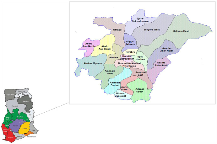
The study was conducted in the Ashanti region located in the middle belt of Ghana. The region is situated between 0.15-2.251W and 5.50-7.46N (Figure 1) surrounded by 5 of the 16 political regions of Ghana. It takes up 10.2% of the total land area of Ghana covering 24 389 km2. One hundred and thirty-seven (137) poultry farms all located in the Ashanti region, a public market (Kumasi Central Market), a town (Ayigya township), and 2 public hospitals were chosen for the study.
Figure 1.
Map of Ghana showing Ashanti region (study area) with detailed boundaries of all the districts.
A total of 900 samples were obtained from the various sources. Stool, urine, and blood from 364 patients were randomly sampled from the 2 hospitals as part of routine surveillance screening for public hospitals in the region. 18 Of 276 poultry litter samples were collected from 137 poultry farms located at least 10 000 miles from the selected hospitals and study community environments. One hundred and twenty-three (123) swabbed samples from community-based latrines, market floors and tables, soil, and sewage distributed within 20 miles of the selected hospitals were also collected for P. aeruginosa isolation.
Isolation and identification of P. aeruginosa
Bacteria in the various samples collected were revived in casein soya bean digest broth and isolated on Cetrimide agar. Preliminary identification was then conducted through Gram-staining, test for catalase and oxidase activity and growth at 42°C on nutrient agar. Production of pyocyanin, pyomelanin, pyorubin, and pyoverdine pigments were examined by culturing the isolates on Pseudomonas isolation agar (Alpha Biosciences, Maryland, USA). The presumptive Pseudomonas aeruginosa isolates were confirmed by amplification of the species-specific outer membrane lipoprotein oprL gene. Using the 0.6 µL of a 10 µM forward primer oprL-F (5′-ATG GAA ATG CTG AAA TTC GGC-3′) and 0.6 µL of a 10 µM reverse primer oprL-R (5′-CTT CTT CAG CTC GAC GCG ACG-3′), polymerase chain reaction was carried out using a thermal cycler (Gene Amp, ThermoFisher Scientific, Waltham, MA, USA USA) in a final volume of 25 µL containing 2 µL of DNA template extracted using the boiling lysis method, 12.5 µL of GoTaq master mix (Promega, Madison, USA), 0.75 µL of a 0.5 mM magnesium chloride, 8.55 µL of nuclease free water. For polymerse chain reaction (PCR) amplification, the DNA template was initially denatured at 94°C for 5 minutes, followed by 35 cycles of denaturation at 94°C for 30 seconds, annealing at 64°C for 30 seconds and extension at 72°C for 1 minute. Finally, the products were extended at 72°C for 10 minutes. The PCR products were examined on a 2% w/v agarose gel at 60 V and visualized using a transilluminator (Fotodyne, Hartland, WI, USA).
Antibiotic susceptibility testing
Susceptibility of P. aeruginosa isolates to the selected antipseudomonal antibiotics was determined by the Kirby-Bauer disk diffusion test according to approved methods of the European Committee on Antimicrobial Susceptibility Testing. 19 Eleven antibiotics from 6 classes including aztreonam (ATM-30 µg), imipenem (IPM-10 µg), meropenem (MEM-10 µg), ciprofloxacin (CIP-5 µg), gentamycin (CN-10 µg), levofloxacin (LEV-5 µg), and ticarcillin/clavulanic acid (TIM-85 µg) piperacillin (PIP-100 µg), ticarcillin (TIC-75 µg), ceftazidime (CAZ-30 µg), and cefepime (FEP-30 µg). All antibiotics used were purchased from Oxoid Ltd, Basingstoke, UK. Strains that were resistant to 3 or more antibiotics from any of the 6 classes were identified as multi-drug resistant. P. aeruginosa ATCC 27853 was used as quality control strain. The isolated strains were classified as susceptible or resistant to the antipseudomonals depending on the zone of inhibition diameters when compared to breakpoint values from the European Committee on Antimicrobial Susceptibility Testing. 19
Genotyping of P. aeruginosa isolates by Repetitive-element-based (Enterobacterial repetitive-intergenic consensus based—ERIC) PCR assay
DNA from bacteria was extracted using the boiling lysis method as described by Meacham et al. 20 Pure colonies of P. aeruginosa cultured on 20 mL nutrient agar were transferred into 25 µL of Tris-Ethylenediamine tetraacetic acid (TE) buffer. The suspension was heated at 95°C for 10 minutes to lyse the bacterial cells, cooled at −20°C for 5 minutes to shrink the cells to release the genetic material into the buffer. The cellular debris was then pelleted by centrifuging at 13 000 × g for 5 minutes. The supernatant was then stored at −20°C and used as the template for PCR amplification of the non-coding intergenic repetitive sequences. The non-coding intergenic repetitive sequences in the genome of P. aeruginosa were amplified using 10 µM ERIC1 (5′-ATG TAA GCT CCT GGG GAT TCA C-3′) and ERIC2 (5′-AAG TAA GTG ACT GGG GTG AGC G-3′) primers as described by Versalovic et al. 21 Using 2 µL of extracted DNA as template, PCR was performed in a final reaction volume of 25 µL containing 12.5 µL of Green Taq master mix, 0.75 µL of 0.5 mM magnesium chloride and 8.55 µL of nuclease free water. With an initial denaturation at 94°C for 5 minutes, the reaction continued with 30 cycles of denaturation at 94°C for 1 minute, annealing at 53°C for 1 minute and extended at 72°C for 4 minutes. The products were finally extended at 72°C for 10 minutes. About 5 µL of the amplicon was loaded into a 20 well 1.5% w/v agarose gel in 1X TAE (1 mM EDTA, 40 mM Tris-acetate) and run for 340 minutes at 65 V.
Extraction of plasmids in P. aeruginosa isolates (Alkaline lysis method)
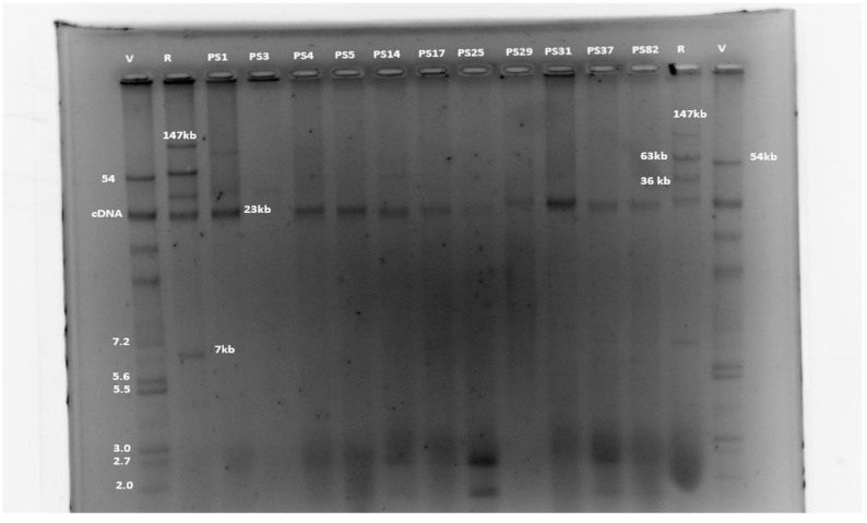
Extra-chromosomal genetic material (plasmids) which may carry antibiotic resistance genes to other bacteria genera were isolated from the P. aeruginosa isolates using alkaline lysis method as described by Kado and Liu. 22 E. coli control strains 39R and V517 with known plasmid sizes and P. aeruginosa isolates were cultured in 2 mL Luria-Bertani (LB) broth for 24 hours at 37°C. A 1.5 mL aliquot of both reference and P. aeruginosa culture grown in LB broth was pelleted by centrifugation at 13 400 × g for 3 minutes. The pellets were re-suspended by vortexing in 20 µL of 10mM:1mM Tris-ethylene diamine tetra acetic acid (TE) buffer; 100 µL of lysis buffer (1%w/v SDS, 2N NaOH) was then added and mixed by repetitive inversions of the 1.5 mL Eppendorf tube. The suspension was incubated at 56°C for 30 minutes in a dry bath. A mixture of 100 µL of phenol:chloroform:isoamylalcohol (25:24:1) was then added to the mixture and vortexed until it turned milky white. The mixture was centrifuged at 13 000 × g for 30 minutes to remove excess protein from the mixture. Forty microliters (40 µL) of the supernatant was transferred into a new Eppendorf tube containing 15 µL of loading dye and stored at 4°C until use. Twenty microliters (20 µL) of the plasmid-dye mix was loaded into a 0.8% w/v agarose gel wells and run at 60 V for 4 hours. The gel was stained in ethidium bromide (0.0002% w/v) and washed for 20 minutes in 1 L sterile distilled water. The plasmid sizes of the isolates were determined from calibration curves constructed from plasmid sizes of control E. coli V517 (54.0, 7.2, 5.6, 5.1, 4.4, 3.0, 2.7 and 2.0 kb) and E. coli R39 (147, 63, 36, and 7 kb) (Figure 4) obtained from Department of Veterinary Disease Biology, University of Copenhagen (KU).
Figure 4.
Electrophoretic gel image showing number and sizes of plasmids in MDR P. aeruginosa isolates.
Abbreviations: cDNA, chromosomal DNA; R 39 = E. coli control strain R39; V 517, E. coli control strain V517.
Analysis of data
Susceptibility data were compared by using Chi-square analysis with GraphPad Prism version 5.0 (Graph Pad Software, San Diego, CA, USA). A level of significance (P-value) <.05 were considered statistically significant. Clonal relatedness of the multidrug resistant P. aeruginosa strains from the various sources were determined by their antibiogram patterns and genomic fingerprint profiles using Gelj version 1.2 software. 23 The discriminatory index-D value, was calculated for each typing method using Simpson’s index of diversity(D). 17 Dendrograms depicting the strain relatedness were generated using Pearson coefficient as a similarity measure and unweighted pair group method with arithmetic averages (UPGMA) cluster analysis as a distance measure. Strains with a threshold linkage value of ⩾98% were assigned the same subtype. Two strains were assigned the same type if they showed identical banding pattern.
N = Total number of strains in the sample population, S = total number of types described
= Number of strains belonging to the jth type
Results
Antibiogram profiles of P. aeruginosa isolates
Based on the morphological, cultural, biochemical, and molecular characteristics of the isolates, a total of 87 (9.6%) P. aeruginosa strains were confirmed from the 900 samples collected. 24 Susceptibility of the P. aeruginosa isolates to all the antipseudomonal agents were in a range of 0% to 90%. Resistance ranged from 7% to 69% (Table 1). Twelve (12), 4, 1, and 21 MDR P. aeruginosa were isolated from the stool, urine, poultry litter, and environmental samples, respectively. Five isolates were resistant to all the antipseudomonal groups investigated. All the P. aeruginosa isolates showed high susceptibility to ceftazidime (90%), piperacillin (85%), meropenem (81.6%), imipenem (72.4%), ticarcillin/clavulanic acid (81.6%), cefepime (81.6%), and levofloxacin (72.4%). About 75% (74.7%) of the P. aeruginosa isolates demonstrated intermediate susceptibility to aztreonam.
Table 1.
Susceptibility profiles of P. aeruginosa isolates to various antipseudomonal agents.
| Antibiotic | Number of P. aeruginosa isolates | |||||||||
|---|---|---|---|---|---|---|---|---|---|---|
| Total isolates (N = 87) | Clinical | Environmental | Poultry litter | |||||||
| (N = 47) | (N = 35) | (N = 5) | ||||||||
| R | % | S | % | R | S | R | S | R | S | |
| CAZ | 9 | 10 | 78 | 90 | 5 | 42 | 3 | 32 | 1 | 4 |
| PIP | 13 | 15 | 74 | 85 | 9 | 38 | 3 | 32 | 1 | 4 |
| MEM | 6 | 7 | 71 | 82 | 3 | 40 | 2 | 30 | 1 | 1 |
| TIM | 16 | 18 | 71 | 82 | 7 | 40 | 5 | 30 | 4 | 1 |
| FEP | 11 | 13 | 71 | 82 | 5 | 42 | 5 | 25 | 1 | 4 |
| IPM | 8 | 9 | 63 | 72 | 5 | 28 | 2 | 31 | 1 | 4 |
| LEV | 17 | 20 | 61 | 70 | 12 | 30 | 5 | 26 | 0 | 5 |
| TIC | 49 | 56 | 38 | 44 | 21 | 26 | 24 | 11 | 4 | 1 |
| CIP | 54 | 62 | 27 | 31 | 30 | 13 | 22 | 11 | 2 | 3 |
| CN | 60 | 69 | 26 | 30 | 34 | 12 | 25 | 10 | 1 | 4 |
| ATM | 22 | 25 | 0 | 0 | 5 | 0 | 15 | 0 | 2 | 0 |
Abbreviations: ATM, Aztreonam; CAZ, Ceftazidime; CI, Ciprofloxacin; CN, Gentamycin; FEP, Cefepime; IPM, Imipinem; LEV, Levofloxacin; MEM, Meropenem; N, number of isolates; TIC, Ticarcillin; TIM, Ticarcillin/Clavulanic acid; PIP, Piperacillin; R, resistant; S, sensitive.
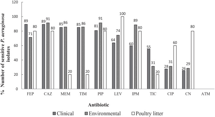
Resistance to aztreonam ranged from 11% to 40% in the clinical, environmental and poultry litter isolates. Gentamicin showed the least activity with 69% of the isolates being resistant. High resistance of the isolates was also observed against ciprofloxacin (62.1%) and ticarcillin (56.3%). The P. aeruginosa isolates showed the least resistance to meropenem (Table 1). Strains isolated from environmental and clinical sources showed high susceptibility to cefepime, ceftazidime, meropenem, piperacillin, ticarcillin/clavulanic acid, levofloxacin, and imipenem (Figure 2). All the poultry litter isolates were susceptible to levofloxacin. Five (5) strains were susceptible to all the antipseudomonal antibiotics. The most frequent pattern of resistance in the isolates were CIP-LEV-CN-TIC, CIP-CN-TIC, and CIP-LEV-CN (Table 2).
Figure 2.
Susceptibility pattern of P. aeruginosa isolates from various sources.
Abbreviations: ATM, Aztreonam 30 µg; CAZ, Ceftazidime 10 µg; CIP, Ciprofloxacin 5 µg; CN, Gentamycin 10 µg; FEP, Cefepime 30 µg; IPM, Imipinem 10 µg; LEV, Levofloxacin 5 µg; MEM, Meropenem 10 µg; PIP, Piperacillin 30 µg; TIC, Ticarcillin 75 µg; TIM, Ticarcillin/Clavulanic acid 85 µg.
Table 2.
Antibiotic resistance pattern of P. aeruginosa isolates from various sources.
| Resistance pattern | Number of isolates | Resistance pattern | Number of isolates |
|---|---|---|---|
| CN-TIC-ATM | 3 | CIP-MEM-IPM-CN-TIC-PIP-TIM-FEP-CAZ-ATM | 2 |
| TIC-TIM-ATM | 3 | CIP-LEV-MEM-IPM-CN-TIC-PIP-FEP-CAZ-ATM | 1 |
| CIP-TIC-CAZ | 3 | CIP-MEM-IPM-CN-TIC-PIP-FEP-ATM | 1 |
| CIP-TIC-TIM | 3 | CIP-CN-TIC-PIP-TIM-FEP-ATM | 1 |
| CN-TIC-CAZ | 3 | CIP-LEV-CN-TIC-PIP-TIM-ATM | 1 |
| CN-TIC-TIM | 3 | CIP-LEV-CN-TIC-PIP-TIM-CAZ | 1 |
| CIP-CN | 2 | CIP-LEV-CN-TIC-TIM-CAZ-ATM | 1 |
| CIP-TIC | 2 | CIP-CN-TIC-FEP-CAZ-ATM | 1 |
| CN-TIC | 2 | CIP-CN-TIC-PIP-TIM-FEP | 1 |
| TIC-ATM | 2 | CIP-LEV-CN-TIC-PIP-FEP | 1 |
| CIP-LEV | 2 | MEM-IPM-TIC-PIP-TIM-ATM | 1 |
| CIP-PIP | 2 | CIP-CN-TIC-FEP-ATM | 1 |
| CN-PIP | 2 | CIP-LEV-IPM-CN-TIC | 1 |
| MEM-CN | 2 | CIP-LEV-CN-TIC | 6 |
| TIC-PIP | 2 | CIP-CN-TIC-FEP | 1 |
| TIC-TIM | 2 | CIP-CN-TIC-TIM | 1 |
| CN | 1 | CIP-IPM-CN-TIM | 1 |
| CIP | 1 | CIP-LEV-CN-CAZ | 1 |
| TIC | 1 | CIP-TIC-CAZ-ATM | 1 |
| ATM | 1 | CN-TIC-TIM-ATM | 1 |
| IPM | 1 | CIP-CN-TIC | 6 |
| CIP-CN-ATM | 3 | CIP-LEV-CN | 4 |
| Number of strains susceptible to all the antipseudomonal antibiotics | 5 | ||
Abbreviations: ATM, Aztreonam 30 µg; CAZ, Ceftazidime 10 µg; CIP, Ciprofloxacin 5 µg; CN, Gentamycin 10 µg; FEP, Cefepime 30 µg; IPM, Imipinem 10 µg; LEV, Levofloxacin 5 µg; MEM, Meropenem 10 µg; PIP, Piperacillin 30 µg; TIC, Ticarcillin 75 µg; TIM, Ticarcillin/Clavulanic acid 85 µg.
There was no significant difference in the in vitro activity within the carbapenem group (meropenem and imipenem; P = .47) and cephalosporin group (ceftazidime and cefepime; P = .51) of antibiotics. The difference in sensitivity within the penicillin group (piperacillin, ticarcillin, ticarcillin/clavulanic acid) was significant (P < .05). Piperacillin and ticarcillin/clavulanic acid were active in most strains of P. aeruginosa compared to ticarcillin. Among the quinolone group, levofloxacin exerted greater activity than ciprofloxacin (P < .05).
Co-resistance and cross-resistance of P. aeruginosa isolates
All the P. aeruginosa isolates that showed resistance to levofloxacin were also resistant to ciprofloxacin. The levels of levofloxacin-gentamicin and ciprofloxacin-gentamycin co-resistance were 94% and 81%, respectively (Table 3). Majority of meropenem and imipenem resistant isolates (63%-83%) demonstrated co-resistance to ciprofloxacin, gentamicin, ticarcillin, piperacillin, and aztreonam but remained susceptible to levofloxacin. β-lactam resistant strains also exhibited co-resistance to ciprofloxacin and gentamicin. Quinolone (levofloxacin and ciprofloxacin) resistant isolates exhibited high susceptibility (88%-96%) to the carbapenem group of β-lactams. Carbapenems also showed high activity in strains that were resistant to gentamicin and other β-lactam antipseudomonal groups.
Table 3.
Co-resistance and cross-resistance in P. aeruginosa isolates.
| N | Number of resistant isolates (%) | |||||||||||
|---|---|---|---|---|---|---|---|---|---|---|---|---|
| CIP | LEV | MEM | IPM | CN | TIC | PIP | TIM | FEP | CAZ | ATM | ||
| CIP | 54 | - | 17 (31) | 4 (7) | 6 (11) | 44 (81)* | 33 (61) | 9 (17) | 10 (19) | 10 (19) | 9 (17) | 13 (24) |
| LEV | 17 | 17 (100) | - | 1 (6) | 2 (12) | 16 (94)* | 11 (65) | 3 (18) | 2 (12) | 2 (12) | 4 (24) | 2 (12) |
| MEM | 6 | 4 (67)* | 1 (17) | - | 5 (83)* | 5 (83)* | 5 (83)* | 5 (83)* | 3 (50) | 4 (67)* | 3 (50) | 5 (83)* |
| IPM | 8 | 6 (75)* | 2 (25) | 5 (63)* | - | 6 (75)* | 6 (75)* | 5 (63)* | 4 (50) | 4 (50) | 3 (38) | 5 (63)* |
| CN | 60 | 44 (73)* | 16 (27) | 5 (8) | 6 (10) | - | 34 (57)* | 9 (15) | 11 (18) | 10 (17) | 8 (13) | 14 (23) |
| TIC | 49 | 33 (67)* | 11 (22) | 5 (10) | 6 (12) | 6 (12) | - | 10 (20) | 15 (31) | 10 (20) | 9 (18) | 18 (37) |
| PIP | 13 | 9 (69)* | 3 (23) | 5 (38) | 5 (38) | 9 (69)* | 10 (77)* | - | 6 (46) | 7 (54)* | 4 (31) | 6 (46) |
| TIM | 16 | 10 (63)* | 2 (13) | 3 (19) | 4 (25) | 11 (69) | 15 (94)* | 6 (38) | - | 4 (25) | 4 (25) | 9 (56)* |
| FEP | 11 | 10 (91)* | 2 (19) | 4 (36) | 4 (36) | 10 (91)* | 10 (91)* | 7 (64)* | 4 (36) | - | 4 (36) | 7 (64)* |
| CAZ | 9 | 9 (100)* | 4 (44) | 3 (33) | 3 (33) | 8 (89)* | 9 (100)* | 4 (44) | 4 (44) | 4 (44) | - | 6 (67)* |
| ATM | 22 | 13 (59)* | 2 (9) | 5 (23) | 5 (23) | 14 (64)* | 18 (82)* | 6 (27) | 9 (41) | 7 (32) | 6 (27) | - |
Abbreviations: ATM, Aztreonam 30 µg; CAZ, Ceftazidime 10 µg; CIP, Ciprofloxacin 5 µg; CN, Gentamycin 10 µg; FEP, Cefepime 30 µg; IPM, Imipinem 10 µg; LEV, Levofloxacin 5 µg; MEM, Meropenem 10 µg; N, number of resistant isolates; PIP, Piperacillin 30 µg; TIC, Ticarcillin 75 µg; TIM, Ticarcillin/Clavulanic acid 85 µg; -, not applicable.
High cross resistance.
Clonal relationship of MDR P. aeruginosa isolates
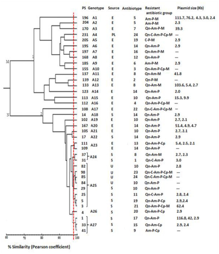
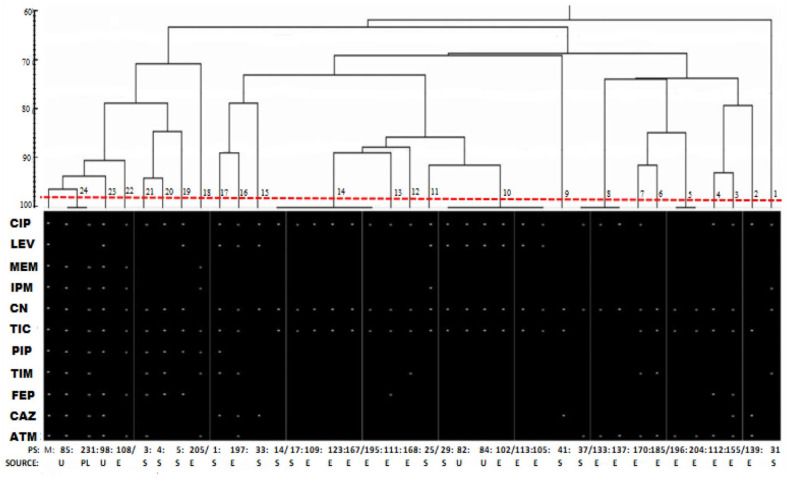
The genetic relatedness among the P. aeruginosa isolates was assessed based on an electrophoretic fingerprint pattern of the genome of the various MDR strains by amplification of conserved repeat regions (ERIC-PCR) of the bacteria genome. Two (2) to 9 bands with molecular weight ranging from 161 to 850 bp was observed. Two distinct clusters (M and N) were formed by the 38 MDR strains (Figure 5). Cluster M further differentiated into 2 sub-clusters (M1 and M2). The 21 environmental MDR P. aeruginosa isolates were distributed among clusters M and N. The poultry litter isolate also shared similarity with environmental strains from cluster M. Cluster N comprised all the clinical MDR isolates (16) and 28.5% (6/21) of the environmental isolates. At a similarity of 98% (Pearson coefficient), a total of 27 subtypes (A1-A27) were generated (D = 0.9559) comprising 23 distinct subtypes and 4 subtype groups. A total of 24 antibiotypes of P. aeruginosa were discovered from the 38 MDR isolates with 5 clusters and 19 distinct strains (Figure 3; D = 0.9502). Two (2) distinct clones from stool and urine samples of 4 different patients were identified genotypically, clone I (PS84 and PS29) and clone II (PS98 and PS85).
Figure 5.
Cluster analysis of enterobacterial repetitive intergenic consensus PCR (ERIC-PCR) fingerprinting of 38 multidrug resistant P. aeruginosa isolates generated by Gelj v.1.2 with corresponding antimicrobial susceptibility patterns and plasmid profile.
Abbreviations: Am, aminoglycoside; C, carbapenem; Cp, cephalosporin; E, environment; M, Cluster I; N, Cluster II; P, penicillin; PS, Pseudomonas aeruginosa isolate; Qn, quinolone; S, stool; U, urine.
Figure 3.
Dendrogram derived from antipseudomonal resistance profiles using Gelj ver.1.2 with Dice coefficient and UPGMA.
Abbreviations: E, environment; M, antibiotic resistance marker strain; PS 1-231, Pseudomonas aeruginosa isolate; PL, poultry litter; S, stool; U, urine.
Plasmid profile
Agarose gel electrophoresis of the plasmid DNA revealed that 25 of the MDR P. aeruginosa strains harbored 1 to 5 plasmids with sizes ranging from 2 to 116.8 kb (Figure 4). Thirteen (13) of the isolates had no plasmids. Nearly 35% had 1 plasmid, 18.4% had 2 plasmids, and 10.5% had 3 plasmids and only 1 isolate habored 5 plasmids.
Discussion
We compared the genotypes, plasmid profile and antimicrobial susceptibility patterns of P. aeruginosa isolated from the environment, poultry farms, and clinical samples of patients from 2 district hospitals in the Ashanti region of Ghana. Our results showed that, the most active agents from 6 antipseudomonal classes were Ceftazidime (90%), Piperacillin (85%), Meropenem, Cefipeme, and Ticarcillin/Clavulanic acid (81.6%). This suggests that these antipseudomonals remain effective in the management of P. aeruginosa infections. The high activity of these antibiotics against P. aeruginosa may be due to the infrequent use of these antibiotics both in agriculture, community, and clinical settings in the region. These antibiotics cannot be obtained without prescriptions; hence their general frequency of use is low in the population. Equally, high susceptibility of clinical P. aeruginosa isolates to meropenem and ceftazidime confirms the reports of Feglo and Opoku. 25
Seventy-five percent (75%, n = 87) of the P. aeruginosa isolates were resistant to more than a single antipseudomonal agent. Gentamycin (69%), Ciprofloxacin (62.1%), Ticarcillin (56.3%), and aztreonam (25%) showed the highest rate of resistance. These findings were relatable to high resistance rates of P. aeruginosa to ticarcillin and aztreonam reported in Brazil by Pitondo-Silva et al 26 Similarly, high ciprofloxacin and gentamicin resistance has been reported in Gram-negative isolates from the southern sector of Ghana. The high ciprofloxacin and gentamicin resistance in the current study is however inconsistent with previous antibiogram reports by Feglo and Opoku 25 and Addo, 27 from other hospitals in the Ashanti and Greater Accra regions of Ghana. This may be so because of the increased use of veterinary medicines containing aminoglycoside and quinolone derivatives. 14 Also, a general increase in the accessibility of consumers to these antibiotics in community pharmacies and hospitals due to frequent prescribing of these antibiotics may account for the change in resistance profiles of P. aeruginosa to these antibiotics. This finding therefore suggests the ineffectiveness of these antibiotics in treatment of P. aeruginosa infections.
Nearly half (43.6%) of the isolates were multi-drug resistant (resistant to antibiotics from ⩾3 antipseudomonal groups). These findings compared to a related study by Addo 27 who reported 13.04% MDR from wounds of patients indicate variations in the prevalence of MDR P. aeruginosa strains within the region. This may be due to increased antibiotic resistance selection pressure in varied areas within the region. High quinolone-aminoglycoside (ciprofloxacin-gentamicin) cross-resistance was also observed in the current study. Carbapenem resistant isolates of P. aeruginosa were found to show cross-resistance to antibiotics from multiple classes including ciprofloxacin, gentamicin, ticarcillin, piperacillin, and aztreonam. These findings are congruent with a muticentric study on P. aeruginosa isolates carried out in 136 hospitals in Spain and Iran.28,29 Using the method by Kado and Liu for plasmid profile analysis, plasmids were detected in 65% (n = 38) of the MDR strains with sizes ranging from 2.0 to 116.8 kb. This is the first report evaluating the occurrence of plasmid in P. aeruginosa strains from the region.
The unique antibiogram profiles, plasmid distribution and cross resistance profiles of the MDR strains suggests a combination of multiple unrelated resistance mechanisms among the isolates. Also, significant differences in the antipseudomonal activity of antibiotics from the same class (quinolone and penicillin groups) implies regulation of resistance by different mechanisms.30,31
P. aeruginosa isolated from the 3 sampling sites belonged to 3 sub-clusters at 78% similarity. This indicates some genetic relatedness between the P. aeruginosa strains from stool, urine, and environmental samples. ERIC-PCR cluster analysis revealed considerable heterogeneity among these P. aeruginosa strains (Figure 5) which suggests that most of the isolates originated from different sources rather than from a single source and being disseminated among the study environments. We could find a total of 27 clonal patterns from 38 MDR strains. This indicates that P. aeruginosa is capable of rapid changes or variations which is in good agreement with the high plasticity and complexity of the large P. aeruginosa genome to reflect its evolutionary adaptations. 32
Interestingly, all the clinical isolates were genotypically distributed to the same cluster indicating a close genetic relationship between them. Some environmental strains showed some similarity with clinical strains suggesting possible exchange of resistant bacteria between the patients and the environment. Two clonal strains of P. aeruginosa (PS84 from urine and PS29 from stool) which showed >98% genetic similarity, had the same antibiogram and plasmid profile suggesting clonality and possible transfer between the 2 patients. A clone (PS98 and PS85) of 2 strains isolated from urine samples of 2 different patients who visited the same hospital showed resistance to all the 6 antipseudomonal groups. This finding is particularly alarming considering the possible dissemination of these strains in the region.
Conclusion
In the present study, MDR P. aeruginosa was isolated from clinical, environmental and poultry litter samples and characterized based on their antibiograms, plasmid profiles and genotypic relatedness. There was low prevalence (9.6%) of P. aeruginosa in the clinical, environmental, and poultry litter samples from the Ashanti Region of Ghana. There is however, an appreciable surge in the number of MDR P. aeruginosa strains in the clinical and environmental samples. Ceftazidime, piperacillin, meropenem, imipenem, ticarcillin/clavulanic acid, cefepime, and levofloxacin remain highly active against P. aeruginosa while gentamicin, ciprofloxacin, and ticarcillin are less effective. There is high cross-resistance within the quinolone group as well as co-resistance among ciprofloxacin, gentamicin, ticarcillin, piperacillin, aztreonam, and levofloxacin. Plasmids which may confer increased antipseudomonal resistance were detected in 65% of the MDR strains with sizes ranging from 2.0 to 116.8 kb. Five isolates were resistant to all the antipseudomonal groups while 2 clonal strains of P. aeruginosa were identified among the MDR strains. It is therefore necessary to regularize routine surveillance and mandatory screening for antimicrobial resistance in pathogenic bacteria associated with nosocomial and community acquired infections in the Ashanti region of Ghana.
Acknowledgments
We are grateful to the officials and management of the various hospitals and clinics, poultry farmers, and farm workers within Ashanti region of Ghana for their cooperation and assistance during the conduct of this study.
Footnotes
Funding: The author(s) received no financial support for the research, authorship, and/or publication of this article.
Declaration of Conflicting Interests: The author(s) declared no potential conflicts of interest with respect to the research, authorship, and/or publication of this article.
Author Contributions: HO performed the experimental work and processed the experimental data. VEB supervised the study and drafted the first manuscript. CD analyzed the data and designed figures and tables. YDB provided analysis of the data and a revision of the first manuscript. CA conceived and designed the project; coordinated the drafting and review of manuscript. All authors read and approved the final manuscript.
Ethical Clearance/Approval: Ethical clearance for the study was obtained from the Committee on Human Research Publications and Ethics (CHRPE), Kwame Nkrumah University of Science and Technology (KNUST), Kumasi. In addition, written consent was obtained from farm participants and patients.
ORCID iD: Yaw Duah Boakye  https://orcid.org/0000-0002-2134-0034
References
- 1. Aslam B, Wang W, Arshad MI, et al. Antibiotic resistance: a rundown of a global crisis. Infect Drug Resist. 2018;11:1645-1658. [DOI] [PMC free article] [PubMed] [Google Scholar]
- 2. World Health Organization. Antimicrobial resistance: global report on surveillance. WHO; 2014:3. Accessed June 15, 2016. http://apps.who.int/iris/bitstream/10665/112642/1/9789241564748_eng.pdf [Google Scholar]
- 3. Centre for Disease Dynamics Economics and Policy. State of the World’s Antibiotics. CDDEP; 2015:1-66. [Google Scholar]
- 4. Boamah VE, Agyare C, Odoi H, Dalsgaard A. Antibiotic practices and factors influencing the use of antibiotics in selected poultry farms in Ghana. J Antimicrob Agents. 2016;2:1-6. [Google Scholar]
- 5. Agoba EE. Antibiotic Use in Selected Fish Farms in the Ashanti Region Ghana. Master of Philosophy thesis submitted to the Department of Pharmaceutics Kwame Nkrumah University of Science and Technology; 2015:1-45. [Google Scholar]
- 6. von Wintersdorff CJ, Penders J, van Niekerk JM, et al. Dissemination of antimicrobial resistance in microbial ecosystems through horizontal gene transfer. Front Microbiol. 2016;7:173. [DOI] [PMC free article] [PubMed] [Google Scholar]
- 7. Davies J, Spiegelman GB, Yim G. The world of sub-inhibitory antibiotic concentrations. Curr Opin Microbiol. 2006;9:445-453. [DOI] [PubMed] [Google Scholar]
- 8. Bengtsson-Palme J, Kristiansson E, Larsson DGJ. Environmental factors influencing the development and spread of antibiotic resistance. FEMS Microbiol Rev. 2018;42:68-80. [DOI] [PMC free article] [PubMed] [Google Scholar]
- 9. Streeter K, Katouli M. Pseudomonas aeruginosa: a review of their pathogenesis and prevalence in clinical settings and the environment. Infect Epidemiol Med. 2016;2:25-32. [Google Scholar]
- 10. Krieg NR, Holt JG. Bergey’s Manual of Systematic Bacteriology. Vol. 2. Williams and Wilkins Publishers; 2005:25-250. [Google Scholar]
- 11. Mahmmudi Z, Gorzin AA. Biofilm of Pseudomonas aeruginosa in nosocomial infection. J Mol Biol Res. 2017;7:29. [Google Scholar]
- 12. Carmeli Y, Troillet N, Eliopoulos GM, Samore MH. Emergence of antibiotic-resistant Pseudomonas aeruginosa: comparison of risks associated with different antipseudomonal agents. Antimicrob Agents Chemother. 1999;43:1379-1382. [DOI] [PMC free article] [PubMed] [Google Scholar]
- 13. Dodd CER. Pseudomonas: introduction. In: Batt C, Patel P, eds. Encyclopedia of Food Microbiology. 2nd ed. Elsevier; 2014;244-247. [Google Scholar]
- 14. Pattison M, McMullin PF, Bradbury JM, Alexander DJ. Poultry Diseases. 6th ed. Saunders Elsevier; 2008:160-163. [Google Scholar]
- 15. Mesaros N, Nordmann P, Plésiat P, et al. Pseudomonas aeruginosa: resistance and therapeutic options at the turn of the new millennium. Clin Microbiol Infect. 2007;13:560-578. [DOI] [PubMed] [Google Scholar]
- 16. Harrison F, Buckling A. High relatedness selects against hypermutability in bacterial metapopulations. Proc R Soc B Biol Sci. 2007;274:1341-1347. [DOI] [PMC free article] [PubMed] [Google Scholar]
- 17. Achtman M. A phylogenetic perspective on molecular epidemiology. In: Tang YW, Sussman M, Liu D, et al., eds. Molecular Medical Microbiology. Academic Press; 2002;486-502. [Google Scholar]
- 18. Odoi H. Isolation and Characterization of Multi-Drug Resistant Pseudomonas aeruginosa From Clinical Environmental and Poultry Litter Sources in Ashanti Region of Ghana. Master of Philosophy thesis Kwame Nkrumah University of Science and Technology. KNUSTSpace; 2017. Accessed May 27, 2021. http://dspace.knust.edu.gh/handle/123456789/10233 [Google Scholar]
- 19. EUCAST. The European Committee on Antimicrobial Susceptibility Testing; 2015. www.eucast.org. Accessed March 9, 2015.
- 20. Meacham KJ, Zhang L, Foxman B, Bauer RJ, Marrs CF. Evaluation of genotyping large numbers of Escherichia coli isolates by Enterobacterial repetitive intergenic consensus-PCR. J Clin Microbiol. 2003;41:5224-5226. [DOI] [PMC free article] [PubMed] [Google Scholar]
- 21. Versalovic J, Koeuth T, Lupski JR. Distribution of repetitive DNA sequences in eubacteria and application to fingerprinting of bacterial genomes. Nucleic Acids Res. 1991;19:6823-6831. [DOI] [PMC free article] [PubMed] [Google Scholar]
- 22. Kado CI, Liu ST. Rapid procedure for detection and isolation of large and small plasmids. J Bacteriol. 1981;145:1365-1373. [DOI] [PMC free article] [PubMed] [Google Scholar]
- 23. Heras J, Domínguez C, Mata E, et al. GelJ – a tool for analyzing DNA fingerprint gel images. BMC Bioinformatics. 2015;16:270. [DOI] [PMC free article] [PubMed] [Google Scholar]
- 24. Odoi H, Boamah VE, Boakye YD, Agyare C. Prevalence and phenotypic and genotypic resistance mechanisms of multidrug-resistant Pseudomonas aeruginosa strains isolated from clinical, environmental, and poultry litter samples from the Ashanti region of Ghana. J Environ Public Health. 2021;2021:1-12. [DOI] [PMC free article] [PubMed] [Google Scholar]
- 25. Feglo P, Opoku S. AmpC beta-lactamase production among Pseudomonas aeruginosa and Proteus mirabilis isolates at the Komfo Anokye teaching Hospital Kumasi Ghana. J Microbiol Antimicrob. 2014;6:13-20. [Google Scholar]
- 26. Pitondo-Silva A, Martins VV, Fernandes AFT, Stehling EG. High level of resistance to aztreonam and ticarcillin in Pseudomonas aeruginosa isolated from soil of different crops in Brazil. Sci Total Environ. 2014;473-474:155-158. [DOI] [PubMed] [Google Scholar]
- 27. Addo FA. Characterization of Antimicrobial Resistant Pseudomonas spp. and Other Bacteria From Infected Wound. Master of Philosophy thesis submitted to the University of Ghana Legon; 2015:1-72. [Google Scholar]
- 28. Japoni A, Alborzi A, Kalani M, Nasiri J, Hayati M, Farshad S. Susceptibility patterns and cross-resistance of antibiotics against Pseudomonas aeruginosa isolated from burn patients in the south of Iran. Burns. 2006;32:343-347. [DOI] [PubMed] [Google Scholar]
- 29. Bouza E, Garcia-Garrote F, Cercenado E, Marin M, Diaz MS. Pseudomonas aeruginosa: a survey of resistance in 136 hospitals in Spain. The Spanish Pseudomonas aeruginosa Study Group. Antimicrob Agents Chemother. 1999;43:981-982. [DOI] [PMC free article] [PubMed] [Google Scholar]
- 30. Douglas L, Mayers Jack D, Sobel MARC, Ouellette Keith S, Kaye DM. Antimicrobial Drug Resistance, Mechanism of Drug Resistance. Vol. 1. Springer International Publishing; 2017:745-749. [Google Scholar]
- 31. Sefton AM. Mechanisms of antimicrobial resistance their clinical relevance in the new millennium. Drugs. 2002;62:557-566. [DOI] [PubMed] [Google Scholar]
- 32. Cristina N, Eusébio L. Identification and Genotyping of Pseudomonas aeruginosa in Bronchiectasis. Thesis submitted to the Institute of Molecular Pathology and Immunology of University of Porto; 2013:1-83. [Google Scholar]


