

Abstract
A 64-year-old Thai woman underwent coronary artery bypass grafting (CABG) using saphenous vein grafts (SVG) for completely occluded left anterior descending artery (LAD) and mitral valve replacement with mechanical valve about a year ago. She presented with unstable angina. Three-dimensional computed tomography angiography (3DCTA) showed occlusion of all the grafts. The left subclavian artery had 99% stenosis. The patient underwent redo CABG via a minimally invasive direct approach. The chest was entered through the left fifth intercostal space. The right gastroepiploic artery (RGEA) and a small length of SVG were harvested. The RGEA was extended using the SVG with an end-to-end anastomosis and used to graft the LAD without cardiopulmonary bypass. The patient’s postoperative course was uneventful. Postoperative 3DCTA revealed patent RGEA-SVG graft. Minimally invasive direct coronary artery bypass to LAD with RGEA is a useful alternative approach for redo CABG in patients with not much choice of conduits.
Keywords: Minimally invasive coronary artery bypass grafting, Right gastroepiploic artery, Left anterior descending artery, Redo surgery, Poor conduits
Introduction
Redo coronary artery bypass grafting (CABG) can be a very difficult and challenging situation for the cardiac surgeons [1]. Compared to primary CABG, redo cases are associated with a much higher morbidity and mortality. The risk becomes much higher if they have had mitral valve replacement done earlier. Keeping all these issues in mind a minimally invasive direct approach was found to be very appropriate. Moreover, heart rotation, comparable to the median sternotomy, can be achieved while approaching the left anterior descending (LAD) coronary artery. Even though the operating space is limited, but it is usually sufficient to anastomose the right gastroepiploic artery (RGEA) in situ to the LAD. Some groups have reported good long-term patency rates with RGEA [2]. When other conduits cannot be used, or are not available, RGEA is a good alternative conduit for LAD lesion. Herein, we report a redo CABG patient who underwent minimally invasive direct coronary artery bypass (MIDCAB) using RGEA.
Case report
A 64-year-old woman underwent CABG using saphenous vein graft (SVG) for totally occluded LAD and obtuse marginal artery, along with mitral valve replacement for rheumatic mitral stenosis using a mechanical valve about a year ago. She presented with recurrence of unstable angina. A three-dimensional computed tomography angiography (3DCTA) showed occlusion of all the grafts. The branches of circumflex artery were small and not graftable.
The left subclavian artery had 99% stenosis at its origin from the arch. Laboratory evaluation revealed that her serum creatinine level was elevated at 1.8 mg/dL. Despite optimal medical treatment after admission, her angina could not be controlled. She required a redo CABG. The previous operating records mentioned that saphenous veins (SV) were harvested from both the thighs and they were of very poor quality.
The anesthesiologist intubated using a double-lumen endotracheal tube, in order to give the surgeons one-lung ventilation during the operation. The patient was placed in a right semi-lateral position. The chest was entered through the fifth left intercostal space. There were no adhesions between the pericardium and the left lung. A pericardiotomy was made and the LAD was inspected, which was found to be graftable (Fig. 1).
Fig. 1.
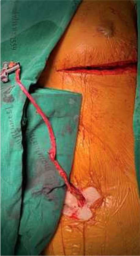
The right gastroepiploic artery graft was extended using a short segment of saphenous vein with an end-to-end anastomosis
A 15-cm midline epigastric incision, just below the xiphoid process, was made to open the abdomen and the RGEA was harvested using a harmonic scalpel (Harmonic Scalpel; Ethicon Endo-Surgery, Cincinnati, OH). Simultaneously, a short segment of the long SV was harvested from the right lower limb. The RGEA measured about 3 mm in diameter while the saphenous vein was about 3.5 mm in diameter. The SV segment was anastomosed to the RGEA. The RGEA-SVG was then introduced into the pericardial space by tunneling through the diaphragm and pericardium. A suitable area in the LAD was then stabilized using the octopus evolution tissue stabilizer (Medtronic, Watford, UK). The RGEA-SVG was then anastomosed to the LAD (Fig. 2).
Fig. 2.

In situ right gastroepiploic artery extended by the saphenous vein that was anastomosed to the left anterior descending artery with an end-to-side anastomosis without cardiopulmonary bypass
The patient’s postoperative course was uneventful. Postoperative 3DCTA demonstrated a patent RGEA-SVG graft (Fig. 3).
Fig. 3.

Postoperative three-dimensional computed tomography angiography showing a patent graft
Discussion
We performed MIDCAB to LAD using the RGEA, extended using a short segment of SV, for redo CABG. The left internal mammary artery (LIMA) could not be used because the left subclavian was severely stenosed. Preoperative computed tomography (CT) scan showed the heart to be quite closely adherent to the sternum. Harvesting the right internal mammary artery (RIMA) would have been difficult because of previous sternotomy. This patient also had mild chronic kidney disease. The right radial artery (RA) had to be spared for creation of an arterio-venous fistula for haemodialysis, should it be needed in future. The SVs had already been harvested from both the thighs for the first surgery. Overall, this patient had poor conduits for LAD revascularization.
Increased risk of mortality in redo CABG has been reported. Christenson et al. suggested that in redo CABGs with unstable angina, impaired preoperative left ventricular function, renal insufficiency, insulin-dependent diabetes, and an interval shorter than 1 year from the initial operation were independent risk factors for early mortality [1]. This patient had 3 out of 5 reported risk factors.
Suma recommended the use of RGEA for LAD, when LIMA was not available [2]. His group reported long-term patency of the RGEA for 10 years in another report [3]. They concluded that RGEA occlusion occurred when target vessel had mild stenosis. Suzuki et al. reported excellent patency rates of 97.8%, 94.7%, and 90.2% early postoperatively and at 5 and 8 years postoperatively, respectively [4].
Akita et al. examined 517 patients who underwent CABG with an in situ semi-skeletonized RGEA. The cumulative patency rate of the RGEA was 79.3% at 10 years. A multivariable analysis showed that an minimum lumen diameter (MLD) (hazard ratio 4.43, 95% confidence interval 3.25–6.82; P < 0.001) was an independent risk factor of RGEA occlusion. A time-dependent receiver operating characteristic (ROC) curve analysis identified that an MLD < 1 mm was set as the cutoff value for graft occlusion [5]. Patients with an MLD > 1 mm had a 10-year patency rate of 89.8%.
However, some meta-analyses reported inferior results compared with other conduits [6]. Suma et al. speculated that this result was due to the less experience in using RGEA in redo coronary artery surgery. At our institute, the RGEA is commonly used as the third arterial graft. This patient’s LAD was totally occluded and so was a good candidate for RGEA [2].
No study has reported the long-term patency rates of end-to-end composite graft using RGEA with SV. Kamiya et al. reported the results of end-to-end composite graft with RGEA and RA. The early patency rate of the gastroepiploic arterial composite graft was 98.3% [7]. However, evidence regarding the long-term patency of composite grafts with RGEA has not been reported.
Some reports described the usefulness of the MIDCAB approach for redo cases. Voutilainen et al. reported that among 25 patients who underwent MIDCAB with RGEA, one underwent redo procedure and five underwent RGEA extended with SVG. The early patency of all RGEA without competitive flow was 95% [8].
Nakagawa et al. reported 4 redo CABG performed with MIDCAB and RGEA. However, the target vessel was the right coronary artery (RCA) in all 4 cases. All cases were done without cardiopulmonary bypass and blood transfusion. The patency of grafts was confirmed by a postoperative angiogram. They suggested MIDCAB for the RCA, employing the RGEA via a subxiphoid incision, provides excellent revascularization in redo CABG cases [9].
Nishi et al. reported 19 redo CABG cases. Median sternotomy (on-pump cardiac arrest) was performed on 13 patients with occluded graft to LAD. They attempted to avoid median sternotomy when patients had patent graft to the LAD. Six patients were operated via left thoracotomy [10].
Conclusion
In patients with paucity of conduits, RGEA comes as a life-saving conduit to graft the LAD. Redo CABG with MIDCAB approach prevents the risk of re-sternotomy and even left ventricle rupture, in patients who have had their mitral valve replaced along with CABG earlier. MIDCAB to LAD using RGEA-SV composite graft is a useful alternative approach to redo CABG in patients with poor conduits.
Funding
None.
Declarations
Ethics approval
Not applicable.
Human and animal rights statement
This article does not contain any studies involving animals performed by any of the authors. All procedures performed in studies involving human participants were in accordance with the ethical standards of the institutional and/or national research committee and with the 1964 Helsinki Declaration and its later amendments or comparable ethical standards.
Informed consent
Obtained from all individual participants involved in the study.
Conflict of interest
The authors declare no competing interests.
Footnotes
Publisher's note
Springer Nature remains neutral with regard to jurisdictional claims in published maps and institutional affiliations.
References
- 1.Christenson JT, Schmuziger M, Simonet F. Reoperative coronary artery bypass procedures: risk factors for early mortality and late survival. Eur J Cardiothorac Surg. 1997;11:129–133. doi: 10.1016/s1010-7940(96)01030-5. [DOI] [PubMed] [Google Scholar]
- 2.Suma H. The right gastroepiploic artery graft for coronary artery bypass grafting: a 30-year experience. Korean J Thorac Cardiovasc Surg. 2016;49:225–231. doi: 10.5090/kjtcs.2016.49.4.225. [DOI] [PMC free article] [PubMed] [Google Scholar]
- 3.Suma H, Tanabe H, Takahashi A, et al. Twenty years experience with the gastroepiploic artery graft for CABG. Circulation. 2007;116:I188–I191. doi: 10.1161/CIRCULATIONAHA.106.678813. [DOI] [PubMed] [Google Scholar]
- 4.Suzuki T, Asai T, Nota H, et al. Early and long-term patency of in situ skeletonized gastroepiploic artery after off-pump coronary artery bypass graft surgery. Ann Thorac Surg. 2013;96:90–95. doi: 10.1016/j.athoracsur.2013.04.018. [DOI] [PubMed] [Google Scholar]
- 5.Akita S, Tajima K, Kato W, et al. The long-term patency of a gastroepiploic artery bypass graft deployed in a semiskeletonized fashion: predictors of patency. Interact Cardiovasc Thorac Surg. 2019;28:868–875. doi: 10.1093/icvts/ivy346. [DOI] [PubMed] [Google Scholar]
- 6.Benedetto U, Raja SG, Albanese A, Amrani M, Biondi-Zoccai G, Frati G. Searching for the second best graft for coronary artery bypass surgery: a network meta-analysis of randomized controlled trials. Eur J Cardiothorac Surg. 2015;47:59–65. doi: 10.1093/ejcts/ezu111. [DOI] [PubMed] [Google Scholar]
- 7.Kamiya H, Watanabe G, Takemura H, Tomita S, Nagamine H, Kanamori T. Total arterial revascularization with composite skeletonized gastroepiploic artery graft in off-pump coronary artery bypass grafting. J Thorac Cardiovasc Surg. 2004;127:1151–1157. doi: 10.1016/j.jtcvs.2003.09.057. [DOI] [PubMed] [Google Scholar]
- 8.Voutilainen S, Verkkala K, Järvinen A, et al. Minimally invasive coronary artery bypass grafting using the right gastroepiploic artery. Ann Thorac Surg. 1998;65:444–448. doi: 10.1016/S0003-4975(97)01129-6. [DOI] [PubMed] [Google Scholar]
- 9.Nakagawa H, Nabuchi A, Terada H, et al. Minimally invasive direct coronary artery bypass surgery with right gastroepiploic artery for redo patients. Ann Thorac Cardiovasc Surg. 2015;21:378–381. doi: 10.5761/atcs.oa.14-00286. [DOI] [PMC free article] [PubMed] [Google Scholar]
- 10.Nishi H, Mitsuno M, Yamamura M, et al. Safe approach for redo coronary artery bypass grafting − preventing injury to the patent graft to the left anterior descending artery. Ann Thorac Cardiovasc Surg. 2021;16:253–258. [PubMed] [Google Scholar]