Figure 5:

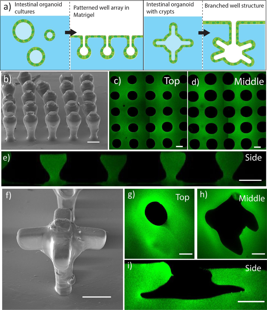
a)Printed thioester features were reproducibly degraded in the presence of Matrigel to cast mold close-packed and overhanging structures reminiscent of the intestinal organoid features. b) SEM image of printed crypt structures consisting of a 150 μm ball on top of a 50 μm wide by 150 μm tall stem printed with a 50 μm spacing. Scale bar 100 μm. Printed organoid features were patterned into Matrigel and a tile scan was taken of the c) stem and d) ball regions of the well. e) Z-stack images of each well were taken with laser scanning confocal microscopy and used to construct an orthogonal view of the patterned array along the XZ plane. Scale bar 100 μm. f) SEM image of branched crypt structure shows defined overhang features. scale bar 100 μm. Printed organoid features were patterned into Matrigel and images were taken of the g) stem and h) branched crypt regions of the well. i) Z-stack images of each well were taken with laser scanning confocal microscopy and used to construct an orthogonal view of the patterned array along the XZ plane. Scale bar 100 μm. Matrigel hydrogels were labeled with FITC fluorophore after patterning. For more details, please see Patterned hydrogel staining and image analysisin methods
