Abstract
Background
The frequency of asymptomatic severe acute respiratory syndrome coronavirus 2 (SARS-CoV-2) infections is unclear and may be influenced by how symptoms are evaluated. In this study, we sought to determine the frequency of asymptomatic SARS-CoV-2 infections in a prospective cohort of health care workers (HCWs).
Methods
A prospective cohort of HCWs, confirmed negative for SARS-CoV-2 exposure upon enrollment, were evaluated for SARS-CoV-2 infection by monthly analysis of SARS-CoV-2 antibodies as well as referral for polymerase chain reaction testing whenever they exhibited symptoms of coronavirus disease 2019 (COVID-19). Participants completed the standardized and validated FLU-PRO Plus symptom questionnaire scoring viral respiratory disease symptom intensity and frequency at least twice monthly during baseline periods of health and each day they had any symptoms that were different from their baseline.
Results
Two hundred sixty-three participants were enrolled between August 25 and December 31, 2020. Through February 28, 2021, 12 participants were diagnosed with SARS-CoV-2 infection. Symptom analysis demonstrated that all 12 had at least mild symptoms of COVID-19, compared with baseline health, near or at time of infection.
Conclusions
These results suggest that asymptomatic SARS-CoV-2 infection in unvaccinated, immunocompetent adults is less common than previously reported. While infectious inoculum doses and patient factors may have played a role in the clinical manifestations of SARS-CoV-2 infections in this cohort, we suspect that the high rate of symptomatic disease was due primarily to participant attentiveness to symptoms and collection of symptoms in a standardized, prospective fashion. These results have implications for studies that estimate SARS-CoV-2 infection prevalence and for public health measures to control the spread of this virus.
Keywords: SARS-CoV-2, COVID-19, symptoms, patient-reported outcomes, prospective study
Defining the frequency of asymptomatic severe acute respiratory syndrome coronavirus 2 (SARS-CoV-2) infections has been elusive. Estimates of asymptomatic infection range from 4% to 80% [1], and a recent systematic review that incorporated large national serosurveillance studies concluded that at least one-third of SARS-CoV-2 infections are asymptomatic [2].
Variability in ascertaining and defining asymptomatic infections is likely due to several factors, including the patient population studied, the manner in which symptoms are assessed, the range of symptoms queried, the frequency and duration of symptom assessment, and whether the study is conducted prospectively or retrospectively. Presymptomatic infection, identified in the initial outbreaks of SARS-CoV-2, and perhaps earliest among the Diamond Princess passengers, further complicates the accurate assessment of asymptomatic infection [3, 4]. Of note, the majority of studies that have estimated asymptomatic/symptomatic ratios have not evaluated symptoms in a prospective, rigorous way [1, 5].
The Prospective Assessment of SARS-CoV-2 Seroconversion (PASS) study was initiated in August of 2020 to prospectively evaluate the clinical and immunological responses to SARS-CoV-2 infection and vaccination in a cohort of generally healthy adult health care workers (HCWs) at the Walter Reed National Military Medical Center (WRNMMC). Here, we present our examination of SARS-CoV-2 infection and self-reported symptoms of participants enrolled in the study over a 6-month time period.
METHODS
Study Participants
Details of the PASS study protocol, including details of the inclusion/exclusion criteria, have been published [6]. Briefly, inclusion criteria included being generally healthy, ≥18 years old, and employed at WRNMMC. Exclusion criteria included history of coronavirus disease 2019 (COVID-19), SARS-CoV-2 immunoglobulin (IgG) seropositivity, and being immunocompromised at screening. The study started in August 2020, with rolling enrollment and monthly research clinic visits to obtain serum for longitudinal SARS-CoV-2 antibody testing. The subset of participants included for analysis in this paper were those enrolled between August 25, 2020, and December 31, 2020. The protocol was approved by the Uniformed Services University Institutional Review Board.
Prospective Collection of Viral Respiratory Symptoms
Study participants were sent a daily email reminder to complete a validated viral respiratory infection (VRI) patient-reported outcome symptom questionnaire (FLU-PRO Plus) each day they experienced any symptoms, and at least twice a month during baseline periods of health. FLU-PRO Plus is a patient-reported outcome instrument, developed to standardize symptoms of viral respiratory infection in clinical research [7] and modified for COVID-19 infection. FLU-PRO Plus measures the severity, frequency, and duration of 34 symptoms organized within the following symptom domains: nasal, throat, eye, chest, gastrointestinal, body/systemic, and sense (taste/smell). The severity of each symptom is measured on a scale from 0 to 4 (0 = not at all, 1 = a little bit, 2 = somewhat, 3 = quite a bit, 4 = very much) for all symptoms except for vomiting and diarrhea, which are scored in terms of frequency per day (0 = 0 times, 1 = 1 time, 2 = 2 times, 3 = 3 times, 4 = 4 or more times). Mean scores in each domain are summed for a total FLU-PRO Plus symptom score of 0–28. Participants were also asked if they felt at their usual state of health each time they completed a questionnaire. Asymptomatic infection was defined as no increase in total FLU-PRO Plus symptom score from each individual’s baseline values.
Duration of Prospective Symptom Collection
Data included in this study were obtained from symptom questionnaires completed from August 25, 2020, until February 28, 2021. The prospective collection of VRI symptoms was stopped after this date due to concerns that participant fatigue with receiving daily email reminders about symptoms beyond month 6 of the study would result in nonrobust data and/or decreased data acquisition.
Diagnosis of SARS-CoV-2 Infection
Serum samples were obtained from participants monthly and tested for SARS-CoV-2 IgG antibodies. Participants were also asked to report to the WRNMMC COVID-19 testing center for SARS-CoV-2 polymerase chain reaction (PCR) testing by nasopharyngeal swab every time they had symptoms of a possible VRI. Participants were considered to be infected if they developed IgG seroconversion to SARS-CoV-2 spike protein or tested positive for SARS-CoV-2 by PCR test.
Antibody Testing
Serum samples were tested monthly for IgG antibodies against SARS-CoV-2 spike protein, as well as for IgG antibodies against the spike proteins of the seasonal coronaviruses HKU1, OC43, 229E, and NL63, using a microsphere-based multiplex immunoassay (MMIA) built using Luminex xMAP–based technology, as previously described (Supplementary Methods) [8].
SARS-CoV-2 Sequencing and Bioinformatics
See the Supplementary Methods.
Statistical Analyses
The Mann-Whitney test was used for unpaired comparisons, with Bonferroni correction applied for analyses with multiple comparisons. Rates of asymptomatic infections were compared with a hypothesized value using a 1-sample binomial test.
RESULTS
Study Participants’ Demographics
A total of 263 participants were enrolled in the study between August 25, 2020, and December 31, 2020, with 11 participants withdrawing before February 28, 2021 (Supplementary Table 1). The mean follow-up period per participant (range) was 135.5 (62–187) days.
Of these 263 participants, 182 (69.2%) were female and 81 (30.8%) were male, with a mean age (interquartile range [IQR]) of 41 (19–69) years. The racial composition was 71.1% White, 12.9% Black, 10.3% Asian, 0.4% Native Hawaiian and other Pacific Islander, and 3.4% multiple racial ethnicities. Of the 263 study participants, 12 (4.6%) were diagnosed with a SARS-CoV-2 infection. Of these, 9 (75%) were female and 3 (25%) were male, with a mean age (IQR) of 37 (23–58) years (Table 1).
Table 1.
Demographics of All Study Participants, the Subset of SARS-CoV-2-Infected Participants, and the Subset of Symptomatic Non-COVID-19 Participants (Total FLU-PRO Plus Score ≥3)
| All | COVID-19 | Symptomatic Non-COVID-19 | |
|---|---|---|---|
| n/N (%) | n/N (%) | n/N (%) | |
| Gender | |||
| Female | 182/263 (69.2) | 9/12 (75) | 31/38 (81.6) |
| Male | 81/263 (30.8) | 3/12 (25) | 7/38 (18.4) |
| Ethnicity | |||
| Non-Hispanic | 243/263 (92.4) | 11/12 (91.7) | 34/38 (89.5) |
| Hispanic | 15/263 (5.7) | 0/12 (0) | 4/38 (10.5) |
| Not reported | 5/263 (1.9) | 1/12 (8.3) | 0/38 (0) |
| Race | |||
| White | 187/263 (71.1) | 8/12 (66.7) | 31/38 (81.6) |
| Black | 34/263 (12.9) | 2/12 (16.7) | 5/38 (13.2) |
| Asian | 27/263 (10.3) | 1/12 (8.3) | 0/38 (0) |
| ≥2 | 9/263 (3.4) | 1/12 (8.3) | 1/38 (2.6) |
| Native Hawaiian and other Pacific Islander | 1/263 (0.4) | 0/12 (0) | 0/38 (0) |
| Not reported | 5/263 (1.9) | 0/12 (0) | 1/38 (2.6) |
| Age | |||
| Mean age (range), y | 41 (19–69) | 37 (23–58) | 37 (19–64) |
Abbreviations: COVID-19, coronavirus disease 2019; SARS-CoV-2, severe acute respiratory syndrome coronavirus 2.
Study participants represented a broad cross-section of hospital employees, with nurses (33.5%), physicians (25.9%), and physical/occupational/recreational therapists (11.4%) comprising the bulk of the cohort (Supplementary Figure 1). Of the 12 SARS-CoV-2-positive participants, 6 were nurses, 3 were physicians, 2 were laboratory scientists/technicians, and 1 was other hospital staff. None of the infected participants belonged to the same household or the same department within the hospital.
SARS-CoV-2 Infections in Study Participants
Twelve participants were diagnosed with SARS-CoV-2 infection before February 28, 2021. Ten were diagnosed by positive PCR, and 2 on the basis of seroconversion with negative PCR testing. All 12 developed SARS-CoV-2 spike IgG binding antibodies (bAb). Participant B was diagnosed on the basis of developing sustained spike IgG bAb and exhibited no significant changes in antibody levels to the seasonal coronaviruses. Participant G was classified as infected despite exhibiting only transient seroconversion because this individual reported a SARS-CoV-2 exposure just a few days before the onset of symptoms and also exhibited no significant changes in antibody levels to the 4 seasonal coronaviruses. One additional participant developed a SARS-CoV-2 spike IgG bAb response just above the threshold of positivity. This individual developed nasal, throat, and systemic symptoms in the month before seroconversion but was not classified as having a SARS-CoV-2 infection as the detection of SARS-CoV-2 spike IgG bAb was associated with the development of high antibody levels against HCoV-229E and HCoV-NL63, and the symptoms were thus ascribed to a seasonal HCoV infection. Moreover, this person’s SARS-CoV-2 IgG bAb decreased below the positivity cutoff by the subsequent monthly collection.
Eluents of nasopharyngeal swabs were available from 6 of 12 participants for sequencing. All samples produced coding complete genomes. Variant B.1.298 was identified in 2 participants, and variants B.1.349, B.1.261, B.1.240, and B.1.243 were each identified in 1 participant (Supplementary Table 2).
All Infected Participants Exhibited Increases in Symptom Scores at or Near the Time of Infection
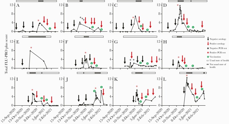
Prospective, standardized collection of baseline symptoms shows that participants often have minimal symptoms present even in their usual state of health (mean FLU-PRO Plus score when in “usual state of health” for all participants = 0.4). All 12 of the SARS-CoV-2-infected individuals experienced mild disease that did not require hospitalization. Of these 12 participants, all reported increases in FLU-PRO Plus scores compared with their baseline scores (Figure 1), resulting in a 100% rate of symptomatic infections (95% CI, 73.5%–100%). One-sample binomial tests demonstrate that this finding would be highly unlikely if the true rate of asymptomatic infection was 50% (P < .001) or even 30% (P = .014).
Figure 1.
Time course of total FLU-PRO Plus scores in SARS-CoV-2-infected participants (n = 12). Between August 25, 2020, and February 28, 2021, study participants completed a standardized and validated FLU-PRO Plus symptom questionnaire scoring viral respiratory disease symptom intensity at least twice monthly and every day they experienced any symptoms different from their baseline. FLU-PRO Plus measures scores (0–4) for 34 symptoms organized in 7 domains; mean scores in each symptom domain are summed for a total FLU-PRO Plus symptom score (0–28). Participants were also asked if they felt at their usual state of health each time they completed a symptoms questionnaire; the state of health is represented by a bar above the graph (open bar: usual state of health; crosshatch bar: not usual state of health). Black arrow, negative SARS-CoV-2 serology test; red arrow, positive SARS-CoV-2 serology test; black asterisk, negative SARS-CoV-2 PCR test; red asterisk, positive SARS-CoV-2 PCR test; green circle, COVID-19 vaccination. Abbreviations: COVID-19, coronavirus disease 2019; PCR, polymerase chain reaction; SARS-CoV-2, severe acute respiratory syndrome coronavirus 2.
Peak total FLU-PRO Plus score increased to a mean value of 6.52, significantly greater than the mean baseline FLU-PRO Plus score of 0.47 (P < .0001). Details of serology, PCR tests, and COVID-19 vaccination are provided in Supplementary Table 3. Of the 10 participants with a positive PCR test, a peak in total FLU-PRO Plus scores occurred within 1 week of the positive PCR test. Importantly, all 10 documented increases in total FLU-PRO Plus scores before participant awareness of COVID-19 diagnosis, as results of PCR testing typically took 3 days. Both of the participants with negative PCR tests (participants B and G) sought repeated PCR testing for COVID-19 because of symptoms, and both exhibited seroconversion within a month of their symptomatic illness (Figure 1).
Heterogeneity and Variability in Patterns of Symptom Expression in Response to SARS-CoV-2 Infection
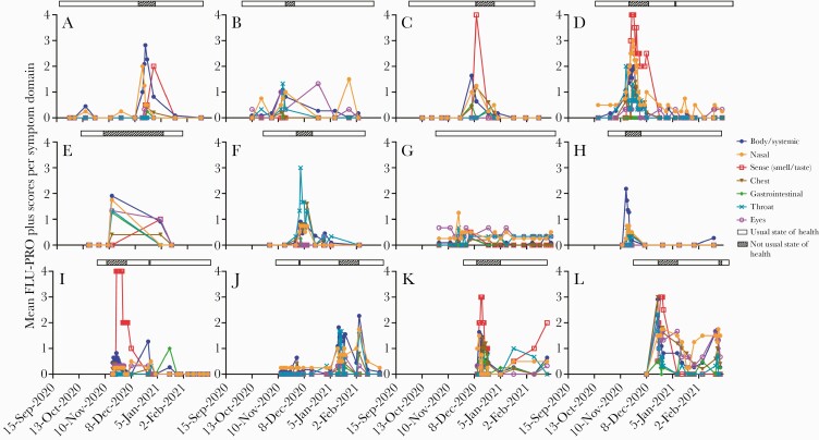
SARS-CoV-2-infected patients exhibited substantial variability with regards to the order, severity, and duration of symptoms experienced in each domain (Figure 2). Highest scores were reported by 4 participants (A, E, H, J) in the body/systemic domain, 4 (C, D, I, K) in the sense (smell/taste) domain, 2 (B, F) in the throat domain, 1 (G) in the nasal domain, and 1 (L) with equal peak scores in the body/systemic, nasal, and sense domains. The number of participants who reported a score of ≥3 in specific domains was 1 (D) for the nasal domain, 1 (F) for the throat domain, 1 (L) for the chest domain, and 5 (C, D, I, K, L) for the sense (smell/taste) domain (Supplementary Figure 2).
Figure 2.
Mean FLU-PRO Plus scores per symptom domain for SARS-CoV-2-infected participants (n = 12). FLU-PRO Plus mean scores for each of the 7 domains are represented in blue (body/systemic), orange (nasal), red (sense—smell/taste), brown (chest), green (gastrointestinal), turquoise (throat), and fuchsia (eyes); the state of health is represented by a bar above the graph (open bar: usual state of health; crosshatch bar: not usual state of health). Abbreviation: SARS-CoV-2, severe acute respiratory syndrome coronavirus 2.
Symptom Scores Were Greater Among Individuals With SARS-CoV-2 Infection Than Among Those who Tested Negative for SARS-CoV-2 Infection
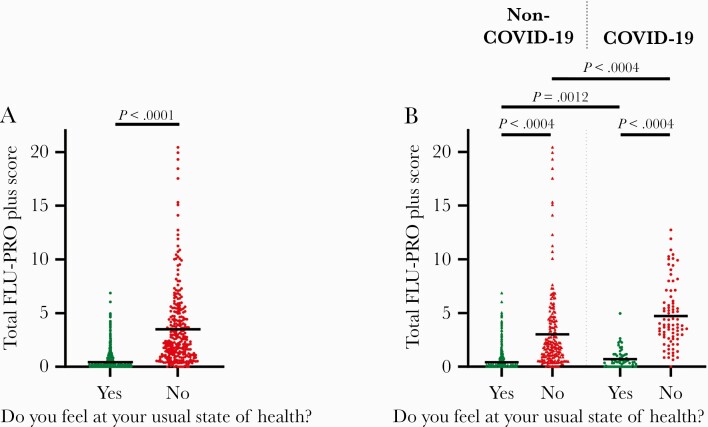
A review of all symptom questionnaires showed that a self-reported “unusual state of health” was associated with a mean total FLU-PRO Plus score (range) of 3.5 (0–20.44), significantly greater than the mean total FLU-PRO Plus score (range) of 0.4 (0–6.87) associated with a “usual state of health” (P < .0001) (Figure 3A). Participants who were never diagnosed with SARS-CoV-2 infection reported a mean total FLU-PRO Plus score (range) of 3.0 (0–20) when feeling not in their usual state of health, compared with a mean total score (range) of 4.7 (0–13) reported by SARS-CoV-2-infected individuals when not in their usual state of health (Figure 3B). Similarly, peak scores experienced by SARS-CoV-2-infected individuals within 2 weeks of diagnosis were greater than the peak scores reported by uninfected individuals.
Figure 3.
Comparison of total FLU-PRO Plus scores between “usual state of health” and “not usual state of health.” A, Total FLU-PRO Plus scores in all PASS study participants when feeling in their “usual state of health” (green) or “not usual state of health” (red; n = 263). B, Total FLU-PRO Plus scores reported when feeling in “usual state of health” (green) or “not usual state of health” (red) for non-COVID participants (n = 251) and COVID-19-positive participants (n = 12). Horizontal bars = means. Statistical significance was determined using a 2-tailed Mann-Whitney U test, and a P value cutoff for significance was set at .05 for (A) and 0.0125 for (B) (Bonferroni correction applied for multiple comparisons). Abbreviations: COVID-19, coronavirus disease 2019; PASS, Prospective Assessment of SARS-CoV-2 Seroconversion.
Extensive Overlap in Symptoms Experienced by Those With SARS-CoV-2 Infection and Among Those Uninfected Individuals who Reported Total FLU-PRO Plus Scores of ≥3
We compared the symptoms reported by SARS-CoV-2-negative participants who reported a total FLU-PRO Plus score of ≥3 (defined as symptomatic non-COVID-19) with the symptoms reported by the 12 participants who acquired SARS-CoV-2 infection (COVID-19 positive). A cutoff of 3 was chosen as this value was reported <1% of the time when study participants felt in their usual state of health (Supplementary Figure 3).
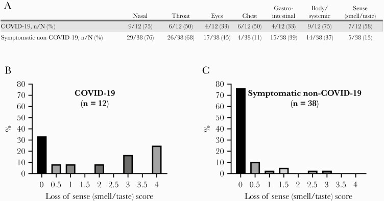
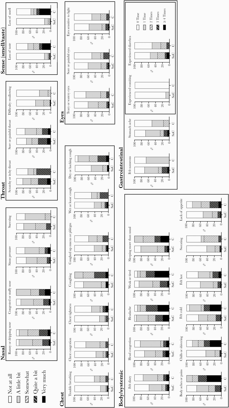
When comparing COVID-19-positive participants (n = 12) with symptomatic non-COVID-19 participants (n = 38), the percentages of individuals who experienced FLU-PRO Plus symptom domain scores of ≥1 were similar when considering the nasal (75% vs 76%, respectively), throat (50% vs 68%, respectively), and gastrointestinal (33% vs 39%, respectively) domains. More COVID-19-positive participants reported scores of ≥1 in the chest (50% vs 11%), body/systemic (75% vs 37%), and sense (58% vs 13%) domains, while ill participants without COVID-19 had more frequent scores of ≥1 in the eyes domain (45% vs 33%) (Figure 4A). As with other studies [9–11], the greatest differentiating symptom was in the sense (smell/taste) domain (Figure 4B, C). Nonetheless, the discriminatory capacity of loss of sense appears low, as 32% of COVID-19-positive individuals experienced no loss of sense, whereas 24% of symptomatic non-COVID-19 individuals did (Figure 4B, C). Analysis on the basis of individual symptoms demonstrates extensive overlap between clinical manifestations in COVID-19-positive and symptomatic non-COVID-19 individuals (Figure 5; Supplementary Table 4). Specifically, ≥70% of both COVID-19-positive and symptomatic non-COVID-19 participants experienced runny or dripping nose, congested or stuffy nose, sinus pressure, sore or painful throat, sneezing, coughing, head congestion, feeling weak or tired, sleeping more than usual, body aches or pains, nausea, and stomach ache (Supplementary Table 4).
Figure 4.
Mean FLU-PRO Plus scores per symptom domain in COVID-19-positive participants and symptomatic non-COVID-19 participants. A, Percentage of COVID-19-positive (n = 12) and symptomatic non-COVID-19 (n = 38) participants who experienced mean FLU-PRO Plus symptom scores of ≥1 for each of the symptom domains. Mean FLU-PRO Plus severity scores for the loss of sense (smell/taste) domain in (B) COVID-19-positive participants (n = 12) and (C) symptomatic non-COVID-19 participants (n = 38). Abbreviation: COVID-19, coronavirus disease 2019.
Figure 5.
Symptom presence and severity reported for each of the 34 symptoms listed in the FLU-PRO Plus symptoms questionnaire by COVID-19-positive participants and by participants in the symptomatic non-COVID-19 subset. The severity of each symptom as measured on a scale from 0 to 4 (0 = not at all, 1 = a little bit, 2 = somewhat, 3 = quite a bit, 4 = very much) for all symptoms except for vomiting and diarrhea, which were scored in terms of frequency per day (0 = 0 time, 1 = 1 time, 2 = 2 times, 3 = 3 times, 4 = 4 or more times). For COVID-positive participants, peak mean FLU-PRO Plus scores within each symptom domain were determined within the 2-week time period surrounding the time of diagnosis (for those with positive PCR tests) or within the 2-week time period when participants initially went for COVID-19 testing if diagnosed by seroconversion alone. For symptomatic non-COVID-19 participants, peak FLU-PRO Plus scores were obtained within the 2-week time period of the greatest total FLU-PRO Plus symptom score. Abbreviations: C, coronavirus disease 2019; COVID-19, coronavirus disease 2019; PCR, polymerase chain reaction; SARS-CoV-2, severe acute respiratory syndrome coronavirus 2; SnC, symptomatic non–coronavirus disease 2019.
DISCUSSION
The principal findings of this study are that (1) completely asymptomatic infections are likely rare in unvaccinated individuals when symptoms are comprehensively, frequently, and prospectively evaluated with standardized, validated measurement tools and (2) the symptoms of mild COVID-19 are not sufficiently distinctive to enable clinical differentiation of SARS-COV-2 infection from other viral upper respiratory infections.
Defining the frequency of asymptomatic SARS-CoV-2 infection has been an elusive goal since the onset of the pandemic. Challenges in enumerating asymptomatic infections include defining what “asymptomatic” means in the setting of some symptoms even in a state of usual health, individuals forgetting mild symptoms due to retrospective recall bias, patient minimization of symptoms or using different words to describe symptoms, questions that focus only on a subgroup of symptoms (eg, respiratory symptoms) and inadequate time of follow-up for assessment of symptoms after diagnosis. In this study, we sought to overcome many of these obstacles by following a cohort of uninfected individuals prospectively, establishing a baseline of each participant’s symptom score when in their usual state of health, and having participants self-report their symptoms every day they had any symptoms that could be due to a viral respiratory infection using an appropriately developed and validated symptom tool. In addition to encouraging participants to report for nasopharyngeal PCR testing whenever they felt symptomatic, we sought to capture all potential asymptomatic cases by monitoring for SARS-CoV-2 spike protein IgG seroconversion.
Using this approach, we observed that all 12 participants prospectively diagnosed with SARS-CoV-2 infection by December 31, 2020, exhibited increases in self-reported symptom scores compared with their baselines (noting that many participants have mild symptoms at baseline). While the total number of SARS-CoV-2 infections captured was relatively small, the finding that all 12 experienced symptomatic disease strongly suggests that completely asymptomatic infection is less common than symptomatic infection. One-sample binomial tests demonstrate that if the true rate of asymptomatic infection is 50%, then the likelihood that 12 of 12 individuals would report symptoms is 0.024%. If the true rate of asymptomatic infection is 30%, then the likelihood that 12 of 12 individuals would all be symptomatic is still only 1.4%.
Thus, the results of this study suggest that completely asymptomatic infections occur at a much lower frequency in unvaccinated, generally healthy adults than reported in most other studies. A meta-analysis of 79 studies that assessed for symptoms at >1 time point estimated that 20% of individuals infected with SARS-CoV-2 remain asymptomatic throughout infection, though the uncertainty interval was wide, at 3% to 67% [12]. Another meta-analysis focusing only on studies that conducted sufficient follow-up to account for presymptomatic infection concluded that 17% of individuals have asymptomatic infection [13]. As Meyerowitz et al. have noted, factors such as incomplete symptom assessment, limited duration of symptom monitoring, and deficiencies in symptom recollection/recall bias with no baseline assessment before infection can result in overestimates of the true proportion of asymptomatic SARS-CoV-2 infections [1]. These factors may explain the differences observed between our study and other studies conducted using retrospective symptom collection. Indeed, the results of our study are more in line with those of others who performed symptom assessment longitudinally. For example, the Centers for Disease Control and Prevention (CDC) and Public Health–Seattle and Kings County found that while individuals are often asymptomatic at the time of infection, most proceed to develop symptoms. In that study, Arons et al. conducted serial point-prevalence PCR testing and symptom monitoring in residents and staff of a skilled nursing facility after identification of a co-resident with COVID-19. While 56% of residents who tested positive for SARS-CoV-2 were asymptomatic at the time of testing, 88% of those went on to develop symptoms at a median of 4 days later, resulting in only 5.2% of individuals reporting no symptoms with SARS-CoV-2 infection [3].
In addition to prospective reporting of symptoms, another factor in assessing symptoms is the variability in methods of self-assessment of symptoms. We hypothesize that recognition of symptoms due to pulmonary changes is dependent in part on patient attentiveness and patient willingness to share symptoms. Another factor may be how questions are asked to patients by clinicians and patients’ understanding of those questions and their response options. In the PASS study, participants were highly engaged and very attuned to monitoring their symptoms. Furthermore, the content validity of FLU-PRO Plus (the relevance of the questions themselves, patient understanding of the questions and response options, and measurement properties of derived scores) have been evaluated in a variety of viral respiratory tract diseases. Self-reporting bias, and most specifically recall bias, is a major issue in most observational studies and can affect the accuracy of self-reported symptoms in many ways: telescoping (lengthening of the recall period), reverse telescoping (shortening of the recall period), or memory decay (often resulting in under-reporting) [14, 15]. In order to overcome recall bias in our study, we shortened the recall period by asking the participants to complete the FLU-PRO Plus symptom questionnaire each day they experienced any symptoms as the instrument was designed with a recall period for daily administration and at least twice a month during baseline periods of health.
With regards to symptoms, our study observed extensive overlap between symptoms of COVID-19 and those of non-COVID-19 upper respiratory tract infections. This is not surprising as the host response to viral respiratory infection may be similar regardless of causative pathogen. Runny nose, sinus pressure, and sore throat occurred in >70% of infected participants and in >70% of symptomatic SARS-CoV-2-negative individuals. These results differ substantially from infections reported to the CDC in the first half of 2020, in which runny nose and sore throat were mentioned in only 6.1% and 20% of cases [16]. We suspect that this may be due to recall bias in settings where symptoms are not collected prospectively, rather than evolution of symptoms over time.
Notably, loss of smell or taste, which was the most distinctive symptom in patients with COVID-19, did not occur with sufficient frequency or specificity to enable diagnosis of SARS-CoV-2 on the basis of symptoms alone. The results of our study confirm those of a recent Cochrane review, which found that signs and symptoms are insufficient to rule in or rule out SARS-CoV-2 infection [17]. The heterogeneity of presenting symptoms as well as symptom evolution over time reinforces the need for comprehensive and continual monitoring of symptoms to obtain accurate measures of patient health status.
A strength of this study is the daily symptom assessment approach in a cohort of individuals before and after any COVID-19 diagnosis. To the best of our knowledge, this is the only study of its type to date, as most studies conducting longitudinal symptom assessment have done so after exposure or diagnosis [12]. One limitation with regards to extrapolating this study to all generally healthy adults is that the cohort was made of HCWs. As such, participants may have been exposed to higher infectious doses of virus than typically encountered in the community. Alternatively, they may have had better infection control and prevention strategies than the general public. Another limitation is that the results of this study, which was conducted with a generally healthy cohort of individuals, may not be applicable to individuals with other demographics. Rates of asymptomatic infection may be different in individuals with comorbidities or who are immunocompromised. For example, 1 study found that 40.3% of patients on hemodialysis were asymptomatic when diagnosed with SARS-CoV-2 by PCR screening [18]. Similarly, our study, in which the mean age was 41 years, may not be generalizable to younger or older adult populations. In contrast to our study, investigation of a SARS-CoV-2 outbreak on the aircraft carrier U.S.S. Theodore Roosevelt observed that 55% of PCR-confirmed infections were asymptomatic in a crew that had a mean age of 27 years [19]. Differences in asymptomatic rates between studies could also be related to the presence of different variants, as some variants may be more virulent than others [20]. Of note, none of the lineages identified in our study is a variant of concern that is known to be more virulent. The low N number for each variant in this study does not allow us to correlate viral genotype with differences in symptoms.
In conclusion, our study suggests that completely asymptomatic SARS-CoV-2 infection compared with baseline symptoms in unvaccinated, immunocompetent adults is much less common than previously reported. While it is possible that viral infectious doses and patient-related factors may have played a role in the reported clinical manifestations of SARS-CoV-2 infections in this cohort of HCWs, we suspect that the high symptomatic rate was primarily due to participant attentiveness to symptoms and to collection of symptoms in a standardized, prospective fashion. These results have implications for studies that estimate SARS-CoV-2 infection prevalence as well as for public health measures to control the spread of this virus and its variants.
Supplementary Data
Supplementary materials are available at Open Forum Infectious Diseases online. Consisting of data provided by the authors to benefit the reader, the posted materials are not copyedited and are the sole responsibility of the authors, so questions or comments should be addressed to the corresponding author.
Acknowledgments
Financial support. This study was executed by the Infectious Disease Clinical Research Program (IDCRP), a Department of Defense (DoD) program executed by the Uniformed Services University of the Health Sciences (USUHS) through a cooperative agreement by the Henry M. Jackson Foundation for the Advancement of Military Medicine, Inc. (HJF). This work was supported by the Defense Health Program, United States Department of Defense, under award HU00012120067 (E.M.). Project funding for J.H.P. was in whole or in part with federal funds from the National Cancer Institute, National Institutes of Health, under Contract No. HHSN261200800001E. Genome sequencing and bioinformatic analysis was supported by Armed Forces Health Surveillance Division (AFHSD) Global Emerging Infections Surveillance (GEIS) Branch P0093_21_NM to K.A.B.-L. The funding bodies have had no role in the study design, data collection and analysis, decision to publish, or preparation of the manuscript.
Potential conflicts of interest. S.D.P., T.H.B., and D.T. report that the Uniformed Services University (USU) Infectious Diseases Clinical Research Program (IDCRP), a US Department of Defense institution, and the Henry M. Jackson Foundation (HJF) were funded under a Cooperative Research and Development Agreement to conduct an unrelated phase III COVID-19 monoclonal antibody immunoprophylaxis trial sponsored by AstraZeneca. The HJF, in support of the USU IDCRP, was funded by the Department of Defense Joint Program Executive Office for Chemical, Biological, Radiological, and Nuclear Defense to augment the conduct of an unrelated phase III vaccine trial sponsored by AstraZeneca. Both of these trials were part of the US Government COVID-19 response. Neither is related to the work presented here. All authors have submitted the ICMJE Form for Disclosure of Potential Conflicts of Interest. Conflicts that the editors consider relevant to the content of the manuscript have been disclosed.
Disclaimers. The views expressed in this article are those of the authors and do not necessarily reflect the official policy or position of the Department of Defense, the Navy, the Uniformed Services University, the Henry M. Jackson Foundation, or the US Government. C.H.O., K.M.H.-P., C.A.D., S.R., W.C., A.M.W.M., C.E.A., R.Z.C., K.A.B.-L., T.H.B., C.C.B., E.D.L., and E.M. are military service members or employees of the US Government. This work was prepared as part of their official duties. Title 17 U.S.C. § 105 provides that “Copyright protection under this title is not available for any work of the United States Government.” Title 17 U.S.C. §101 defines a US Government work as a work prepared by a military service member or employee of the US Government as part of that person’s official duties.
Ethics approval and participant consent. This study protocol was approved by the Uniformed Services University of the Health Sciences Institutional Review Board (FWA 00001628; DoD Assurance P60001) in compliance with all applicable federal regulations governing the protection of human participants. Written consent was obtained from all study participants.
References
- 1. Meyerowitz EA, Richterman A, Bogoch II, et al. Towards an accurate and systematic characterisation of persistently asymptomatic infection with SARS-CoV-2. Lancet Infect Dis 2021; 21:e163–9. [DOI] [PMC free article] [PubMed] [Google Scholar]
- 2. Oran DP, Topol EJ.. The proportion of SARS-CoV-2 infections that are asymptomatic: a systematic review. Ann Intern Med 2021; 174:655–62. [DOI] [PMC free article] [PubMed] [Google Scholar]
- 3. Arons MM, Hatfield KM, Reddy SC, et al. Presymptomatic SARS-CoV-2 infections and transmission in a skilled nursing facility. N Engl J Med 2020; 382:2081–90. [DOI] [PMC free article] [PubMed] [Google Scholar]
- 4. Sakurai A, Sasaki T, Kato S, et al. Natural history of asymptomatic SARS-CoV-2 infection. N Engl J Med 2020; 383:885–6. [DOI] [PMC free article] [PubMed] [Google Scholar]
- 5. Oran DP, Topol EJ.. Prevalence of asymptomatic SARS-CoV-2 infection: a narrative review. Ann Intern Med 2020; 173:362–7. [DOI] [PMC free article] [PubMed] [Google Scholar]
- 6. Jackson-Thompson BM, Goguet E, Laing ED, et al. Prospective assessment of SARS-CoV-2 seroconversion (PASS) study: an observational cohort study of SARS-CoV-2 infection and vaccination in healthcare workers. BMC Infect Dis 2021; 21:544. [DOI] [PMC free article] [PubMed] [Google Scholar]
- 7. Powers JH, Guerrero ML, Leidy NK, et al. Development of the Flu-PRO: a patient-reported outcome (PRO) instrument to evaluate symptoms of influenza. BMC Infect Dis 2016; 16:1. [DOI] [PMC free article] [PubMed] [Google Scholar]
- 8. Laing ED, Sterling SL, Richard SA, et al. Antigen-based multiplex strategies to discriminate SARS-CoV-2 natural and vaccine induced immunity from seasonal human coronavirus humoral responses. medRxiv 2021.02.10.21251518 [Preprint]. 12 February 2021. Available at: 10.1101/2021.02.10.21251518. Accessed 7 February 2022. [DOI] [Google Scholar]
- 9. Mika J, Tobiasz J, Zyla J, et al. Symptom-based early-stage differentiation between SARS-CoV-2 versus other respiratory tract infections—Upper Silesia pilot study. Sci Rep 2021; 11:13580. [DOI] [PMC free article] [PubMed] [Google Scholar]
- 10. Dixon BE, Wools-Kaloustian KK, Fadel WF, et al. Symptoms and symptom clusters associated with SARS-CoV-2 infection in community-based populations: results from a statewide epidemiological study. PLoS One 2021; 16:e0241875. [DOI] [PMC free article] [PubMed] [Google Scholar]
- 11. Nuertey BD, Ekremet K, Haidallah AR, et al. Performance of COVID-19 associated symptoms and temperature checking as a screening tool for SARS-CoV-2 infection. PLoS One 2021; 16:e0257450. [DOI] [PMC free article] [PubMed] [Google Scholar]
- 12. Buitrago-Garcia D, Egli-Gany D, Counotte MJ, et al. Occurrence and transmission potential of asymptomatic and presymptomatic SARS-CoV-2 infections: a living systematic review and meta-analysis. PLoS Med 2020; 17:e1003346. [DOI] [PMC free article] [PubMed] [Google Scholar]
- 13. Byambasuren O, Cardona M, Bell K, et al. Estimating the extent of asymptomatic COVID_19 and its potential for community transmission: systematic review and meta-analysis. Off J Assoc Med Microbiol Infect Dis Canada 2020; 5:223–34. [DOI] [PMC free article] [PubMed] [Google Scholar]
- 14. Althubaiti A. Information bias in health research: definition, pitfalls, and adjustment methods. J Multidiscip Healthc 2016; 9:211–7. [DOI] [PMC free article] [PubMed] [Google Scholar]
- 15. Short ME, Goetzel RZ, Pei X, et al. How accurate are self-reports? Analysis of self-reported health care utilization and absence when compared with administrative data. J Occup Environ Med 2009; 51:786–96. [DOI] [PMC free article] [PubMed] [Google Scholar]
- 16. Stokes EK, Zambrano LD, Anderson KN, et al. Coronavirus disease 2019 case surveillance - United States, January 22-May 30, 2020. MMWR Morb Mortal Wkly Rep 2020; 69:759–65. [DOI] [PMC free article] [PubMed] [Google Scholar]
- 17. Struyf T, Deeks JJ, Dinnes J, et al. Signs and symptoms to determine if a patient presenting in primary care or hospital outpatient settings has COVID-19 disease. Cochrane Database Syst Rev 2020; 7:CD013665. [DOI] [PMC free article] [PubMed] [Google Scholar]
- 18. Clarke C, Prendecki M, Dhutia A, et al. High prevalence of asymptomatic COVID-19 infection in hemodialysis patients detected using serologic screening. J Am Soc Nephrol 2020; 31:1969–75. [DOI] [PMC free article] [PubMed] [Google Scholar]
- 19. Kasper MR, Geibe JR, Sears CL, et al. An outbreak of COVID-19 on an aircraft carrier. N Engl J Med 2020; 383:2417–26. [DOI] [PMC free article] [PubMed] [Google Scholar]
- 20. Danchin A, Timmis K.. SARS-CoV-2 variants: relevance for symptom granularity, epidemiology, immunity (herd, vaccines), virus origin and containment? Environ Microbiol 2020; 22:2001–6. [DOI] [PMC free article] [PubMed] [Google Scholar]
Associated Data
This section collects any data citations, data availability statements, or supplementary materials included in this article.


