Abstract
Background
The antibacterial activities of aqueous leaf extracts of Moringa oleifera, Vernonia amygdalina, Azadirachta indica and Acalypha wilkesiana against multidrug resistance (MDR) Staphylococcus aureus associated with skin and soft tissue infections were investigated.
Methods
Staphylococcus aureus (n = 183) from the skin and soft tissue infections with evidence of purulent pus, effusions from aspirates, wounds, and otorrhea were biotyped, and evaluated for biofilm production. The phenotypic antibiotic resistance and MDR strains susceptibility to plant leaves extract were determined using disc diffusion and micro-broth dilution assays respectively. The correlation of plant extract bioactive components with inhibitory activities was determined.
Results
High occurrence rate of S. aureus were recorded among infant and adult age groups and 13.2% mild biofilm producers from the wound (p < 0.05). Of 60.2% MDR strains with overall significant MARI of more than 0.85 (p < 0.05), high resistant rates to linozidine (92.7%; 95% CI:7.27–10.52), ofloxacin (94.2%; 95% CI:6.09–8.15), chloramphenicol (91.2%; 95% CI:6.11–8.32), gentamicin (97.3%; 95% CI:6.20–8.22), ciprofloxacin (92.7%; 95% CI: 5.28–7.99) and vancomycin (86.6%; 95% CI:6.81–9.59) were observed. Vernonia amygdalina and Azadirachta indica showed significant antimicrobial activity at 100 mg/ml and 75 mg/ml, with low susceptibility of less than 10% to 25 mg/ml, 50 mg/ml, and 75 mg/ml Moringa oleifera. Alkaloids, saponin and terpenoids were significant in Moringa oleifera, Acalypha wilkesiana, Azadirachta indica and Vernonia amygdalina leaves extracts (p < 0.05). High inhibitory concentrations at IC50; 3.23, 3.75 and 4.80 mg/ml (p = 0.02, CI: − 0.08 – 11.52) and IC90; 12.9, 7.5, and 9.6 mg/ml (p = 0.028, CI: 2.72–23.38) were shown by Acalypha wilkesiana, Vernonia amygdalina and Moringa oleifera respectively. Comparative outcome of the plant extracts showed Acalypha wilkesiana, Vernonia amygdalina and Moringa oleifera to exhibit significant inhibition activities (p < 0.05) compared to other extracts. Significant median inhibitory concentration (15.3 mg/ml) of Azadirachta indica were observed (p < 0.01) and strong associations of phytochemical compounds of Azadirachta indica (eta = 0.527,p = 0.017), Vernonia amygdalina (eta = 0.123,p = 0.032) and Acalypha wilkesiana (eta = 0.492,p = 0.012) with their respective inhibitory values.
Conclusion
Observed high occurrence rate of skin and soft tissue infections caused by biofilm-producing MDR S. aureus requires alternative novel herbal formulations with rich bioactive compounds from Moringa oleifera, Acalypha wilkesiana, Azadirachta indica and Vernonia amygdalina as skin therapeutic agents.
Supplementary Information
The online version contains supplementary material available at 10.1186/s12906-022-03527-y.
Keywords: S. aureus, Biofilm, Antibiotic resistance, Plant extracts, Skin and soft tissue infections
Background
One of the most common causes of skin and soft tissue infections (SSTIs) is Staphylococcus aureus [1] and increases the risk of more invasive systemic infections including bacteremia, septicemia and osteomyelitis [2, 3]. The severity of Staphylococci infection ranges from mild skin abscess, superficial tissue infection to life-threatening diseases [4]. Global epidemiological reports have shown SSTIs are usually aggravated by Staphylococcus aureus biofilm, leading to extensive antibiotic resistance, thereby limiting available treatment options [1, 3]. Secretion of extracellular polymeric substance (called biofilm) minimizes therapeutic drug activities and enhances colonization due to polysaccharide intercellular adhesin, a major component of staphylococci biofilm [5, 6]. Formation of Staphylococci biofilm, particularly in wound infection and skin abscess, usually increases severity and chances of bloodstream infection, thereby aiding the SSTIs morbidity mostly among the In-patients [4]. Persistent antibiotic resistance to β-lactams [7], fluoroquinolones [8], and cephalosporins [9], provide a high magnitude of skin morbidity and infection burden.
Due to the poor efficacy of antibiotics against resistant S. aureus in SSTIs, selected plant extracts such as Moringa oleifera, Vernonia amygdalina, Azadirachta indica and Acalypha wilkesiana showed high antimicrobial activities as alternative skin therapy used mainly by numerous infected individuals as local concoction with undocumented successes. These plant extracts are natural, safe, easily accessible, non-toxic with little or no side effects, and with a significant level of phytochemicals expressing higher functional antimicrobial activity against SSTIs than synthetic drugs [10].
Ethnobotanical relevance of leaves parts of Moringa oleifera (L) Millsp [11], Vernonia amygdalina Del. [12], Azadirachta indica Juss [13] and Acalypha wilkesiana Mueli. Arg [14] were most used plant parts for medicinal purposes because of their rich and functional bioactive compounds such as anthocyanin, glycosides, coumarins, flavonoids, phenols, saponins, tannins and terpenoids [15]. Use of organic solvents (methanol, ethanol, dichloromethane, and acetone) for plant extraction influenced the antibacterial activities of the extracts due to solvents bacteriostatic or bacteriocidal activities [16, 17], but the aqueous extract of the plant leaves is preferably utilized for the investigation of the anti-staphylococci activities due to high polarity of water as a solvent for plant bioactive compounds and non-inhibitory potential to the organism [18]. To date, anti-staphylococci potential of these plant aqueous extracts from leaves part against SSTIs has not been well explored for scientific evidence. Therefore, the present study investigates the antibacterial activities of leaf extract of Moringa oleifera, Vernonia amygdalina, Azadirachta indica and Acalypha wilkesiana against multi-resistant Staphylococcus aureus associated with skin and soft tissue infections.
Methods
Isolate collections
Staphylococci isolates from purulent pus (n = 58), effusions from aspirates (n = 34), wounds (n = 55), and otorrhea (n = 36) among out-patients diagnosed with skin and soft tissue infections were selected. Ethical permission from the Health Research Ethics Committees with protocol approval (FMCA/470/HREC/09/2017; NHREC/08/10–2015) was obtained. Each isolate was phenotypically characterized on mannitol salt and Baird-Parker agars and further Gram-stained and biotyped using API as previously described [19].
Biofilm estimation
The level of biofilm produced from biomass was evaluated in three replicates using standard inoculum of 0.5 McFarland turbidity adjusted to the absorbance of 0.01. Each isolate of 500 μl was distributed in a 96-well plate (Corning, New York, NY, United States) and incubated at 37 °C for 24 h. After incubation, the wells were gently washed three times with phosphate buffer saline (PBS), then 500 μl of 0.2% crystal violet was added to each well and further incubated for 20 min at room temperature. All the wells were washed three times with PBS, and 500 μl of 95% ethanol was added to each well. The intensity of the developed colour was measured at absorbance of 595 nm to evaluate the amount of biofilm biomass, which is proportional to the absorbance value [20]. According to the proportion of biofilm biomass, the biofilm level was classified as strong, mild, and weak as previously described [21].
Antibiogram
Antibiotic susceptibility pattern of each isolate was determined using disc diffusion method according to Bauer et al. [22] on Mueller–Hinton agar plates. Briefly, a standardized inoculum of 0.5 McFarland turbidity was prepared from pure overnight isolates and tested against antibiotic discs of linozidine (LZD, 30 μg), vancomycin (VA, 30 μg), fosfomycin (FOX, 30 μg), erythromycin (E, 15 μg); trimethoprim/sulfamethoxazole (SXT, 25 μg), ofloxacin (OFX, 10 μg), ceftriaxone (CRO, 30 μg), amoxicillin (AMC, 10 μg), gentamicin (CN, 10 μg), ciprofloxacin (CIP, 10 μg), ceftazidime (CAZ, 30 μg) and tetracycline (TE, 30 μg). Zones of inhibitions were measured after aerobic incubation at 37 °C for 24 h and interpreted according to CLSI guidelines [23]. The multi-antibiotic resistance index (MARI) was determined [19], in which the number of antibiotics an isolate is resistant to is divided by the total number of antibiotics used. Resistance to at least one agent in three or more antimicrobial classes was defined as multidrug-resistant (MDR) and non-MDR as susceptibility to all antibiotics [24].
Plant collection and authentication
Four plant samples were selected from previous ethnopharmacological studies, based on the frequency of these plant leaves (extracts) for the local treatment of skin infections and the high rate of their citation, as described in Table 1. In addition, these plant leaves are commonly known by their antibacterial efficacy among the locals for treating septic wounds and other skin infections (but not reported in the literature). Fresh leaves of each plant from a single population were collected from the premises of the Covenant University, Ota, Nigeria in February, 2021 with permission from the University Floral management. Taxonomic identification was done by Dr. A. S Oyelakin [25] and deposited with voucher numbers; Moringa oleifera (L) Millsp (FHA0025), Acalypha wilkesiana Mueli. Arg (FHA007), Azadirachta indica Juss (FHA0084) and Vernonia amygdalina Del. (FHA0083) at the Federal University of Agriculture, Abeokuta (FUNAAB) Herbarium, Abeokuta, Nigeria.
Table 1.
Ethnobotanical surveys of selected plants
| Plant (Scientific name) | Family | Yielded (g) | Ethnobotanical relevance | Ref |
|---|---|---|---|---|
| Vernonia amygdalina Del. | Asteraceae | 7.5 | Skin infections and diarrhoea | [12, 26] |
| Acalypha wilkesiana Mueli. Arg | Euphorbiaceae | 12.9 |
Skin infection, antihelmithics, Nosocomial infections |
[14, 27] |
| Moringa oleifera (L) Millsp | Moringaceae | 9.6 |
Wound infection, Anti-inflammatory, cytotoxic, |
[11, 28, 29] |
| Azadirachta indica Juss | Meliaceae | 11.1 |
Wound therapy, antivirals, anti-inflammatories, Antiseptics, antihelmintics Antibiofilm, |
[13, 30, 31] |
Aqueous extraction
Obtained leaves were dried at room temperature, crushed and blended to powdery form. An aqueous extraction was carried out using cold extraction process as previously described [32]. Briefly, powdered leaves were homogenized in separate 500 ml sterile distilled water and maintained at 35 °C for 3 days. Subsequently, the aqueous mixtures were filtered and kept in the oven at 40 °C for 2–3 days. The filtered extracts were concentrated using the rotary evaporator at 70 °C for 2–4 h, and stored at 2–4 °C in sterile bottles.
Phytochemical analysis
Qualitative phytochemical analysis was carried out to detect the presence of anthocyanin, alkaloid, glycosides, coumarins, flavonoids, phenols, quinons, saponins, tannins and terpenoids in all four extracts following standard procedures described by Chinnadurai et al [33] with slight modifications. Briefly, for anthocyanin detection, 1 ml of 2 N sodium hydroxide (NaOH) was added to 2 ml of filtrates and heated for 5 min at 100 °C in a beaker on a hot plate and 2 ml of Glacial acetic acid (CH3COOH) and a few drops of 5% ferric chloride was added to 0.5 ml of the filtered extracts, under the layer of 1 ml of concentrated H2SO4 for detection of glycosides. The detection of coumarins follows the addition of 1 ml of 10% sodium hydroxide to 1 ml of the extracts and flavonoids was determined by adding 5 ml dilute ammonia solution (NH3) to 2 ml of the aqueous filtrates followed by addition of concentrated H2SO4. Phenol was determined following addition of 2 ml distilled water and few drops of 10% ferric chloride to 1 ml of filtered extracts. Saponin was detected by adding 2 ml sterile distilled water to 2 ml of filtrates, shaken vigorously lengthwise for 5 min and 1 cm formation of foam was observed. Only 2 ml 5% ferric chloride (FeCl3) was added to 1 ml of the filtrates and observed for a greenish-black precipitation to detect tannins and 2 ml chloroform was added to 0.5 ml of the filtered extracts for detection of terpenoids.
Antimicrobial activity of the aqueous extracts
MDR strains among the Staphylococci were selected for the extract susceptibility assay according to the previously described method [34]. Briefly, 0.5 McFarland turbid inoculum was evenly spread on the surface of sterile Mueller–Hinton agar plates, and sterile paper disc previously soaked in a known concentration of extract (20 mg/ml per disc) was gently and firmly placed. Produced inhibition zones were measured after 24 h incubation at 37 °C and compared with the control disc containing only sterile physiological saline. The inhibitory concentration (IC) of each extract was determined using varying dilutions of the aqueous plant extract at concentrations ranging between 0.5 to 64 mg/ml. Serial dilutions of equal volume of 100 μl each of the extract and the nutrient broth was prepared in a sterile microtitre plate and 100 μl of standardized inoculum was added to all the wells. Separate plates for each extract were incubated aerobically at 37 °C for 18–24 h. Test extract control (extract and the growth medium without inoculum) and the organism control (the growth medium, physiological saline and the inoculum) were included. The lowest concentration (the highest dilution) of the extract with no turbidity (no visible bacterial growth) compared with the control tubes were regarded as the inhibitory concentrations.
Data analysis
The level of significance of the occurrence of suspected S. aureus from the clinical samples obtained from various age groups and rates of biofilm production was evaluated with ANOVA at 95 and 99% confidence intervals. To ascertain the differences in rates of the MDR and non-MDR strains, Wilcoxon Signed Rank Test was used to compare at 95% probability level and T-test to estimate the significance of resistance of the antibiotic among the S. aureus. Eta-square was calculated as a descriptive measure of the strength of association between phytochemical compounds (independent) and antimicrobial activities of plant extracts (dependent) and further interpreted the Pearson’s coefficient taking the significance at p < 0.05. Significance of IC50 and IC90 were evaluated with the phytochemical compound taking the p-value < 0.05 at the confident interval of 95%.
Results
Prevalence rate of S. aureus and degree of biofilm production
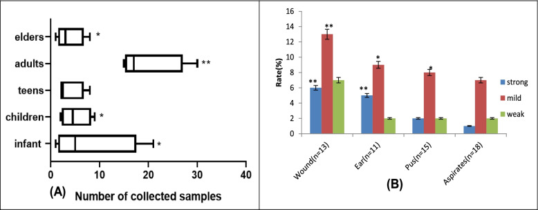
A significance occurrence rate of suspected S. aureus in SSTIs was observed among the infant, children, adult, and elderly age groups (p < 0.05) with less number from the teens. The highest median of estimated clinical samples collected was observed among the adult group (Fig. 1A). All the samples harbor S. aureus with the potential to produce biofilm regarding the accumulation of the biomass biofilm estimations in all the clinical samples. Significantly high and mild biofilm-producers were observed in wound infections (13.2%) and more than 8.0% rate in ear, pus and aspirates (p < 0.05 and p = 0.01) (Fig. 1B).
Fig. 1.
A Occurrence rate of suspected S. aureus in clinical samples according to the subjects’ age distribution B Degree of biofilm production by S. aureus in various collected clinical samples (key: **p = 0.01; *p < 0.05)
Multidrug resistance pattern of the S. aureus isolates
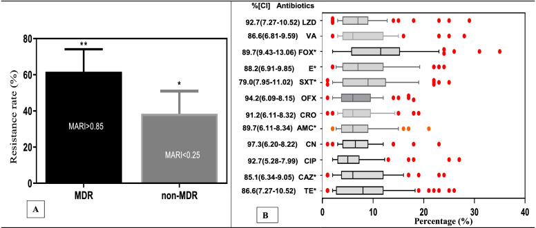
From the clinical isolates of S. aureus, more than 60% were multidrug-resistant strains (showing significant resistance to more than three antibiotic classes) with an overall MARI of more than 0.85 compared to non-MDR strains (p < 0.05) (Fig. 2A). The MDR strains showed more than 90% resistant rates to linozidine (92.7%; 95% CI:7.27–10.52), ofloxacin (94.2%; 95% CI:6.09–8.15), chloramphenicol (91.2%; 95% CI:6.11–8.32), gentamicin (97.3%; 95% CI:6.20–8.22), ciprofloxacin (92.7%; 95% CI: 5.28–7.99) and vancomycin (86.6%; 95% CI: 6.81–9.59). Significant median resistance rates of more than 10% were shown to fosfomycin, erythromycin, trimethoprim/sulfamethoxazole, amoxicillin, ceftazidime, tetracycline (p < 0.05) (Fig. 2B).
Fig. 2.
A Overall multi-drug resistance (MDR) pattern and multi antibiotic resistance indices (MARI) B Box plot evaluation of the antibiotic resistance pattern of S. aureus (*p < 0.05; linozidine (LZD); vancomycin (VA); fosfomycin (FOX), erythromycin (E); trimethoprim/sulfamethoxazole (SXT); ofloxacin (OFX); ceftriaxone (CRO); amoxicillin (AMC); gentamicin (CN); ciprofloxacin (CIP); ceftazidime (CAZ), tetracycline (TE))
Susceptibility rates of S. aureus to plant extracts
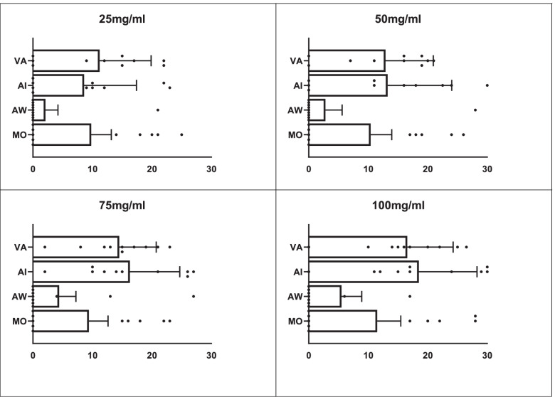
Susceptibility rates of the S. aureus to various dilutions of the plant extracts revealed both Vernonia amygdalina and Azadirachta indica exhibited significant antimicrobial activity at 100 mg/ml and 75 mg/ml. Low susceptibility of less than 10% to Moringa oleifera, was recorded at 25 mg/ml, 50 mg/ml and 75 mg/ml. In contrast, very low susceptibility to Acalypha wilkesiana was observed at all the dilutions (Fig. 3).
Fig. 3.
Susceptibility rates of S. aureus to Moringa oleifera (MO), Acalypha wilkesiana (AW), Azadirachta indica (AI) and Vernonia amygdalina (VA) at various dilutions of 100 mg/ml, 75 mg/ml, 50 mg/ml and 25 mg/ml
Phytochemical composition and antibacterial activities of the plant extracts
The primary estimation of phytochemical composition of the aqueous leave extracts from the plants revealed significant level of alkaloid, flavonoids, phenol, saponin, tannins and terpenoids in Moringa oleifera, Acalypha wilkesiana, Azadirachta indica and Vernonia amygdalina leaves extracts (p < 0.05). Anthocyanin and quinones, glycosides, and coumarins were not detected in Moringa oleifera, Vernonia amygdalina, and Acalypha wilkesiana respectively (Table 2). Significant inhibitory concentrations of 4.8, 3.23, 11.10 and 3.75 mg/ml were shown at IC50 (p = 0.02, CI: − 0.08 – 11.52) and 9.6, 12.9, 22.2 and 7.5 mg/ml at IC90 (p = 0.028, CI: 2.72–23.38) by Moringa oleifera, Acalypha wilkesiana, Azadirachta indica and Vernonia amygdalina respectively against the MDR-S. aureus (Table 2).
Table 2.
Phytochemical compounds and antimicrobial activities of plant extracts
| Plant extract | Anthocyanin | Alkaloid | Glycosides | Coumarins | Flavonoids | Phenols | Quinons | Saponins | Tannins | Terpenoids | Inhibitory Concentration | |
|---|---|---|---|---|---|---|---|---|---|---|---|---|
| IC50 (mg/ml) | IC90 (mg/ml) | |||||||||||
| Moringa oleifera | – | ++ | + | + | ++ | + | – | +++ | + | +++ | 4.80 | 9.6 |
| Acalypha wilkesiana | ++ | + | ++ | – | + | ++ | + | ++ | ++ | ++ | 3.23 | 12.9 |
| Azadirachta indica | + | ++ | + | ++ | +++ | ++ | + | ++ | + | ++ | 11.10 | 22.2 |
| Vernonia amygdalina | + | +++ | – | + | ++ | ++ | +++ | ++ | ++ | ++ | 3.75 | 7.5 |
| p-value | 0.391 | 0.638 | 0.95 | 0.95 | 0.761 | 0.391 | 0.058 | 0.015 | 0.391 | 0.031 | 0.02 | 0.028 |
| 95%CI | −0.08 – 11.52 | 2.72–23.38 | ||||||||||
Comparative evaluation of extracts inhibitory concentration (IC) and association with phytochemical compounds
Comparative outcome of the plant extracts showed Acalypha wilkesiana, Vernonia amygdalina and Moringa oleifera to exhibit significant inhibitory activities (p < 0.05). Highest and significant median IC (15.3 mg/ml) of Azadirachta indica were observed compared to other plant extracts (p < 0.01). Significant but low inhibitory concentrations of less than 10 mg/ml were shown by Moringa oleifera, Acalypha wilkesiana and Vernonia amygdalina against the strains (p < 0.05) (Fig. 4A). High and significant associations of phytochemical compounds of Azadirachta indica (eta = 0.527, p = 0.017), Vernonia amygdalina (eta = 0.123, p = 0.032) and Acalypha wilkesiana (eta = 0.492, p = 0.012) with their respective IC values were recorded but mild association with Moringa oleifera was observed (Fig. 4B).
Fig. 4.
A comparative evaluation of the inhibitory concentrations of the plant extracts (*p = 0.05; **p = 0.01) B correlation analysis of the phytochemical compounds with the antimicrobial activity
Discussion
Staphylococcus aureus infection of the furuncles, carbuncles, skin abscesses, and wounds are common skin and soft tissue infections (SSTIs) [3], which usually begin with minor boils and may progress to severe disease conditions associated with subcutaneous tissues, muscle, bones and bloodstream. The study reveals a significant rate of S. aureus infectivity in all age groups due to its contagious potential and transmission through skin contact, particularly with animals [19, 35], which is frequently mediated by several adhesins [19]. Biofilm is a major component involved in S. aureus invasion. Adherence of S. aureus through biofilm, initiates colonization of exposed or broken skin or soft tissue surfaces influenced by the hydrophobic and hydrophilic interactions [36, 37]. Continue spread or dispersal of biofilm encased S. aureus in wound or abscess further enhance tissue tropism, extracellular matrix invasion, tissue necrosis and possibly bloodstream infection in cases of severe or immunocompromised conditions [38, 39]. In addition, produced biofilm reduces or prevents the diffusion of antimicrobial drugs, hindering access of the drug to pathogen cells, which is adopted tools to resist antibiotic activity [40, 41].
Evidence of antimicrobial resistance (AMR) among the SSTIs caused by Staphylococcus aureus is a major challenge to the treatment and clinical management of abscesses, wound, non-purulent skin and soft tissue infections [42, 43]. Recent reports have shown an increasing prevalence of the SSTIs caused by S. aureus across the globe [44, 45]. A high rate of S. aureus with MARI of more than 0.85 suggests increasing emergence of community-associated Staphylococci with high MDR pattern to ofloxacin, gentamicin, ciprofloxacin and vancomycin. This further portrays a gradual loss of potency of the available antibiotics for purulent skin and soft tissue infection. This occurrence further suggests the impending emergence of MDR strains, leading to a high spread and pandemic of community-associated SSTIs and debilitating skin morbidity.
The high AMR pattern of S. aureus associated with SSTIs presents a worrisome situation that requires urgent and improved strategic intervention. There is a need to investigate commonly used indigenous plants leaves for antimicrobial properties as therapeutic options for the treatment of SSTIs. Significant anti-staphylococci activities of low concentrations of Vernonia amygdalina and Azadirachta indica extracts indicate promising natural antimicrobial products that could provide important opportunities for developing new drug leads for the treatment of SSTIs [46, 47]. Despite low susceptibility to aqueous leaf extracts of Moringa oleifera and Acalypha wilkesiana, these extracts produce significant anti-staphylococci activities with inhibition of S. aureus isolated from wounds, purulent abscess and severe otorrhea. Vernonia amygdalina and Azadirachta indica leaves have been reported to be valuable medicinal plant parts with high content of complex active components with functional pharmacological activities showing evident of anti-staphylococci activity [48, 49]. Evidence of significant anti-staphylococci effect of Vernonia amygdalina, Azadirachta indica, Moringa oleifera and Acalypha wilkesiana extracts at concentration of 25 to 100 mg/ml, further emphasizes the ethnopharmacological relevance of these plants as excellent candidates to overcome S. aureus infection associated with hospital- and community-acquired skin and soft tissue infections. The significant median inhibitory activity of Acalypha wilkesiana, Vernonia amygdalina and Moringa oleifera to MDR-S. aureus, reveal contribution of phytochemical composition of these plant extracts by facilitating the inhibition of staphylococci replication through inactivation of the DNA synthesis [50]. The anti-staphylococci effect could be attributed to expression of alkaloids, saponin, tannin and terpenoids which are reported to cause precipitation of cell wall proteins in Gram positive bacterial, inhibition of microtubules that are essential cytoskeleton, promoting binding to proteins receptors resulting to cell death [51, 52]. The observed inhibitory activities depend on the plant phylogenetic and taxonomic composition that make them suitable alternative anti-staphylococci agents for therapeutic use in cases of SSTIs [15].
Recorded significant associations of phytochemical compounds with respective inhibitory values further prove the anti-staphylococci potential of these plant extracts through the interaction and synergistic activities of detected bioactive compounds. Phenols and flavonoids causes deprivation of metabolized iron or inhibition of hydrogen bonding with vital proteins and microbial enzymes (mostly hyaluranidase and staphylokinase), resulting in complex compounds [53]. According to a previous report, high-level glycosides, which are condensed products of sugars with varieties of organic hydroxyl compounds or its derivatives, could effectively inhibit the efflux pump activity of S. aureus, which is one of the potential resistance mechanisms [54]. Saponin disrupts the outer phospholipidic membrane carrying the structural lipopolysaccharide components of Gram positive bacterial (S. aureus) cell wall; leading to permeability of lipophilic solutes rendering the cell wall inactive [55]. In contrast, porins that constitute a selective barrier to the hydrophilic solutes are hindered, causing loss of osmotic activity of the cell membrane, resulting in bacterial cell death [55]. Action of tannins to inactivate microbial adhesions, enzyme synthesis (usually coagulase, staphylokinase and exoenzymes), cell envelope transport proteins and mineral uptake provide effective inhibitory activity [56]. Significant level of alkaloids provides promising anti-staphylococci activity due to its ability to inhibit the extra-outer membrane, that acts as a barrier for other compound(s) to diffuse into the bacterial cytosol and act similarly as DNA intercalating agents or topoisomerase inhibitors which adversely affect DNA replication and supercoiling [57].
Recorded antibacterial activities of the plant extracts against multi-antibiotic resistance S. aureus substantially corroborate the functional properties of the phytochemicals as a good source for effective lead compounds for anti-staphylococci drug candidates. Aqueous extract of these plants has shown high-level anti-staphylococci activities that could be harnessed to develop an antibacterial agent for SSTIs. Formulating these extracts with skin ointment, wound wash, antiseptics, and surgical wound dressing pad would accelerate effective healing process involving coagulation, epithelization, collagenation, wound contraction and reduce septic skin inflammation [58].
Conclusion
The observed high occurrence of skin and soft tissue infections indicates increasing morbidity and continuous dissemination of biofilm-producing MDR-S. aureus. There is need to develop the significant phytochemical compounds such as alkaloids, phenolic, terpeniods and flavonoids in aqueous leave extracts of Moringa oleifera, Acalypha wilkesiana, Azadirachta indica and Vernonia amygdalina which are suitable potential substances for new anti-staphylococci agent. Further studies are recommended to effectively process and develop the extract into novel herbal formulations as skin therapeutic agents.
Supplementary Information
Acknowledgements
Authors kindly appreciate the staff of the Department of Medical Microbiology, Federal Medical Centre, Abeokuta for the collection and storage of the bacterial isolates and staff of Pure and Applied Botany, Federal University of Agriculture, Abeokuta, Nigeria, for plant authentication of the selected plants.
Authors’ contributions
Akinduti Paul A- conceptualization; methodology; data collection; sample analysis; data analysis; validation; data curation; writing – the initial draft; revisions; student supervision; project leadership; project management; and funding acquisition. EV and OT- source for the plant, performed aqueous extract and phytochemical analysis. YD- data collection; sample analysis; data analysis; validation; data curation. BTT- conceptualization; methodology; data collection; sample analysis; data analysis; validation; data curation; writing – the initial draft; writing – revisions; student supervision; project leadership; project management. All authors read and approved the final manuscript.
Funding
Publication of this article was supported by the CUCRID, Covenant University, Ota, Nigeria. The afore-mentioned support agents had no role in study design, data collection, and analysis, interpretation of data, the decision to publish, or preparation of the manuscript.
Availability of data and materials
All data generated or analyzed during this study are included in this article and its supplementary information files.
Declarations
Ethics approval and consent to participate
Ethical permission for the study was obtained from the Health Research Ethics Committees with protocol approval (FMCA/470/HREC/09/2017; NHREC/08/10–2015), and informed consent of the participant was also sought. All methods were carried out in accordance with relevant guidelines and regulations as approved by the Ethics Committee and with appropriate and published references.
Consent for publication
Not applicable.
Competing interests
The authors declare that they have no competing interests
Footnotes
Publisher’s Note
Springer Nature remains neutral with regard to jurisdictional claims in published maps and institutional affiliations.
References
- 1.Olaniyi R, Pozzi C, Grimaldi L, Bagnoli F. Staphylococcus aureus-Associated Skin and Soft Tissue Infections: Anatomical Localization, Epidemiology, Therapy and Potential Prophylaxis. Curr Top Microbiol Immunol. 2017;409:199–227. doi: 10.1007/82_2016_32. [DOI] [PubMed] [Google Scholar]
- 2.Jin T, Mohammad M, Pullerits R, Ali A. Bacteria and host interplay in Staphylococcus aureus septic arthritis and sepsis. Pathogens. 2021;10(2):158. doi: 10.3390/pathogens10020158. [DOI] [PMC free article] [PubMed] [Google Scholar]
- 3.Bouvet C, Gjoni S, Zenelaj B, Lipsky BA, Hakko E, Uçkay I. Staphylococcus aureus soft tissue infection may increase the risk of subsequent staphylococcal soft tissue infections. Int J Infect Dis. 2017;60:44–48. doi: 10.1016/j.ijid.2017.05.002. [DOI] [PubMed] [Google Scholar]
- 4.Cupane L, Pugacova N, Berzina D, Cauce V, Gardovska D, Miklaševics E. Patients with Panton-Valentine leukocidin positive Staphylococcus aureus infections run an increased risk of longer hospitalisation. Int J Mole Epidemiol Gen. 2012;3(1):48. [PMC free article] [PubMed] [Google Scholar]
- 5.Kwiecinski J, Kahlmeter G, Jin T. Biofilm Formation by Staphylococcus aureus Isolates from Skin and Soft Tissue Infections. Curr Microbiol. 2015;70:698–703. doi: 10.1007/s00284-014-0770-x. [DOI] [PubMed] [Google Scholar]
- 6.Rohde H, Frankenberger S, Zähringer U, Mack D. Structure, function and contribution of polysaccharide intercellular adhesin (PIA) to Staphylococcus epidermidis biofilm formation and pathogenesis of biomaterial-associated infections. Eur J Cell Biol. 2010;89(1):103–111. doi: 10.1016/j.ejcb.2009.10.005. [DOI] [PubMed] [Google Scholar]
- 7.Mzee T, Kazimoto T, Madata J, Masalu R, Bischoff M, Matee M, Becker SL. Prevalence, antimicrobial susceptibility and genotypic characteristics of Staphylococcus aureus in Tanzania: a systematic review. Bull Natl Res Centre. 2021;45(1):1–21. [Google Scholar]
- 8.Candel FJ, Peñuelas M. Delafloxacin: design, development and potential place in therapy. Drug Design, Development and Therapy. 2017. p. 881. [DOI] [PMC free article] [PubMed] [Google Scholar]
- 9.Corcione S, Lupia T, De Rosa FG. Novel cephalosporins in septic subjects and severe infections: present findings and future perspective. Front Med. 2021;8:617378. doi: 10.3389/fmed.2021.617378. [DOI] [PMC free article] [PubMed] [Google Scholar]
- 10.Akinyemi KO, Oladapo O, Okwara CE, et al. Screening of crude extracts of six medicinal plants used in South-West Nigerian unorthodox medicine for anti-methicillin resistant Staphylococcus aureus activity. BMC Complement Altern Med. 2005;5:6. 10.1186/1472-6882-5-6. [DOI] [PMC free article] [PubMed]
- 11.Enan G, Al-Mohammadi AR, Mahgoub S, Abdel-Shafi S, Askar E, Ghaly MF, El-Gazzar N. Inhibition of Staphylococcus aureus LC 554891 by Moringa oleifera seed extract either singly or in combination with antibiotics. Molecules. 2020;25(19):4583. doi: 10.3390/molecules25194583. [DOI] [PMC free article] [PubMed] [Google Scholar]
- 12.Teia FK, Osman MA, Hussien FA, Reihan AM, Daffalla MA, Almagboul AZ. Antimicrobial activity of extracts of Vernonia amygdalina leaves from cultivated mother plants and progeny. Arabian Jf Med Aromatic Plants. 2021;7(1):141–150. [Google Scholar]
- 13.Galeane MC, Martins CH, Massuco J, Bauab TM, Sacramento LV. Phytochemical screening of Azadirachta indica A. Juss for antimicrobial activity. Afr J Microbiol Res. 2017;11(4):117–122. [Google Scholar]
- 14.Adeleye IA, Ogunniyi AA, Omonigbehin EA. Antimicrobial activity of some local herbs on common skin pathogens. Biosci Res Jl. 2021;15(3):1-6.
- 15.Poddar S, Sarkar T, Choudhury S, Chatterjee S, Ghosh P. Indian traditional medicinal plants: A concise review. Int J Botany Stud. 2020;5(5):174–190. [Google Scholar]
- 16.Casagrande M, Zanela J, Júnior AW, Busso C, Wouk J, Iurckevicz G, et al. Influence of time, temperature and solvent on the extraction of bioactive compounds of Baccharis dracunculifolia: In vitro antioxidant activity, antimicrobial potential, and phenolic compound quantification. Ind Crop Prod. 2018;125:207–219. [Google Scholar]
- 17.Borges A, José H, Homem V, Simões M. Comparison of Techniques and Solvents on the Antimicrobial and Antioxidant Potential of Extracts from Acacia dealbata and Olea europaea. Antibiotics. 2020;9(2):48. doi: 10.3390/antibiotics9020048. [DOI] [PMC free article] [PubMed] [Google Scholar]
- 18.Njume C, Afolayan AJ, Samie A, Ndip RN. In-vitro anti-Helicobacter pylori activity of acetone, ethanol and methanol extracts of the stem bark of Combretum molle (Combretaceae) J Med Plant Res. 2011;5(14):3210–3216. [Google Scholar]
- 19.Akinduti AP, Osiyemi JA, Banjo TT, Ejilude O, El-Ashker M, Adeyemi AG, et al. Clonal diversity and spatial dissemination of multiantibiotics resistant Staphylococcus aureus pathotypes in Southwest Nigeria. PLoS One. 2021;16(2):e0247013. doi: 10.1371/journal. [DOI] [PMC free article] [PubMed] [Google Scholar]
- 20.Lamret F, Varin-Simon J, Velard F, Terryn C, Mongaret C, Colin M, et al. Staphylococcus aureus strain-dependent biofilm formation in bone-like environment. Front Microbiol. 2021;2426:2-5. [DOI] [PMC free article] [PubMed]
- 21.Stepanovic S, Vukovic D, Hola V, et al. Quantification of biofilm in microtiter plates: overview of testing conditions and practical recommendations for assessment of biofilm production by staphylococci. APMIS. 2007;115(8):891–899. doi: 10.1111/j.1600-0463.2007.apm_630.x. [DOI] [PubMed] [Google Scholar]
- 22.Bauer AW, Kirby WM, Sherris JC, Turck M. Antibiotic susceptibility testing by a standardized single disk method. Am J Clin Pathol. 1966;45(4):493–496. [PubMed] [Google Scholar]
- 23.Clinical and Laboratory Standards Institute . Performance standards for antimicrobial susceptibility testing, sixteenth informational supplement, document M100-S20. Wayne: CLSI; 2018. [Google Scholar]
- 24.Magiorakos A-P, Srinivasan A, Carey RB, Carmeli Y, Falagas ME, Giske CG, Harbarth S, Hindler JF, Kahlmeter G, Olsson-Liljequist B, et al. Multidrug-resistant, extensively drug-resistant and pandrug-resistant bacteria: An international expert proposal for interim standard definitions for acquired resistance. Clin Microbiol Infect. 2012;18:268–281. doi: 10.1111/j.1469-0691.2011.03570.x. [DOI] [PubMed] [Google Scholar]
- 25.Ajuru MG, Williams LF, Ajuru G. Qualitative and quantitative phytochemical screening of some plants used in ethnomedicine in the Niger Delta Region of Nigeria. Journal of Food and Nutrition Sciences. 2017;5(5):198-205.
- 26.Oboh FO, Masodje HI. Nutritional and Antimicrobial Properties of Vernonia amygdalina Leaves. Int J Biomed Health Sci. 2021;5(2).
- 27.Osibote EA, Adesida SA, Nwafor S, Iluobe H. Chemical Composition and Antimicrobial Activities of Essential Oil from the Leaves of Acalypha wilkesiana on Pathogenic Microorganisms. Egyptian Academic Journal of Biological Sciences, G. Microbiology. 2021;13(1):19–28. [Google Scholar]
- 28.Ali A, Garg P, Goyal R, Khan A, Negi P, Li X, et al. An efficient wound healing hydrogel based on a hydroalcoholic extract of Moringa oleifera seeds. S Afr J Bot. 2021:7–8.
- 29.Fernandes Â, Bancessi A, Pinela J, Dias MI, Liberal Â, Calhelha RC, et al. Nutritional and phytochemical profiles and biological activities of Moringa oleifera Lam. edible parts from Guinea-Bissau (West Africa) Food Chem. 2021;341:128229. doi: 10.1016/j.foodchem.2020.128229. [DOI] [PubMed] [Google Scholar]
- 30.Raihan T, Azad AK, Ahmed J, Shepon MR, Dey P, Chowdhury N, Suhani S. Extracellular metabolites of endophytic fungi from Azadirachta indica inhibit multidrug-resistant bacteria and phytopathogens. Future Microbiol. 2021;16(8):557–576. doi: 10.2217/fmb-2020-0259. [DOI] [PubMed] [Google Scholar]
- 31.Lahiri D, Nag M, Dutta B, Mukherjee I, Ghosh S, Dey A, Ray RR. Catechin as the Most Efficient Bioactive Compound from Azadirachta indica with Antibiofilm and Anti-quorum Sensing Activities Against Dental Biofilm: an In Vitro and In Silico Study. Appl Biochem Biotechnol. 2021;193(6):1617–1630. doi: 10.1007/s12010-021-03511-1. [DOI] [PubMed] [Google Scholar]
- 32.Ingle KP, Deshmukh AG, Padole DA, Dudhare MS, Moharil MP, Khelurkar VC. Phytochemicals: Extraction methods, identification and detection of bioactive compounds from plant extracts. J Pharmacognosy Phytochem. 2017;6(1):32–36. [Google Scholar]
- 33.Chinnadurai V, Viswanathan P, Kalimuthu K, Vanitha A, Ranjitha V, Pugazhendhi A. Comparative studies of phytochemical analysis and pharmacological activities of wild and micropropagated plant ethanol extracts of Manihot esculenta. Biocatalysis Agricultural Biotechnol. 2019;19:101166. [Google Scholar]
- 34.Mushore J, Matuvhunye M. Antibacterial properties of Mangifera indica on Staphylococcus aureus. African journal of clinical and experimental microbiology. 2013;14(2):62-74.
- 35.Wetzels SU, Strachan CR, Conrady B, Wagner M, Burgener IA, Virányi Z, Selberherr E. Wolves, dogs and humans in regular contact can mutually impact each other’s skin microbiota. Sci Rep. 2021;11(1):1–11. doi: 10.1038/s41598-021-96160-7. [DOI] [PMC free article] [PubMed] [Google Scholar]
- 36.Hao Z, Guo Y, Rao L, Yu J, Zhan Q, Xu Y, et al. Deletion of SarX Decreases Biofilm Formation of Staphylococcus aureus in a Polysaccharide Intercellular Adhesin (PIA)-Dependent Manner by Downregulating spa. Infect Drug Resist. 2021;14:2241. doi: 10.2147/IDR.S305650. [DOI] [PMC free article] [PubMed] [Google Scholar]
- 37.Neopane P, Nepal HP, Shrestha R, Uehara O, Abiko Y. In vitro biofilm formation by Staphylococcus aureus isolated from wounds of hospital-admitted patients and their association with antimicrobial resistance. Int J Gen Med. 2018;11:25–32. doi: 10.2147/IJGM.S153268. [DOI] [PMC free article] [PubMed] [Google Scholar]
- 38.Foster N. Immunity to bacterial pathogens of pigs and chickens. In Advancements and Technologies in Pig and Poultry Bacterial Disease Control. 2021. pp. 79–115. [Google Scholar]
- 39.Cheung GY, Bae JS, Otto M. Pathogenicity and virulence of Staphylococcus aureus. Virulence. 2021;12(1):547–569. doi: 10.1080/21505594.2021.1878688. [DOI] [PMC free article] [PubMed] [Google Scholar]
- 40.Khelissa SO, Abdallah M, Jama C, Faille C, Chihib NE. Bacterial contamination and biofilm formation on abiotic surfaces and strategies to overcome their persistence. J Mater Environ Sci. 2017;8(9):3326–3346. [Google Scholar]
- 41.Idrees M, Sawant S, Karodia N, Rahman A. Staphylococcus aureus biofilm: Morphology, genetics, pathogenesis and treatment strategies. Int J Environ Res Public Health. 2021;18(14):7602. doi: 10.3390/ijerph18147602. [DOI] [PMC free article] [PubMed] [Google Scholar]
- 42.Darboe S, Dobreniecki S, Jarju S, Jallow M, Mohammed NI, Wathuo M, et al. Prevalence of Panton-Valentine leukocidin (PVL) and antimicrobial resistance in community-acquired clinical Staphylococcus aureus in an urban Gambian hospital: a 11-year period retrospective pilot study. Front Cell Infect Microbiol. 2019;9:170. doi: 10.3389/fcimb.2019.00170. [DOI] [PMC free article] [PubMed] [Google Scholar]
- 43.Watkins RR, Holubar M, David MZ. Antimicrobial resistance in methicillin-resistant Staphylococcus aureus to newer antimicrobial agents. Antimicrob Agents Chemother. 2019;63(12):e01216–e01219. doi: 10.1128/AAC.01216-19. [DOI] [PMC free article] [PubMed] [Google Scholar]
- 44.Klein S, Menz MD, Zanger P, Heeg K, Nurjadi D. Increase in the prevalence of Panton–Valentine leukocidin and clonal shift in community-onset methicillin-resistant Staphylococcus aureus causing skin and soft-tissue infections in the Rhine-Neckar Region, Germany, 2012–2016. Int J Antimicrob Agents. 2019;53(3):261–267. doi: 10.1016/j.ijantimicag.2018.10.026. [DOI] [PubMed] [Google Scholar]
- 45.Poulakou G, Lagou S, Tsiodras S. What's new in the epidemiology of skin and soft tissue infections in 2018? Curr Opin Infect Dis. 2019;32(2):77–86. doi: 10.1097/QCO.0000000000000527. [DOI] [PubMed] [Google Scholar]
- 46.Ugboko HU, Nwinyi OC, Oranusi SU, Fatoki TH, Omonhinmin CA. Antimicrobial importance of medicinal plants in Nigeria. Sci World J. 2020;2020:9–11. doi: 10.1155/2020/7059323. [DOI] [PMC free article] [PubMed] [Google Scholar]
- 47.Joshi B, Panwar GS, Singh SK. Vernonia amygdalina Delile-a promising anti-cancerous and ethnomedicinal shrub. J Non-Timber Forest Products. 2019;26(4):225–228. [Google Scholar]
- 48.Gutiérrez-Morales A, Velázquez-Ordoñez V, Khusro A, Salem AZ, Estrada-Zúñiga ME, Salem MZ, et al. Anti-staphylococcal properties of Eichhornia crassipes, Pistacia vera, and Ziziphus amole leaf extracts: Isolates from cattle and rabbits. Microb Pathog. 2017;113:181–189. doi: 10.1016/j.micpath.2017.10.015. [DOI] [PubMed] [Google Scholar]
- 49.Wiest JM, Carvalho HH, Avancini CAM, Gonçalves AR. Anti-staphylococci activity in extracts from plants presenting medicinal and spice indicative. Revista Brasileira de Plantas Medicinais. 2009;11(2):209–215. [Google Scholar]
- 50.Simoes M, Bennett RN, Rosa EA. Understanding antimicrobial activities of phytochemicals against multidrug resistant bacteria and biofilms. Nat Prod Rep. 2009;26(6):746–757. doi: 10.1039/b821648g. [DOI] [PubMed] [Google Scholar]
- 51.Fyhrquist P. Traditional medicinal uses and biological activities of some plant extracts of African Combretum Loefl., Terminalia L. and Pteleopsis Engl. Species. 2007. [Google Scholar]
- 52.Alamgir ANM. Pharmacognostical Botany: Classification of medicinal and aromatic plants (MAPs), botanical taxonomy, morphology, and anatomy of drug plants. In Therapeutic Use of Medicinal Plants and Their Extracts. 2017;1:177-293.
- 53.Kothari M, Jain DK. Quantification of Phytoconstituents and Isolation of Flavonoids from Thespesia populnea Bark Extracts. Pharmaceutical Biosciences J. 2018:39–45.
- 54.Nazemiyeh H, Rahman M, Gibbons S, Nahar L, Delazar A, Ghahramani M, Talebpour A, Sarker S. Assessment of the antibacterial activity of phenylethanoid glycosides from Phlomis lanceolata against multiple-drug-resistant strains of Staphylococcus aureus. J Nat Med. 2008;62:91–95. doi: 10.1007/s11418-007-0194-z. [DOI] [PubMed] [Google Scholar]
- 55.Usman H, Kaigama AU, Ibisagba OO, Fulata AM, Ahmed IA. Phytoconstituents evaluation and antimicrobial efficacy of the crude flavonoids and saponins rootbark extracts of Terminalia avicennioides and Ficus polita. J Herbmed Pharmacol. 2018;7(2):10-15.
- 56.Farha AK, Yang QQ, Kim G, Li HB, Zhu F, Liu HY, et al. Tannins as an alternative to antibiotics. Food Biosci. 2020:100751.
- 57.Rossi R, Ciofalo M. An Updated Review on the Synthesis and Antibacterial Activity of Molecular Hybrids and Conjugates Bearing Imidazole Moiety. Molecules. 2020;25(21):5133. doi: 10.3390/molecules25215133. [DOI] [PMC free article] [PubMed] [Google Scholar]
- 58.Ahmad W, Alam SS, Aquil Z. Herbo-medicinal formulation; Marham-e-raal: A potent ointment for acute and chronic wounds–A review. Turkish J Plastic Surg. 2019;27(2):77. [Google Scholar]
Associated Data
This section collects any data citations, data availability statements, or supplementary materials included in this article.
Supplementary Materials
Data Availability Statement
All data generated or analyzed during this study are included in this article and its supplementary information files.


