Abstract
Objective:
To evaluate the dentoskeletal features of the “Habsburg jaw” by analyzing the skull of Joanna of Austria.
Materials and Methods:
The skull, the panoramic radiograph, and the lateral cephalogram of Joanna of Austria were analyzed. The cephalometric values of Joanna were compared to cephalometric standards for adult female subjects.
Results:
The analysis of the dentition on the dry skull and on the panoramic radiograph showed a generalized horizontal alveolar bone resorption with severe bone loss that was interpreted as a sign of severe periodontal disease with respect to the young age (31 years). The cephalometric analysis revealed the presence of a skeletal Class III disharmony associated with maxillary retrusion and normal sagittal position of the mandible. The maxilla exhibited a reduction in the sagittal dimension while the mandible presented with increased dimensions both in total mandibular length (Co-Gn) and in the mandibular body (Go-Gn). The skeletal open bite contributed to the lack of mandibular protrusion though in presence of increased mandibular sagittal dimensions.
Conclusion:
Joanna of Austria appeared to be affected by a peculiar type of “Habsburg jaw” as the Class III skeletal disharmony was due to a retrognathic maxilla rather than to a prognathic mandible.
Keywords: Cephalometrics, Class III malocclusion
INTRODUCTION
Among the many attributes identifying the Habsburg dynasty, the hereditary overgrown mandible (“Habsburg jaw”) associated with mandibular prognathism and Class III malocclusion has always captured the most attention. This skeletal craniofacial feature is clearly visible in many of the portraits of the members of the Habsburg family from the Renaissance and after, and it can be seen also on coinage of the period.1–4
Mandibular prognathism is assumed to be a polygenic trait in the vast majority of cases.5 In a few families, this phenotype, and perhaps a syndrome with a broader spectrum of facial anomalies, seems to be determined by a single dominant gene with very low frequency (McKusick No *176700).4 The phenotype (Class III malocclusion) is known to have occurred independently in several European noble families. A wide range of environmental factors can contribute to the development of mandibular prognathism, but the observation of familial aggregation suggests the hypothesis that heredity plays a substantial role in the etiology. Genetic analysis of families with the Class III phenotype supports the hypothesis of polygenic inheritance.4–7 Recently, Cruz et al.8 in a study on 55 Brazilian families with mandibular prognathism concluded that there is a major gene that influences the expression of the trait with clear signs of Mendelian inheritance.
It is generally accepted that the genetic flaw of mandibular prognathism entered the Habsburg family early in the fifteenth century, descending into both the Spanish and Austrian/German Royal lines for at least two centuries, before seemingly disappearing. The very high degree of intermarriage between the various branches of the Habsburgs greatly contributed to the increase condition's prevalence. This deformity, however, was so far observed only in paintings, statues, coins, and philately.1–4,9 The only exception is the study by Vicek and Smahel10 who analyzed the craniofacial features of four members of the Czech branch of the Habsburg family. However, none of the members evaluated in this study were affected by the so-called “Habsburg jaw.”
The possibility of studying the skull of Joanna of Austria, the wife of Francesco I de' Medici, gave us the opportunity to describe for the first time the dentoskeletal features of the “Habsburg jaw.”
Historical Background
Joanna of Austria, the youngest daughter of the Emperor Ferdinand I of Habsburg, was born in Prague in 1547. As sister of the incoming Emperor Maximilian II (1527–1576), she was married to Francesco I (1541–1587), the son of Cosimo de' Medici (1389–1464), the Grand Duke of Tuscany. Joanna was triumphantly welcomed in Florence in 1565 as Grand Duchess of Tuscany. Many works of art were realized to celebrate the wedding, including the Vasarian Corridor, the Neptune Fountain, and many frescoes in Palazzo Vecchio. According to the archive documents and to the iconographic sources, Joanna was not a pretty woman: she limped and she had a childish look. Joanna delivered six daughters, one of whom, Maria (1575–1642), was destined to become Queen of France. Joanna's marriage to Francesco was an unhappy union, as Francesco had hopelessly fallen in love with Venetian noblewoman and adventurer Bianca Cappello (1548–1587). Bianca's husband died in 1572 and Joanna died in 1578. There were rumors that Joanna had been poisoned by Bianca Cappello's brother, with the intention to expedite the wedding between Bianca and Francesco.
According to documents of the time, in April 1578 Joanna, who was pregnant for the eighth time, died of uterine rupture at the age of 31 years. The autopsy confirmed that the fetus had passed in the abdominal cavity as the uterus had been torn up. The surgeons were astonished to see that Joanna's lower spine had just a hint of an “S” shape and they attributed her difficult deliveries to this defect.11 Joanna was buried in the New Sacristy in the Basilica of San Lorenzo in Florence. She was exhumed in 1857, when all the corpses of the Medici Grand Dukes were definitively buried in the crypt of the Medici Chapels. In 1947, Joanna's corpse was studied by a group of anthropologists,12 who wanted to analyze the skeletons of the Medici to provide evidence of the latest anthropological tendencies. On that occasion, the skulls and the corpses were scalped in order to measure the bones.
Almost 60 years later in 2003, members of the Medici Project, an historic-medical and paleo-pathological research project organized by the Universities of Pisa and Florence (Prof. G. Fornaciari; Prof. D. Lippi), opened the coffins of 12 members of the grand ducal branch of the Medici family buried in the Medici Chapel of San Lorenzo Church in Florence, Italy. Their bones, completely neat, smooth, and depilated in 1947, were enveloped in paper and they appeared spoiled by the flood of the River Arno in 1966. Joanna's skeleton was preserved in a small zinc cask where the anthropologists placed it in 1947.
The anthropological description of Joanna's skeleton showed an anthropological age of 25–35 years, 1.57 m of height, middle-low skull and orbits, narrow face and nose.13
Muscular insertions confirmed a limited physical activity. A serious scoliosis of the lumbar spine with deformity of the pelvis and a bilateral congenital subluxation of the hip was shown. The pelvis bore clear evidence of the numerous difficult deliveries, in the enormous retro-pubic spaces and the deep preauricular sulci of the sacrum. The most visible deformity, however, occurred in the serious S-scoliosis of the lumbar spine, which, together with the great deformity of the pelvis, explained the difficult deliveries and the rupture of the uterus.14
Joanna's skull was characterized by a visible hyperostosis, with a thickening of 1 cm and enamel defects on the tooth surface which were interpreted as amelogenesis imperfecta.15,16
MATERIALS AND METHODS
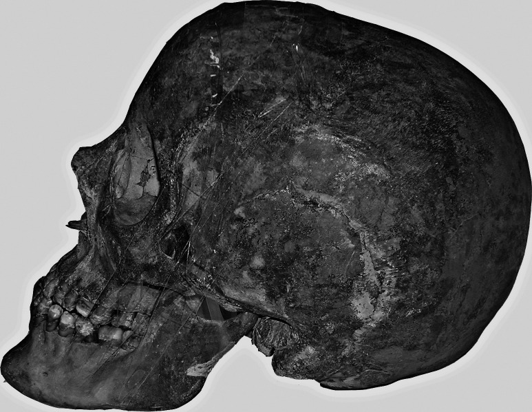
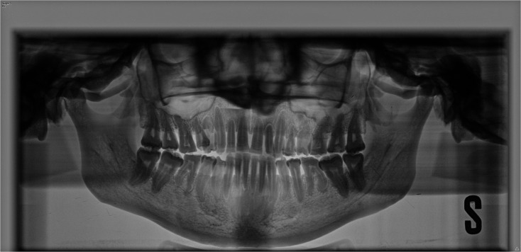
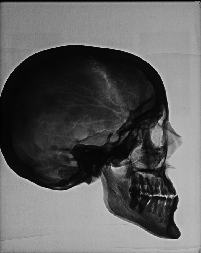
The skull, the panoramic radiograph and the lateral cephalogram of Joanna of Austria were analyzed (Figures 1 through 4). On the skull and on the panoramic radiograph the macroscopic features of the dentition were evaluated (Figures 1 through 3). A customized digitization regimen and analysis provided by cephalometric software (Viewbox 3.0., dHAL Software, Kifissia, Greece) was utilized for the cephalometric analysis on the digital lateral cephalogram (Figure 4). The customized cephalometric analysis contained measurements from the analyses of Downs,17 Steiner,18 Björk,19 and Jacobson,20 and it comprised 20 variables, 12 angular and 8 linear. The lateral cephalogram was taken with the mandible stabilized into a simulated position in centric occlusion (Figures 2 and 4). The magnification of the lateral cephalogram was standardized to an 8% enlargement. The cephalometric values of Joanna were compared to cephalometric standards for adult female subjects.21
Figure 1.
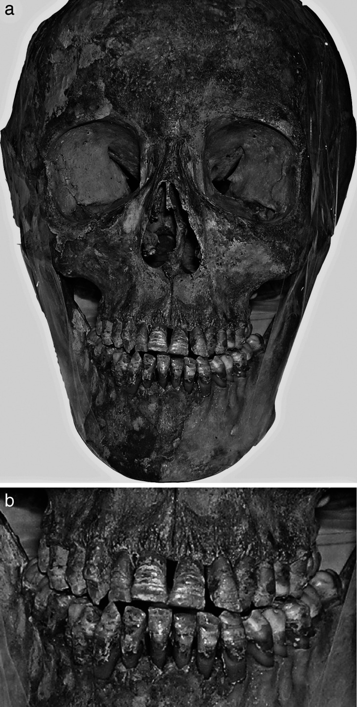
(a) The dry skull of Joanna of Austria (frontal view). (b) Detail of the dentition. Both images used with the permission of the Soprintendenza Speciale al Polo Museale Fiorentino, Firenze, Italy – Medici Project.
Figure 2.
The dry skull of Joanna of Austria (lateral view). Image used with the permission of the Soprintendenza Speciale al Polo Museale Fiorentino, Firenze, Italy – Medici Project.
Figure 3.
Panoramic radiograph of Joanna of Austria. Image used with the permission of the Soprintendenza Speciale al Polo Museale Fiorentino, Firenze, Italy – Medici Project.
Figure 4.
Lateral cephalogram of Joanna of Austria. Image used with the permission of the Soprintendenza Speciale al Polo Museale Fiorentino, Firenze, Italy – Medici Project.
RESULTS
The analysis of the dry skull and of the panoramic radiograph (Figures 1 through 3) showed that all the permanent teeth were present. The visual analysis of the permanent teeth on the skull revealed the presence of enamel defects organized in horizontal grooves that were more evident on vestibular surfaces of the upper and lower incisors and canines. The incisor edges presented with marked signs of abrasion probably related to the anterior occlusal trauma. The upper right permanent canine exhibited a more marked enamel loss on the buccal surface probably because of a traumatic event to the skull. The upper right second premolar showed a palatine malposition and a 90-degree rotation with a contraction of the ipsilateral hemiarch. The upper right second molar showed a loss of hard tissue on the occlusal surface at the mesial and distal buccal cusps. A generalized horizontal alveolar bone resorption with severe bone loss was evident. The first molars showed furcation exposure. The panoramic radiograph (Figure 3) confirmed the generalized severe horizontal bone loss that could be interpreted as a sign of severe periodontal disease with respect to the young age.
An asymmetry in the heights of the left mandibular ramus and of the right mandibular ramus measured on the panoramic radiograph was recorded (59 mm and 55 mm, respectively).22 This discrepancy in height was due to the neck of the left condyle that was 4 mm longer than the right one, and was an abnormal shape.
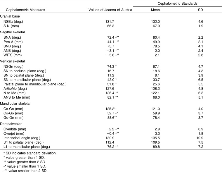
The cephalometric analysis showed a normal inclination of the cranial base (NSBa) with a normal dimension of the anterior cranial base (S-N) (Table 1). As for the sagittal skeletal relationships, a skeletal Class III disharmony was associated with maxillary retrusion and normal sagittal position of the mandible. The maxilla also exhibited a reduction in the sagittal dimension (Ptm-A) with respect to the reference standard. The mandible presented with increased dimensions both in total mandibular length (Co-Gn) and in the mandibular body (Go-Gn). The mandibular ramus (Co-Go), however, was shorter when compared to the cephalometric reference standard. Joanna showed increased vertical skeletal relationships with a posterior inclination of the mandibular plane to the cranial base (SN to mandibular plane) associated with an increased total and lower anterior facial heights (N to Me and ANS to Me). The skeletal open bite contributed to the lack of mandibular protrusion though in presence of increased mandibular sagittal dimensions. The lower incisors were inclined lingually, thus revealing a tendency to dentoalveolar compensation of the Class III relationship.
Table 1.
Cephalometric Values Shown by Joanna of Austria with Respect to Cephalometric Standards
At the occlusal level Joanna showed Class III malocclusion with Class III canine and molar relationships, a negative overjet (−2.2 mm), and anterior open bite (overbite = −0.4 mm). The lower midline was slightly deviated to the left side. As for the transverse relationships, there was a bilateral posterior crossbite. The analysis of the posterior transverse interarch discrepancy23 showed that the maxillary intermolar width measured as the distance between the central fossae of the upper first molars was smaller (43 mm) than the mandibular intermolar width measured as the distance between the tips of the distobuccal cusps of the lower first molars (51 mm), thus revealing a severe negative posterior transverse interarch discrepancy of −8 mm.
DISCUSSION
The Medici Project had the opportunity to re-exhume the body of Joanna of Austria who had been buried in the New Sacristy in the Basilica of San Lorenzo in Florence. As part of this research project, the current study aimed to describe for the first time the dentoskeletal features of the “Habsburg jaw” that affected the Grand Duchess of Tuscany. The Habsburg family has always attracted the interest of plastic surgeons, orthodontists, and oral surgeons because of the presence of mandibular prognathism, and the interest of geneticists because of its occurrence in several successive generations of the family. Examination of the abundant portraits of the Habsburg family shows, in addition to mandibular prognathism, a thick, everted lower lip, a large, often misshapen nose with a prominent dorsal hump, a tendency to flattening of the malar areas, and mild eversion of the lower eyelids.9
The cephalometric analysis showed that Joanna was affected by Class III skeletal disharmony. It has been shown24,25 that both ANB angle and Wits appraisal can be influenced by the length and inclination of the anterior cranial base as well as by the inclination of the occlusal plane to the cranial base. In the case of Joanna both ANB and Wits appraisal were concordant in demonstrating the presence of Class III skeletal disharmony because the length and inclination of the anterior cranial base as well as the inclination of the occlusal plane to the cranial base were within normal values (Table 1). Interestingly, Joanna of Austria exhibited Class III skeletal disharmony that was due to a severe retrusion of the maxilla rather than to mandibular prognathism. The maxilla exhibited a deficiency both in the sagittal and transverse dimensions. The mandible presented with increased dimensions when measured both along total mandibular length (Co-Go) and along the mandibular body (Go-Me). The skeletal open bite, however, masked the mandibular dimensional excess leading to an improvement to the skeletal Class III disharmony with mandibular protrusion within normal limits. It is interesting to note that the dentoskeletal features of Joanna of Austria are similar to those of Class III European white subjects with maxillary retrognathism who demonstrate a tendency toward vertical growth as a possible compensation mechanism of Class III malocclusion.26 On the contrary, Class III subjects with mandibular prognathism tend to exhibit a horizontal facial growth pattern and typically include more pronounced dentoalveolar compensation, that is, proclination of maxillary and retroclination of mandibular incisors.26
The Class III long-face profile that affected Joanna of Austria is clearly testified by the portraits of the noble woman (Figure 5). Joanna appears with a peculiar type of “Habsburg jaw” as the Class III profile was related to the presence of a marked maxillary retrusion rather than to mandibular prognathism. This is in contrast with a preliminary observation16 of the lateral cephalogram of Joanna of Austria that reported the presence of mandibular prognathism. In this preliminary observation, however, no cephalometric analysis had been performed.
Figure 5.
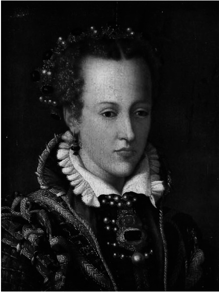
Portrait of Joanna of Austria by Alessandro Allori, 1570, Museo degli Argenti, Firenze, Italy. Image used with the permission of the Soprintendenza Speciale al Polo Museale Fiorentino, Firenze, Italy – Medici Project.
The analysis of the skull and of the panoramic radiograph allowed researchers to identify that Joanna suffered from a severe periodontal disease at the young age of 31 years. The visual analysis of the permanent teeth on the skull revealed the presence of enamel defects organized in horizontal grooves that were more evident on vestibular surfaces of the upper and lower incisors and canines (Figure 1B). At the time of exhumation in 2005, Fornaciari et al.15 already formulated the diagnosis of amelogenesis imperfecta (AI). AI is a genetic disease that affects the enamel of both the deciduous and the permanent teeth. Recent advances in the understanding of the genetics of AI allow for a better interpretation of the peculiar type of AI shown by Joanna. The AIs are a clinically and genetically diverse group of conditions that are caused by mutations in a variety of genes that provide instructions for the expression of proteins for normal enamel formation. All of the X-linked forms of AI (OMIM 300391) with a known molecular basis are associated with mutations in the AMELX gene (located at chromosome Xp22) that codes for the protein amelogenin.27 In addition to amelogenin there are numerous other extracellular matrix components in developing enamel, including proteins such as ameloblastin, enamelin, and proteinases that are required to process the matrix proteins during mineralization.28 It has been shown that mutations in the genes encoding for enamel matrix proteins like enamelin and ameloblastin (4q11-q21) are responsible for autosomal forms of amelogenesis imperfecta.29 In particular, Mårdh et al.30 have shown that the local hypoplastic phenotype resulting from enamelin coding gene mutations that stop protein production from one allele is characterized by horizontal hypoplastic grooves very similar to those shown by Joanna's teeth. It should also be stressed that an association of Class III malocclusions and/or open bites with AI has been recognized.31 It would be interesting, therefore, to analyze the presence of this type of dental anomaly in other members of the Habsburg family, especially in those exhibiting open bite Class III malocclusion.
CONCLUSIONS
The analysis of the craniofacial features of Joanna of Austria helped shed new light on a typical stigma that has been found in many members the Royal House of the Habsburg: the so-called “Habsburg jaw.”
Cephalometric analysis of the lateral cephalogram of Joanna of Austria revealed that the Class III skeletal disharmony was due to a retrognathic maxilla rather than to a prognathic mandible.
The increased vertical skeletal relationship (long face) masked the protrusion of the mandible that showed increased sagittal dimensions.
REFERENCES
- 1.Grabb W. C, Hodge G. P, Dingman R. O, Oneal R. M. The Habsburg jaw. Plast Reconstr Surg. 1968;42:442–445. [PubMed] [Google Scholar]
- 2.Hart G. D. The Habsburg jaw. Can Med Assoc J. 1971;104:601–603. [PMC free article] [PubMed] [Google Scholar]
- 3.Hodge G. P. A medical history of the Spanish Habsburgs. As traced in portraits. J Am Med Assoc. 1977;238:1169–1174. [PubMed] [Google Scholar]
- 4.Wolff G, Wienker T. F, Sander H. On the genetics of mandibular prognathism: analysis of large European noble families. J Med Genet. 1993;30:112–116. doi: 10.1136/jmg.30.2.112. [DOI] [PMC free article] [PubMed] [Google Scholar]
- 5.Xue F, Wong R. W, Rabie A. B. Genes, genetics, and Class III malocclusion. Orthod Craniofac Res. 2010;13:69–74. doi: 10.1111/j.1601-6343.2010.01485.x. [DOI] [PubMed] [Google Scholar]
- 6.Litton S. F, Ackermann L. V, Isaacson R. J, Shapiro B. L. A genetic study of Class III malocclusion. Am J Orthod Dentofacial Orthop. 1970;58:565–77. doi: 10.1016/0002-9416(70)90145-4. [DOI] [PubMed] [Google Scholar]
- 7.El-Gheriani A. A, Maher B. S, El-Gheriani A. S, Sciote J. J, Abu-Shahba F, Al-Azemi R, Marazita M. L. Segregation analysis of mandibular prognathism in Libya. J Dent Res. 2003;82:523–527. doi: 10.1177/154405910308200707. [DOI] [PubMed] [Google Scholar]
- 8.Cruz R. M, Krieger H, Ferreira R, Mah J, Hartsfield J, Jr, Oliveira S. Major gene and multifactorial Inheritance of mandibular prognathism. Am J Med Genet A. 2008;146A:71–77. doi: 10.1002/ajmg.a.32062. [DOI] [PubMed] [Google Scholar]
- 9.Chudley A. E. Genetic landmarks through philately-the Habsburg jaw. Clin Genet. 1998;54:283–284. doi: 10.1034/j.1399-0004.1998.5440404.x. [DOI] [PubMed] [Google Scholar]
- 10.Vicek E, Smahel Z. Contribution to the origin of progeny in middle European Habsburgs: skeletal roentgencephalometric analysis of the Habsburgs buried in Prague. Acta Chir Plast. 1997;39:39–47. [PubMed] [Google Scholar]
- 11.Pieraccini G. La stirpe dei Medici di Cafaggiolo vol 2. Firenze: Nardini Editore; 1986. pp. 32–47. [Google Scholar]
- 12.Genna G. Ricerche antropologiche sulla famiglia dei Medici. Atti Accademia Nazionale dei Lincei Classe di Scienze Fisiche Matematiche e Naturali Serie VIII. 1948;15:589–593. [Google Scholar]
- 13.Aufderheide A. C, Rodriguez-Martin C. Human Paleopathology. Cambridge: Cambridge University Press; 1988. p. 405. [Google Scholar]
- 14.Fornaciari G, Vitiello A, Giusiani S, Giuffra V, Fornaciari A. The “Medici Project”: first results of the explorations of the Medici tombs in Florence (15th–18th centuries) Paleopath Newsl. 2006;133:15–22. [Google Scholar]
- 15.Fornaciari G. Il “Progetto Medici”: primi risultati dello studio paleopatologico dei Granduchi di Toscana (secoli XVI-XVIII) Arch Antropol Etnol. 2009;138:138–157. [Google Scholar]
- 16.Villari N, Fornaciari G, Lippi D, Cerinic M. M, Ginestroni A, Pellicanò G, Mascalchi M. Scenes from the past: the Medici Project: radiographic survey. Radiographics. 2009;29:2101–2114. doi: 10.1148/rg.297085212. [DOI] [PubMed] [Google Scholar]
- 17.Downs W. B. Variations in facial relationships: their significance in treatment and prognosis. Am J Orthod. 1948;34:812–840. doi: 10.1016/0002-9416(48)90015-3. [DOI] [PubMed] [Google Scholar]
- 18.Steiner C. C. Cephalometrics for you and me. Am J Orthod. 1953;39:729–755. [Google Scholar]
- 19.Björk A. Follow-up X-ray study of the individual variation in growth occurring between of 12 and 20 years and its relation to brain case and face development. Am J Orhod. 1955;41:199–255. [Google Scholar]
- 20.Jacobson A. The “Wits” appraisal of jaw disharmony. Am J Orthod. 1975;67:125–138. doi: 10.1016/0002-9416(75)90065-2. [DOI] [PubMed] [Google Scholar]
- 21.Bathia S. N, Leighton B. C. A manual of facial growth. Oxford, NY: Oxford University Press; 1993. pp. 40–375. [Google Scholar]
- 22.Kambylafkas P, Murdock E, Gilda E, Tallents R. H, Kyrkanides S. Validity of panoramic radiographs for measuring mandibular asymmetry. Angle Orthod. 2006;76:388–393. doi: 10.1043/0003-3219(2006)076[0388:VOPRFM]2.0.CO;2. [DOI] [PubMed] [Google Scholar]
- 23.Tollaro I, Baccetti T, Franchi L, Tanasescu C. D. Role of posterior transverse interarch discrepancy in Class II, Division 1 malocclusion during the mixed dentition phase. Am J Orthod Dentofacial Orthop. 1996;110:417–422. doi: 10.1016/s0889-5406(96)70045-8. [DOI] [PubMed] [Google Scholar]
- 24.Hussels W, Nanda R. S. Analysis of factors affecting angle ANB. Am J Orthod. 1984;85:411–423. doi: 10.1016/0002-9416(84)90162-3. [DOI] [PubMed] [Google Scholar]
- 25.Del Santo M., Jr Influence of occlusal plane inclination on ANB and Wits assessments of anteroposterior jaw relationships. Am J Orthod Dentofacial Orthop. 2006;129:641–648. doi: 10.1016/j.ajodo.2005.09.025. [DOI] [PubMed] [Google Scholar]
- 26.Spalj S, Mestrovic S, Lapter Varga M, Slaj M. Skeletal components of class III malocclusions and compensation mechanisms. J Oral Rehabil. 2008;35:629–637. doi: 10.1111/j.1365-2842.2008.01869.x. [DOI] [PubMed] [Google Scholar]
- 27.Wright J. T, Hart P. S, Aldred M. J, Seow W. K, Crawford P. J. M, Hong S. P, Gibson C, Hart T. C. Relationship of phenotype and genotype in X-linked amelogenesis imperfecta. Connect Tissue Res. 2003;44:72–78. [PubMed] [Google Scholar]
- 28.Wright J. T. The molecular etiologies and associated phenotypes of amelogenesis imperfecta. Am J Med Genet A. 2006;140:2547–2555. doi: 10.1002/ajmg.a.31358. [DOI] [PMC free article] [PubMed] [Google Scholar]
- 29.Kim J. W, Seymen F, Lin B. P, Kiziltan B, Gencay K, Simmer J. P, Hu J. C. ENAM mutations in autosomal-dominant amelogenesis imperfecta. J Dent Res. 2005;84:278–282. doi: 10.1177/154405910508400314. [DOI] [PubMed] [Google Scholar]
- 30.Mårdh C. K, Bäckman B, Holgren G, Hu J. C-C, Simmer J. P, Forsman-Semb K. A nonsense mutation in the enamelin gene causes local hypoplastic autosomal dominant amelogenesis imperfecta (AIH2) Human Mol Genet. 2002;11:1069–1074. doi: 10.1093/hmg/11.9.1069. [DOI] [PubMed] [Google Scholar]
- 31.Ravassipour D. B, Powell C. M, Phillips C. L, Hart P. S, Hart T. C, Boyd C, Wright J. T. Variation in dental and skeletal open bite malocclusion in humans with amelogenesis imperfecta. Arch Oral Biol. 2005;50:611–623. doi: 10.1016/j.archoralbio.2004.12.003. [DOI] [PubMed] [Google Scholar]


