Abstract
Objective:
To investigate the dependence of force decay on the initial strain applied to currently available elastic chains.
Materials and Methods:
Eight different elastic chains from eight major companies were tested for force decay over a period of 3 weeks at 50% and 100% strain. They were stored in water and thermocycled between 5°C and 55°C. An Instron 3344 was used for the force measurements.
Results:
Absolute force values at 50% strain varied between 2.3 N and 4.1 N initially, and between 0.9 N and 1.6 N after 21 days. Thus, the force decay of the elastic chains varied from 37% to 75%. At 100% strain, the force values varied between 2.9 N and 4.7 N initially, and between 1.3 N and 2.1 N after 21 days of continuous strain. The force decay varied between 39% and 67%. Most force decays between 24 hours and 21 days were not significant. This information should be taken into consideration when the appropriate elastic chain is selected for clinical use.
Conclusion:
A wide array of elastic chains with various force levels is available. However, differences between products of greater than 100% were measured for force decay over time.
Keywords: Elastic chains, Initial strain
INTRODUCTION
Elastic chains are widely used in combination with fixed orthodontic appliances to close or to prevent the opening of spaces. Their main advantages include the following: ease of use, low price, reduced potential for intraoral trauma, minimal need for patient compliance, and wide array of colors or transparency. Their disadvantages can be seen in inconsistency of force levels over time, absorption of fluids leading to discoloration, and impairment of oral hygiene. Synthetic elastic chains are made from polyurethane, a linear polymer produced through a chemical reaction between diisocyanate and a polyol.1,2 The mechanical properties can vary widely from soft to hard and from elastic to brittle. Since their introduction in the 1960s, manufacturers have tried to overcome their deficiencies by modifying the material composition (Power Chain, Rocky Mountain Orthodontics [RMO], RMO Europe, Strasbourg, France) or the design (Elast-O-Chain, TP Orthodontics Inc, La Porte, Ind). The main focus of developing research has been to deliver light continuous and constant forces over the clinically meaningful time period of 4 to 8 weeks.
Many investigations of the mechanical behavior of elastic chains have looked at different parameters, including force decay over time,3–10 force decay at different levels of activation,3,9,11 simulated space closure,6,12 prestretching of the elastic chains,3,10,13–15 environmental factors and storage media,4,7,14,15 and chain designs.3,12 These studies however are difficult to compare because the experimental designs vary widely. Force decay has been reported to be rapid within the first 24 hours with loss of 50% to 70% of the initial value. Thereafter, a more stable phase has been reported with only minor changes of 10% to 20% up to 4 weeks.2–4,6,12,16–18 This decay in initial force levels was dependent on the storage media; stronger decay was noted in a humid environment compared with dry air.4 Thermocycling during storage slightly increases the residual force levels.6 Acid storage media were found to be more favorable than bases8 and phosphate fluorides have been found to influence force levels.19 Preconditioning of the elastic chains with heat20 led to reduced residual force levels, and preconditioning by prestretching showed an inconclusive behavior, most often leading to marginally (5%) less force decay.14,15,20–22
However, over the past decade, only three evaluations of force decay of elastomeric chains, including altogether four companies, were published in the leading orthodontic journals.8,23,24 Because material development plays a significant role in orthodontic progress, an overview of currently available elastic chains from most major companies was undertaken, especially looking at force decay after different initial loading.
MATERIALS AND METHODS
Eight elastic chains from different companies were included in the study (Table 1). All specimens had a length of 10 ringlets. On each side, half of an additional ringlet was left in place. The sample size for each product was 20. Each product was split into two groups of 10 samples. In one group, the chains were stretched by 50%, and in the other by 100%, of the initial length (Table 1). All chains were tested at the following time intervals: 0, 2, 8, and 24 hours and 7 and 21 days, yielding a total of 960 measurements.
Table 1.
Manufacturers, Product Identification, Length of the Samples, and Reference and Lot Numbers of All Elastic Chains Testeda
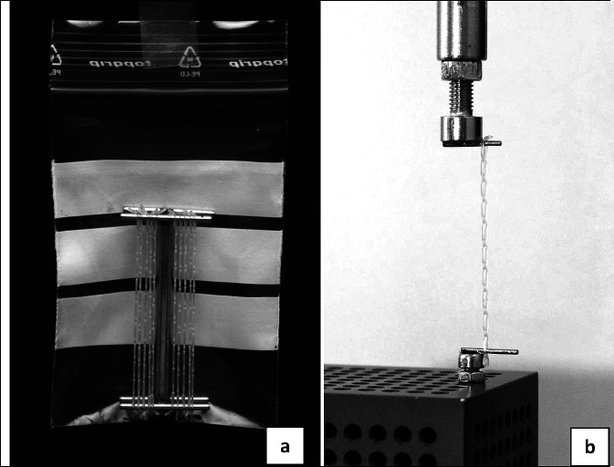
Metal holders were soldered individually for each product, according to the length needed for the appropriate strain. All 10 chains of one group were attached to the metal holders (Figure 1a) and stored in small plastic bags, filled with tap water. During storage, they were exposed to continuous thermocycles between 5°C and 55°C twice an hour in a fully automated CTS Clima chamber (CTS Clima, T-40/25, Lauda Dr R Wobser GmbH & Co KG, Lauda-Köngishofen, Germany). Additionally, temperature cycles were controlled with a thermometer inside one plastic bag.
Figure 1.
Experimental setup with individually soldered metal holders placed in plastic bags and filled with tap water for storage of chains at the determined strain (a). Care was taken to slide the chains on and off the measurement pins of the Instron frame without losing their strain (b).
Measurements were recorded with an Instron 3344 (Instron Corp, Wilmington, Del). The distance between the pins in the Instron measurement frame was altered to the appropriate length, which corresponded to the distance of the metal holder, and care was taken to slide the chains on and off the measurement pins without varying their strain (Figure 1b). After measurements were taken, the chains were again placed in the CTS Clima chamber.
All groups were tested for normality (Shapiro Wilk) and were evaluated by means of Kruskal-Wallis with Dunn's posttest at a significance level of P ≤ .05 using Prism (GraphPad, San Diego, Calif), because normal distribution was not achieved for all groups. In addition, a mean curve consisting of all measurements was calculated for both strains. It served as a baseline for comparison of the relative characteristics of both strains.
RESULTS
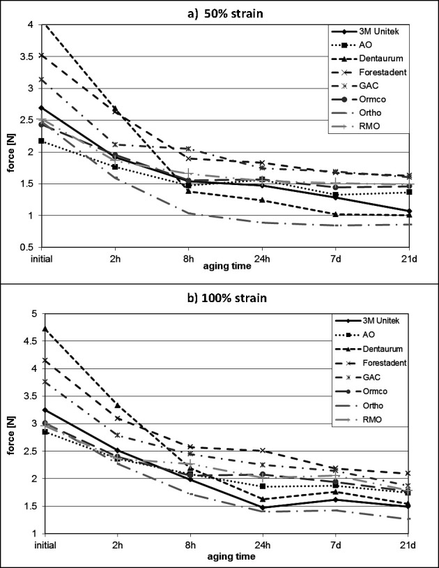
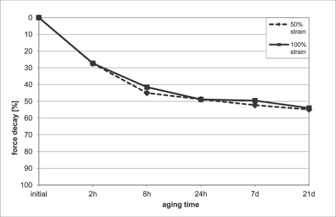
The results are summarized in Table 2. Figure 2 shows the mean curves for each group individually at 50% and 100% strain, respectively, and Figure 3 shows the mean curves calculated from all groups combined, comparing the relative behaviors of the chains under different strains. Standard deviations for all measurements were very low, with only 16 measurements out of 96 showing a standard deviation greater than 5% of the mean value.
Table 2.
Mean, Standard Deviation (in parentheses) and Percentage Force Decay (bottom line) of All Chains at Both Strain Levels for All Time Intervalsa,b
Figure 2.

Force decay curves of all chains at 50% (a) and 100% (b) strain. The chains of American Orthodontics, Ormco, and Rocky Mountain Orthodontics showed the lowest percentage force decay from their initial force levels.
Figure 3.

Percentage of force decay. Comparison of all chains combined at 50% and 100% strain. The percentage of force decay showed no dependency on initial strain.
At 50% strain, initial force values were found between 2.3 N (American Orthodontics, Sheboygan, Wis) and 4.1 N (Dentaurum GmbH & Co KG, Ispringen, Germany), and final force levels after 21 days of continued strain between 0.9 N (Ortho Organizers Inc, Melville, NY) and 1.6 N (Forestadent, Pforzheim, Germany). The percentage of force decay varied from 28% to 70% within the first 24 hours, and from 37% to 75% after 21 days. When significant differences in force decay of the elastic chains of American Orthodontics were examined, Ormco (Orange, Calif) and Rocky Mountain Orthodontics (Denver, Colo) showed the least force reduction. When the groups were combined, the force decay between different time intervals was significant for most measurements on the first day, but was not significant for the measurements between 24 hours and 21 days.
At 100% strain, all force levels were higher compared with respective levels at 50% strain. Initial force values were found between 2.9 N (American Orthodontics) and 4.7 N (Dentaurum), and final force levels after 21 days of continued strain were between 1.3 N (Ortho Organizers) and 2.1 N (Forestadent). The percentage of force decay varied from 31% to 66% within the first 24 hours, and from 39% to 67% after 21 days. Again, the best elastics with regard to force decay were those from American Orthodontics, Ormco, and Rocky Mountain Orthodontics. For all chains combined, force decay between the different time intervals was significant for all measurements on the first day, but was not significant for measurements between 24 hours and 21 days.
When all chains combined at 50% vs all chains combined at 100% strain were compared, significant differences in force decay were noted for the time intervals between 2 hours and 8 hours, and between 24 hours and 7 days. All other time intervals did not show significant differences.
DISCUSSION
Despite ongoing debate on the use of in vitro or in vivo designs, the in vitro design has several advantages when it comes to the characterization of materials. The oral cavity presents an environment that is very difficult to standardize. Variations in microbial flora and enzyme levels, as well as exposure to dietary factors and different functional forces, result in poor validity in terms of the evaluation of specific material properties. This problem was also observed in an earlier study comparing canine retraction vs elastic chain or thread.25 Standard deviations in tooth movement were approximately 40% of mean values, resulting in insignificant differences between the products tested. In comparison, standard deviations of the current study were mostly below 5% of the mean value. For baseline comparisons, it seems therefore more appropriate to compare different products in a constant environment, keeping in mind that the behavior of materials tested in the oral environment may differ from laboratory results.
Methodologically comparative studies of in vitro and in vivo investigations of elastic chains have revealed strong differences between dry and in vivo storage, but only small differences between storage in water or in vivo.4,26 The use of thermocycles, although significant, led to only small differences.6 The effect of prestretching on the force decay response of elastic chains is controversial; most studies have found a slight reduction in force decay.14,15,20–22 To guarantee better standardization in the current investigation, water was chosen as the storage medium, and no prestretching was performed. The use of thermocycles may be considered disadvantageous for standardization. However, because thermocycling was performed in a fully automated oven, and all chains were measured during the same time period, the differences in stress induction were regarded as insignificant.
Results showed a huge variation in initial force levels as well as in force decay over the time intervals. This is consistent with earlier literature on elastic chains.3,4,6,10–12,14,18,22,26–29 When a strain of 50% of the initial length of the chains was applied, initial force values ranged from 2.3 N to 4.1 N, and at 100% from 3 N to 4.7 N. These differences might be considered clinically important and, in the case of forces exceeding 3 N, clinically excessive for movement of a single tooth.30–33 However, at 1 hour of strain, the force levels of all chains were in an acceptable clinical range of 1.6 N to 2.7 N at 50% strain, and 2.3 N to 3.3 N at 100% strain. Of greater importance than initial force levels was the force decay of the chains. Force decay has been shown to exhibit large variations between different products,1–4,10,28 and this was confirmed in the current investigation with similar behavior for the two strains. The lowest force decay after the 3-week period following a 50% and a 100% strain was found for the American Orthodontics chains at 37% and 39%, respectively. When compared with the largest force decay, which was reported with the Dentaurum chain at 75% and 67%, these differences were highly significant and of clinical interest. When force decay was examined over different time intervals, it was found that most changes were insignificant during the time periods of 1 day to 3 weeks. When the groups for one strain were combined, the difference in force decay between 1 day and 3 weeks was not significant for the 50% strain or the 100% strain. The large amount of force decay within the first 24 hours was followed by mostly consistent force levels up to 3 weeks, as is confirmed in the literature.1,3,6,10,28 Longer test periods have revealed only slight reductions in force levels after this time period,9,12,16,17 with the longest test period extending to 100 days.22 Force levels at the clinical control at chairside at 4 to 5 weeks therefore will probably be similar to those at 3 weeks. In the current study, three chains showed markedly less reduction in force; these were the chains from American Orthodontics, Ormco, and Rocky Mountain Orthodontics. For these chains, the reduction in force after 3 weeks was approximately 40% of the initial value. The graphs indicate that regardless of whether the strain was 50% or 100%, the rate of force decay was similar and was not dependent on initial strain. Results showed that within the idealized in vitro environment, highly significant differences in force decay were found for different products. These differences in force decay might be due to linear or cross-linked polymer composition, as well as to thermoplastic or thermoset materials. However, this was not verified in the present study. To clinically apply the most controlled force levels, appropriate products should be selected and initial forces measured to estimate the remaining force levels between 24 hours and subsequent chairside control.
CONCLUSIONS
The force levels of chains from different manufacturers vary widely, with some chains showing excessive initial forces at 50% and 100% strain.
Loss of force over 3 weeks was pronounced during the first 24 hours, with only minor loss noted in the remaining time period up to 3 weeks.
Loss of force, depicted as a percentage of the initial force levels, was dependent only on the type of chain being tested, not on the initial activation.
Three chains were identified as having superior characteristics with respect to loss of force. These had only 40% loss of force after 3 weeks.
REFERENCES
- 1.Billmeyer F. W. Textbook of Polymer Science 3rd ed. New York: John Wiley; 1984. [Google Scholar]
- 2.Wong A. K. Orthodontic elastic materials. Angle Orthod. 1976;46:196–205. doi: 10.1043/0003-3219(1976)046<0196:OEM>2.0.CO;2. [DOI] [PubMed] [Google Scholar]
- 3.Andreasen G. F, Bishara S. E. Comparison of alastik chains and elastics involved with intra-arch molar to molar forces. Angle Orthod. 1970;40:151–158. doi: 10.1043/0003-3219(1970)040<0151:COACWE>2.0.CO;2. [DOI] [PubMed] [Google Scholar]
- 4.Ash J, Nikolai R. Relaxation of orthodontic elastic chains and modules in vitro and in vivo. J Dent Res. 1978;57:685–690. doi: 10.1177/00220345780570050301. [DOI] [PubMed] [Google Scholar]
- 5.Cofflet M, von Fraunhofer J. The Effects of Artificial Saliva and Topical Fluoride Treatment on Degradation of the Elastic Properties of Orthodontic Chains [master's thesis] Louisville, Ky: University of Louisville; 1991. [DOI] [PubMed] [Google Scholar]
- 6.De Genova D. C, McInnes-Ledoux P, Weinberg R, Shaye R. Force degradation of orthodontic elastomeric chains—a product comparison study. Am J Orthod. 1985;87:377–384. doi: 10.1016/0002-9416(85)90197-6. [DOI] [PubMed] [Google Scholar]
- 7.Ferriter J. P, Meyers C. E, Jr, Lorton L. The effect of hydrogen ion concentration on the force-degradation rate of orthodontic polyurethane chain elastics. Am J Orthod Dentofacial Orthop. 1990;98:404–410. doi: 10.1016/S0889-5406(05)81648-8. [DOI] [PubMed] [Google Scholar]
- 8.Kim K. H, Chung C. H, Choy K, Lee J. S, Vanarsdall R. L. Effects of prestreching on force degradation of synthetic elastomeric chain. Am J Orthod Dentofacial Orthop. 2005;128:477–482. doi: 10.1016/j.ajodo.2004.04.027. [DOI] [PubMed] [Google Scholar]
- 9.Lu T. C, Wang W. N. Force decay of elastomeric chain. China Dent J. 1988;7:74–79. doi: 10.1016/S0889-5406(05)81336-8. [DOI] [PubMed] [Google Scholar]
- 10.Wong A. Orthodontic elastic materials. Angle Orthod. 1976;46:196–205. doi: 10.1043/0003-3219(1976)046<0196:OEM>2.0.CO;2. [DOI] [PubMed] [Google Scholar]
- 11.Storie D, von Fraunhofer J, Regennitter F. Degradation and Therapeutic Potential of Fluoride Releasing Orthodontic Elastic [master's thesis] Louisville, Ky: University of Louisville; 1992. [Google Scholar]
- 12.Hershey G, Reynolds W. The plastic module as an orthodontic tooth moving mechanism. Am J Orthod. 1975;67:554–662. doi: 10.1016/0002-9416(75)90300-0. [DOI] [PubMed] [Google Scholar]
- 13.Andreasen G. F, Bishara S. E. Relaxation of orthodontic elastomeric chains and modules in vitro and in vivo. Angle Orthod. 1970;40:319–328. [Google Scholar]
- 14.Brantley W, Salander S, Meyers L, Winders R. Effects of prestretching on force degradation characteristics of plastic modules. Angle Orthod. 1979;49:37–43. doi: 10.1043/0003-3219(1979)049<0037:EOPOFD>2.0.CO;2. [DOI] [PubMed] [Google Scholar]
- 15.Young J, Sandrik J. Influence of preloading on stress relaxation of orthodontic elastic polymers. Angle Orthod. 1979;49:104–109. doi: 10.1043/0003-3219(1979)049<0104:TIOPOS>2.0.CO;2. [DOI] [PubMed] [Google Scholar]
- 16.Baty D. L, Storie D. J, von Fraunhofer J. A. Synthetic elastomeric chains: a literature review. Am J Orthod Dentofacial Orthop. 1994;105:536–542. doi: 10.1016/S0889-5406(94)70137-7. [DOI] [PubMed] [Google Scholar]
- 17.Bishara S. E, Andreasen G. F. A comparison of time-related forces between plastic alastiks and latex elastics. Angle Orthod. 1970;40:319–328. doi: 10.1043/0003-3219(1970)040<0319:ACOTRF>2.0.CO;2. [DOI] [PubMed] [Google Scholar]
- 18.Killiany D. M, Duplessis J. Relaxation of elastomeric chains. J Clin Orthod. 1985;19:592–593. [PubMed] [Google Scholar]
- 19.Von Fraunhofer J. A, Coffelt M. T, Orbell G. M. The effects of artificial saliva and topical fluoride treatments on the degradation of the elastic properties of orthodontic chains. Angle Orthod. 1992;62:265–274. doi: 10.1043/0003-3219(1992)062<0265:TEOASA>2.0.CO;2. [DOI] [PubMed] [Google Scholar]
- 20.Brooks D. G, Hershey H. G. Effects of heat and time on stretched plastic orthodontic modules [abstract] J Dent Res. 1976;55:363. [Google Scholar]
- 21.Chang H. F. Effects of instantaneous prestretching on force degradation characteristics of orthodontic plastic modules. Proc Natl Sci Counc Repub China B. 1987;11:45–53. [PubMed] [Google Scholar]
- 22.Williams J, von Fraunhofer J. A. Degradation of the Elastic Properties of Orthodontic Chains [thesis] Louisville, Ky: University of Louisville; 1990. [Google Scholar]
- 23.Bosquet J, Tuesta O, Flores-Mir C. In vivo comparison of force decay between injection molded and die-cut stamped elastomers. Am J Orthod Dentofacial Orthop. 2006;129:384–389. doi: 10.1016/j.ajodo.2005.09.002. [DOI] [PubMed] [Google Scholar]
- 24.Eliades T, Eliades G, Silikas N, Watts D. C. Tensile properties of orthodontic elastomeric chains. Eur J Orthod. 2004;26:157–162. doi: 10.1093/ejo/26.2.157. [DOI] [PubMed] [Google Scholar]
- 25.Sonis A. L, Van der Plas E, Gianelly A. A comparison of elastomeric auxiliaries versus elastic thread on premolar extraction site closure: an in vivo study. Am J Orthod. 1986;89:73–77. doi: 10.1016/0002-9416(86)90115-6. [DOI] [PubMed] [Google Scholar]
- 26.Kuster R, Ingervall B, Burgin W. Laboratory and intraoral test of the degradation of elastic chains. Eur J Orthod. 1986;8:202–208. doi: 10.1093/ejo/8.3.202. [DOI] [PubMed] [Google Scholar]
- 27.Kovatch J, Lautenschlager D, Keller J. Load extensions-time behavior of orthodontic alastiks. J Dent Res. 1976;55:783–786. doi: 10.1177/00220345760550051201. [DOI] [PubMed] [Google Scholar]
- 28.Rock W, Wilson H, Fisher S. A laboratory investigation of orthodontic elastomeric chains. Br J Orthod. 1985;12:202–207. doi: 10.1179/bjo.12.4.202. [DOI] [PubMed] [Google Scholar]
- 29.Baty D, von Fraunhofer J, Volz J. Force Displacement and Dimensional Stability of Various Colored Elastomeric Chains in Air Distilled Water and Artificial Saliva [master's thesis] Louisville, Ky: University of Louisville; 1992. [Google Scholar]
- 30.Reitan K. Some factors determining the evaluation of forces in orthodontics. Am J Orthod. 1957;43:32–45. [Google Scholar]
- 31.Boester C. H, Johnston L. E. A clinical investigation of the concepts of differential and optimal force in canine retraction. Angle Orthod. 1974;44:113–119. doi: 10.1043/0003-3219(1974)044<0113:ACIOTC>2.0.CO;2. [DOI] [PubMed] [Google Scholar]
- 32.Quinn R. S, Yoshikawa D. K. A reassessment of force magnitude in orthodontics. Am J Orthod. 1985;88:252–260. doi: 10.1016/s0002-9416(85)90220-9. [DOI] [PubMed] [Google Scholar]
- 33.Lotzof L. P, Fine H. A, Cisneros G. J. Canine retraction: a comparison of two preadjusted bracket systems. Am J Orthod Dentofacial Orthop. 1996;110:191–196. doi: 10.1016/s0889-5406(96)70108-7. [DOI] [PubMed] [Google Scholar]


