Abstract
Variant-specific loss of heterozygosity (LOH) analyses may be useful to classify BRCA1/2 germline variants of unknown significance (VUS). The sensitivity and specificity of this approach, however, remains unknown. We performed comparative next-generation sequencing analyses of the BRCA1/2 genes using blood-derived and tumour-derived DNA of 488 patients with ovarian cancer enrolled in the observational AGO-TR1 trial (NCT02222883). Overall, 94 pathogenic, 90 benign and 24 VUS were identified in the germline. A significantly increased variant fraction (VF) of a germline variant in the tumour indicates loss of the wild-type allele; a decreased VF indicates loss of the variant allele. We demonstrate that significantly increased VFs predict pathogenicity with high sensitivity (0.84, 95% CI 0.77 to 0.91), poor specificity (0.63, 95% CI 0.53 to 0.73) and poor positive predictive value (PPV; 0.71, 95% CI 0.62 to 0.79). Significantly decreased VFs predict benignity with low sensitivity (0.26, 95% CI 0.17 to 0.35), high specificity (1.0, 95% CI 0.96 to 1.00) and PPV (1.0, 95% CI 0.85 to 1.00). Variant classification based on significantly increased VFs results in an unacceptable proportion of false-positive results. A significantly decreased VF in the tumour may be exploited as a reliable predictor for benignity, with no false-negative result observed. When applying the latter approach, VUS identified in four patients can now be considered benign. Trial registration number NCT02222883.
Keywords: genetic predisposition to disease, genetic testing, genetic research, germ-line mutation
Introduction
In cancer genetics, individual risk stratification and the choice of targeted therapies are increasingly dependent on the germline mutation status in disease-associated genes such as BRCA1 (MIM: 113705) and BRCA2 (MIM: 600185). Thus, the unambiguous classification of germline variants identified in a routine diagnostic setting is vitally important for the clinical management of the individuals seeking advice. Criteria for BRCA1/2 germline variant classification were continuously standardised, in particular through the work of the Evidence-based Network for the Interpretation of Germline Mutant Alleles (ENIGMA), the International Agency for Research on Cancer (IARC) and the American College of Medical Genetics and Genomics (ACMG).1–4 While common BRCA1/2 germline variants with a minor allele frequency (MAF) ≥1% in the general population are considered benign by default, the classification of rare BRCA1/2 germline variants with a MAF <1% remains challenging, especially for those that cannot be predicted protein-truncating based on their mutation type. For intronic and missense variants, the multifactorial likelihood analysis demonstrated utility for quantitative assessment of variant pathogenicity, a model based on variant location, in silico prediction of variant effect, cosegregation, family cancer history, co-occurrence with a pathogenic variant in the same gene, tumour pathology and case–control information.3 The multifactorial likelihood analysis, however, requires input data that may not be available for all rare BRCA1/2 germline variants.
In tumours with a hereditary disease cause, it is generally suggested that the heterozygous germline inactivation of a predisposition gene may be accompanied by a somatic inactivation of the wild-type allele by another deleterious variant, loss of the wild-type allele or promoter methylation.5 In 473 patients with ovarian cancer (OC (MIM: 167 000)) enrolled in the observational AGO-TR1 trial, we demonstrated that pathogenic germline variants in the BRCA1/2 genes very rarely associate with deleterious somatic variants or promoter methylation.6 In OC, more than 80% of the pathogenic BRCA1/2 germline variants showed significantly increased proportion of reads that support the variant allele (variant allele fractions (VFs)) in the tumour-derived versus blood-derived DNA, indicating loss of the wild-type alleles.6–8 Based on these findings, it was suggested that loss of heterozygosity (LOH) analyses might be useful to classify rare germline variants in the BRCA1/2 genes.9 10 For rare germline variants in (candidate) cancer predisposition genes showing significantly increased VFs in the tumour, a potential role in cancer susceptibility was frequently suggested, as for example in the analyses of 429 patients with OC included in The Cancer Genome Atlas (TCGA) project.7 However, sensitivity along with specificity of LOH analyses for germline variant classification was not assessed so far. Thus, our study aims to quantify the sensitivity and the specificity of LOH analyses and their potential benefit for the classification of rare BRCA1/2 germline variants in a well-characterised study sample of 488 patients with OC enrolled in the observational AGO-TR1 trial (NCT02222883).
Methods
Study sample
A total of 523 consecutive patients with invasive epithelial OC were enrolled. All patients were older than 18 years at study inclusion and provided written informed consent prior to enrolment. Venous blood samples were available from all 523 patients and formalin-fixed paraffin-embedded (FFPE) tumour samples were available from 496 patients. Genomic DNA was isolated from blood samples and from FFPE tumour samples as described previously.6 Briefly, for the isolation of DNA from FFPE tumour samples, H&E-stained 3 µm tissue sections were centrally investigated (Institute of Pathology, University Hospital Bonn, Bonn, Germany); that is, tumour areas containing >80% tumour nuclei were chosen for DNA isolation (see online supplemental materials and methods for details).
jmedgenet-2020-107353supp001.pdf (430.1KB, pdf)
Next-generation sequencing (NGS)
Targeted NGS of blood and tumour samples of 496 patients was performed using a customised gene panel covering the coding regions and exon-flanking sequences (±15 nt) of the BRCA1 (NM_007294.3) and BRCA2 (NM_000059.3) genes.6 The hybridisation capture-based NGS method (Agilent SureSelect XT protocol optimised for 200 ng of genomic DNA) was suitable for the analysis of DNA derived from either blood or FFPE tumour samples. Sequencing was performed on a HiSeq4000 device (Illumina, San Diego, California, USA). NGS analyses with a mean read coverage of at least 100× were considered successful. NGS data derived from both blood and corresponding FFPE tumour samples of 488 individuals achieved this threshold. The clinical characteristics of the 488 individuals were described in the online supplemental table 1. For the 488 individuals included, the mean read coverage was 455× (range 171×–882×) for NGS of blood-derived DNA and 570× (range 110×–1802×) for tumour-derived DNA. Bioinformatic analyses, including variant calling, were carried out using the VARBANK V.2.10–2.24 pipeline of the Cologne Center for Genomics and the DDM1 platform (Sophia Genetics, Saint-Sulpice, Switzerland).
Germline variant classification
We employed criteria based on the ENIGMA and ACMG Guidelines for variant classification.4 Rare variants were defined as variants with a MAF <1% in large outbred control reference groups. Common variants with a MAF above this threshold were generally considered benign and excluded from this investigation. All rare variants in splice regions and non-synonymous single-nucleotide/indel variants were included in this investigation. CNVs were not considered. To determine MAFs, we used Exome Aggregation Consortium (ExAC)11 data of individuals of European, non-Finnish ancestry, excluding samples from TCGA. All rare BRCA1/2 germline variants were classified using a five-tier variant classification system as proposed by the IARC Unclassified Genetic Variants Working Group,12 namely, pathogenic=class 5, likely pathogenic=class 4, variant of uncertain significance (VUS)=class 3, likely benign=class 2 and benign=class 1. For reasons of clarity, class 4/5 are referred to as pathogenic variants and class 1/2 variants as benign variants in the following.
LOH analysis
VFs were derived from VARBANK VCF files by division of the number of reads showing the variant allele and the observed read depth. Fold changes, that is, the ratio of tumour and blood VFs, were computed for each rare germline variant. Fisher’s exact test was applied to assess the significance level of deviating proportions of reads showing a variant allele between blood and tumour sample, with p values <0.05 after correction for multiple testing using the Benjamini-Hochberg approach13 considered significant. A significantly increased VF of a variant in the tumour suggests loss of the wild-type allele. A significantly decreased VF of a variant in the tumour suggests loss of the variant allele. Statistical analyses were performed using SPSS Statistics V.25 and the epiR-Package under R V.3.6.2.
Web resources
OMIM: http://www.omim.org/
ClinVar: https://www.ncbi.nlm.nih.gov/clinvar/
ENIGMA: https://enigmaconsortium.org/
Results
Germline analysis revealed 208 rare variants in 181 of the 488 patients (37.1%). One hundred and fifty-seven patients carried one (32.2%), 21 carried two (4.3%) and 3 patients carried three rare germline variants (0.6%) (online supplemental figure 1A). Of the 208 rare variants, 94 were pathogenic (class 4/5), 90 were benign (class 1/2) and 24 were of unknown significance (VUS, class 3). The combined BRCA1/2 genotypes of the 181 patients with rare variants are illustrated in online supplemental figure 1B). All rare variants were listed in the online supplemental table 2).
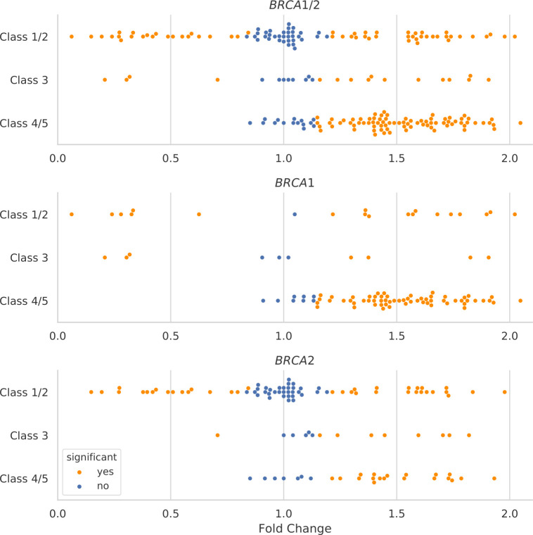
All rare germline variants were also detected in the corresponding tumour samples. Of the 94 pathogenic germline variants, 79 (84.0%) showed significantly increased VF in the tumour suggesting loss of the wild-type alleles, with fold changes ranging from 1.15 to 2.05 (figure 1, online supplemental table 2). The VF differences of the remaining 15 class 4/5 variants (16%) were statistically not significant with fold changes ranging from 0.85 to 1.13. Of note, none of the class 4/5 variants showed a significantly decreased VF in the tumour. Of the 90 class 1/2 variants, 33 (36.7%) showed significantly increased VFs in the tumour with fold changes ranging from 1.22 to 2.02, 34 showed non-significant differences (37.8%, fold changes ranging from 0.87 to 1.16) and for the remaining 23 variants, VFs were significantly decreased in tumour samples (25.6%, fold changes ranging from 0.06 to 0.84) (figure 1, online supplemental table 2).
Figure 1.
Fold change of VF between blood-derived and tumour-derived DNA observed for 208 class 4/5, class 3 and class 1/2 germline variants in the BRCA1/2 genes observed in the overall study sample. Significant differences in VFs between blood-derived and tumour-derived DNA were indicated by orange dots. Non-significant differences in VFs between blood-derived and tumour-derived DNA were indicated by blue dots. VFs, variant fraction.
Variant classification based on significantly increased VFs shows a high sensitivity of 0.84 (95% CI 0.77 to 0.91), but a poor specificity of 0.63 (95% CI 0.53 to 0.73) and a poor positive predictive value (PPV) of 0.71 (95% CI 0.62 to 0.79). For this approach, the positive likelihood ratio (LR+) for pathogenicity is 2.29 (95% CI 1.72 to 3.05). Briefly, variant classification based on significantly increased VFs is hampered by the random distribution of VFs observed for benign variants. At least in a routine diagnostic setting, classification of rare BRCA1/2 germline variants may not be based on significantly increased VFs due to an unacceptable proportion of false-positive results.
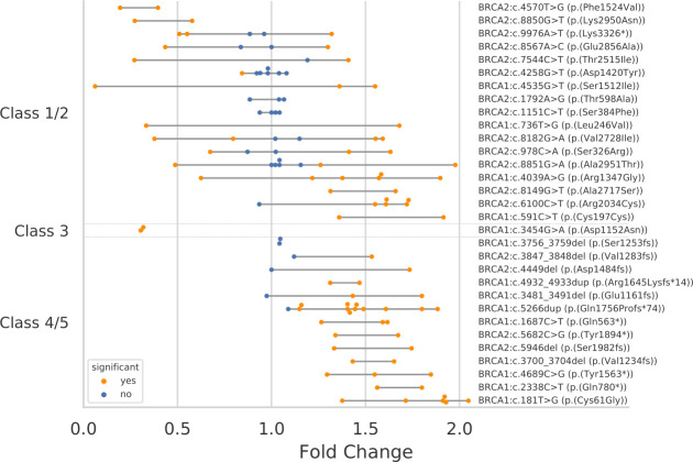
As an alternative approach, a significantly decreased VF of a variant in the tumour, suggesting loss of the variant allele, may be useful to classify a rare BRCA1/2 germline variant as benign. Significantly decreased VFs were specific for benign variants and were not observed for pathogenic germline variants (figure 1). Of the benign variants observed in 90 patients, 17 were recurrent and found at least twice in the sample set. For most of the recurrent benign variants, we found a high variability of fold changes, occasionally ranging from a significant decrease to a significant increase (figure 2). Classification of benign BRCA1/2 germline variants based on significantly decreased VFs results in a low sensitivity of 0.26 (95% CI 0.17 to 0.35) but a high specificity of 1.0 (95% CI 0.96 to 1.00) and a high PPV of 1.0 (95% CI 0.85 to 1.00). For this approach, the LR+ for benignity was 49.07 (95% CI 3.02 to 795.93) after Haldane-Anscombe correction (online supplemental table 3). A significantly decreased VF of a variant in the tumour may be exploited as a reliable predictor for benignity, with no false-negative result observed. This also holds true when analysis were performed for both genes separately (figure 1, online supplemental table 3). When applying this approach to the 24 VUS identified in our study sample, three distinct VUS found in four patients, that is, BRCA1 p.(Val525Ile), BRCA1 p.(Asp1152Asn) and BRCA2 p.(Lys2498del), may be considered benign (figure 2).
Figure 2.
Fold change of VF observed for 31 recurrent class 4/5, class 3 and class 1/2 germline variants in the BRCA1/2 genes observed in the overall study sample. Significant differences in VFs between blood-derived and tumour-derived DNA were indicated by orange dots. Non-significant differences in VFs between blood-derived and tumour-derived DNA were indicated by blue dots. VFs, variant fractions.
Discussion
It was controversially discussed whether the results of LOH analyses may be useful for the classification of rare BRCA1/2 germline variants.7–10 14–18 Information from LOH analyses has not been implemented in the current ENIGMA variant classification system3 19 based on the previously published data16 suggesting that LOH analyses are not sufficiently reliable. Using paired analyses of blood-derived and tumour-derived DNA, we demonstrated that rare germline variants in the BRCA1/2 genes might be classified benign based on significantly decreased VFs in the tumour. This approach reached a specificity of 1.0 (95% CI 0.96 to 1.00), a PPV of 1.0 (95% CI 0.85 to 1.00) and a LR+ of 49.07 (95% CI 3.02 to 795.93). Given the fact that changes in VFs of benign variants occur randomly (figure 2), this approach shows a limited sensitivity of only 0.26 (95% CI 0.17 to 0.36). As of March 2020, more than 6.100 distinct BRCA1/2 germline VUS were listed in the ClinVar database, indicating the need for additional sources for the classification of BRCA1/2 germline variants. We suggest that large-scale comparative germline/tumour NGS analyses with sufficient read depths may significantly reduce the number of VUS, especially for VUS for which data regarding cosegregation, family cancer history, co-occurrence with a pathogenic variant in the same gene and case–control information are not available.3
Limitations of the study
In the overall study sample of patients with OC enrolled in the observational AGO-TR1 study, pathogenic germline mutations in non-BRCA1/2 OC predisposition genes such as RAD51C/D and BRIP1 were observed. However, the prevalence of pathogenic germline mutations in these genes was too low to perform meaningful calculations. Larger studies are required to quantify the sensitivity and the specificity of LOH analyses for the classification of rare germline mutations in additional OC predisposition genes. Moreover, this investigation was focused on patients with OC and FFPE samples with a high tumour content. It remains elusive to which extent our approach may be transferred to breast tumour analyses that are usually associated with lower BRCA1/2 LOH rates20 and probably lower tumour contents in FFPE samples.
Acknowledgments
We would like to thank AstraZeneca Germany for the support of the trial.
Footnotes
JH and PH contributed equally.
Contributors: JH and PH contributed equally to this work; and study concept and design: JH, PH, RKS and EH; acquisition, analysis and interpretation of data: all authors; drafting the manuscript: JH, CE and EH; critical revision of the manuscript for important intellectual content: all authors; accountable for all aspects of the work in ensuring that questions related to the accuracy or integrity of any part of the work are appropriately investigated and resolved: all authors; final approval of the submitted manuscript: all authors; obtained funding: PH; study supervision: PH, RKS and EH; administrative, technical or material support: all authors.
Funding: The study was funded by AstraZeneca Germany and the AGO Research GmbH.
Competing interests: PH: consulting or advisory role: AstraZeneca, Roche/Genentech, Tesaro, Clovis, Pharmamar Lilly and Sotio; research funding: AstraZeneca (Inst); travel, accommodations and expenses: Medac. AB: honoraria: AstraZeneca and Roche; consulting or advisory role: AstraZeneca and Roche. DD is a consultant for AJ Innuscreen GmbH (Berlin, Germany), a 100% daughter company of Analytik Jena AG (Jena, Germany) and receives royalties from product sales (innuCONVERT kits). KK: honoraria: Roche, Pfizer and AstraZeneca. JS: honoraria: AstraZeneca, PharmaMar, Roche and Tesaro; consulting or advisory role: AstraZeneca, Clovis Oncology, Novocure, Roche and Tesaro; research funding: Amgen (Inst), Bayer (Inst), Lilly (Inst) and Novartis (Inst). RKS: honoraria: AstraZeneca; consulting or advisory role: AstraZeneca; research funding: AstraZeneca (Inst); patents, royalties, other intellectual property: University of Cologne. EH: honoraria: AstraZeneca; consulting or advisory role: AstraZeneca; research funding: AstraZeneca (Inst).
Provenance and peer review: Not commissioned; externally peer reviewed.
Supplemental material: This content has been supplied by the author(s). It has not been vetted by BMJ Publishing Group Limited (BMJ) and may not have been peer-reviewed. Any opinions or recommendations discussed are solely those of the author(s) and are not endorsed by BMJ. BMJ disclaims all liability and responsibility arising from any reliance placed on the content. Where the content includes any translated material, BMJ does not warrant the accuracy and reliability of the translations (including but not limited to local regulations, clinical guidelines, terminology, drug names and drug dosages), and is not responsible for any error and/or omissions arising from translation and adaptation or otherwise.
Ethics statements
Patient consent for publication
Not required.
References
- 1. Maxwell KN, Hart SN, Vijai J, Schrader KA, Slavin TP, Thomas T, Wubbenhorst B, Ravichandran V, Moore RM, Hu C, Guidugli L, Wenz B, Domchek SM, Robson ME, Szabo C, Neuhausen SL, Weitzel JN, Offit K, Couch FJ, Nathanson KL. Evaluation of ACMG-Guideline-Based variant classification of cancer susceptibility and Non-Cancer-Associated genes in families affected by breast cancer. Am J Hum Genet 2016;98:801–17. 10.1016/j.ajhg.2016.02.024 [DOI] [PMC free article] [PubMed] [Google Scholar]
- 2. Nykamp K, Anderson M, Powers M, Garcia J, Herrera B, Ho Y-Y, Kobayashi Y, Patil N, Thusberg J, Westbrook M, Topper S, Invitae Clinical Genomics Group . Sherloc: a comprehensive refinement of the ACMG-AMP variant classification criteria. Genet Med 2017;19:1105–17. 10.1038/gim.2017.37 [DOI] [PMC free article] [PubMed] [Google Scholar]
- 3. Parsons MT, Tudini E, Li H, Hahnen E, Wappenschmidt B, Feliubadaló L, Aalfs CM, Agata S, Aittomäki K, Alducci E, Alonso-Cerezo MC, Arnold N, Auber B, Austin R, Azzollini J, Balmaña J, Barbieri E, Bartram CR, Blanco A, Blümcke B, Bonache S, Bonanni B, Borg Åke, Bortesi B, Brunet J, Bruzzone C, Bucksch K, Cagnoli G, Caldés T, Caliebe A, Caligo MA, Calvello M, Capone GL, Caputo SM, Carnevali I, Carrasco E, Caux-Moncoutier V, Cavalli P, Cini G, Clarke EM, Concolino P, Cops EJ, Cortesi L, Couch FJ, Darder E, de la Hoya M, Dean M, Debatin I, Del Valle J, Delnatte C, Derive N, Diez O, Ditsch N, Domchek SM, Dutrannoy V, Eccles DM, Ehrencrona H, Enders U, Evans DG, Farra C, Faust U, Felbor U, Feroce I, Fine M, Foulkes WD, Galvao HCR, Gambino G, Gehrig A, Gensini F, Gerdes A-M, Germani A, Giesecke J, Gismondi V, Gómez C, Gómez Garcia EB, González S, Grau E, Grill S, Gross E, Guerrieri-Gonzaga A, Guillaud-Bataille M, Gutiérrez-Enríquez S, Haaf T, Hackmann K, Hansen TVO, Harris M, Hauke J, Heinrich T, Hellebrand H, Herold KN, Honisch E, Horvath J, Houdayer C, Hübbel V, Iglesias S, Izquierdo A, James PA, Janssen LAM, Jeschke U, Kaulfuß S, Keupp K, Kiechle M, Kölbl A, Krieger S, Kruse TA, Kvist A, Lalloo F, Larsen M, Lattimore VL, Lautrup C, Ledig S, Leinert E, Lewis AL, Lim J, Loeffler M, López-Fernández A, Lucci-Cordisco E, Maass N, Manoukian S, Marabelli M, Matricardi L, Meindl A, Michelli RD, Moghadasi S, Moles-Fernández A, Montagna M, Montalban G, Monteiro AN, Montes E, Mori L, Moserle L, Müller CR, Mundhenke C, Naldi N, Nathanson KL, Navarro M, Nevanlinna H, Nichols CB, Niederacher D, Nielsen HR, Ong K-R, Pachter N, Palmero EI, Papi L, Pedersen IS, Peissel B, Perez-Segura P, Pfeifer K, Pineda M, Pohl-Rescigno E, Poplawski NK, Porfirio B, Quante AS, Ramser J, Reis RM, Revillion F, Rhiem K, Riboli B, Ritter J, Rivera D, Rofes P, Rump A, Salinas M, Sánchez de Abajo AM, Schmidt G, Schoenwiese U, Seggewiß J, Solanes A, Steinemann D, Stiller M, Stoppa-Lyonnet D, Sullivan KJ, Susman R, Sutter C, Tavtigian SV, Teo SH, Teulé A, Thomassen M, Tibiletti MG, Tischkowitz M, Tognazzo S, Toland AE, Tornero E, Törngren T, Torres-Esquius S, Toss A, Trainer AH, Tucker KM, van Asperen CJ, van Mackelenbergh MT, Varesco L, Vargas-Parra G, Varon R, Vega A, Velasco Ángela, Vesper A-S, Viel A, Vreeswijk MPG, Wagner SA, Waha A, Walker LC, Walters RJ, Wang-Gohrke S, Weber BHF, Weichert W, Wieland K, Wiesmüller L, Witzel I, Wöckel A, Woodward ER, Zachariae S, Zampiga V, Zeder-Göß C, Lázaro C, De Nicolo A, Radice P, Engel C, Schmutzler RK, Goldgar DE, Spurdle AB, KConFab Investigators . Large scale multifactorial likelihood quantitative analysis of BRCA1 and BRCA2 variants: An ENIGMA resource to support clinical variant classification. Hum Mutat 2019;40:1557–78. 10.1002/humu.23818 [DOI] [PMC free article] [PubMed] [Google Scholar]
- 4. Richards S, Aziz N, Bale S, Bick D, Das S, Gastier-Foster J, Grody WW, Hegde M, Lyon E, Spector E, Voelkerding K, Rehm HL, ACMG Laboratory Quality Assurance Committee . Standards and guidelines for the interpretation of sequence variants: a joint consensus recommendation of the American College of medical genetics and genomics and the association for molecular pathology. Genet Med 2015;17:405–23. 10.1038/gim.2015.30 [DOI] [PMC free article] [PubMed] [Google Scholar]
- 5. Knudson AG. Mutation and cancer: statistical study of retinoblastoma. Proc Natl Acad Sci U S A 1971;68:820–3. 10.1073/pnas.68.4.820 [DOI] [PMC free article] [PubMed] [Google Scholar]
- 6. Hauke J, Hahnen E, Schneider S, Reuss A, Richters L, Kommoss S, Heimbach A, Marmé F, Schmidt S, Prieske K, Gevensleben H, Burges A, Borde J, De Gregorio N, Nürnberg P, El-Balat A, Thiele H, Hilpert F, Altmüller J, Meier W, Dietrich D, Kimmig R, Schoemig-Markiefka B, Kast K, Braicu E, Baumann K, Jackisch C, Park-Simon T-W, Ernst C, Hanker L, Pfisterer J, Schnelzer A, du Bois A, Schmutzler RK, Harter P. Deleterious somatic variants in 473 consecutive individuals with ovarian cancer: results of the observational AGO-TR1 study (NCT02222883). J Med Genet 2019;56:574–80. 10.1136/jmedgenet-2018-105930 [DOI] [PubMed] [Google Scholar]
- 7. Kanchi KL, Johnson KJ, Lu C, McLellan MD, Leiserson MDM, Wendl MC, Zhang Q, Koboldt DC, Xie M, Kandoth C, McMichael JF, Wyczalkowski MA, Larson DE, Schmidt HK, Miller CA, Fulton RS, Spellman PT, Mardis ER, Druley TE, Graubert TA, Goodfellow PJ, Raphael BJ, Wilson RK, Ding L. Integrated analysis of germline and somatic variants in ovarian cancer. Nat Commun 2014;5:3156. 10.1038/ncomms4156 [DOI] [PMC free article] [PubMed] [Google Scholar]
- 8. Kechin AA, Boyarskikh UA, Ermolenko NA, Tyulyandina AS, Lazareva DG, Avdalyan AM, Tyulyandin SA, Kushlinskii NE, Filipenko ML. Loss of heterozygosity in BRCA1 and BRCA2 genes in patients with ovarian cancer and probability of its use for clinical classification of variations. Bull Exp Biol Med 2018;165:94–100. 10.1007/s10517-018-4107-9 [DOI] [PubMed] [Google Scholar]
- 9. Osorio A, de la Hoya M, Rodríguez-López R, Martínez-Ramírez A, Cazorla A, Granizo JJ, Esteller M, Rivas C, Caldés T, Benítez J. Loss of heterozygosity analysis at the BRCA loci in tumor samples from patients with familial breast cancer. Int J Cancer 2002;99:305–9. 10.1002/ijc.10337 [DOI] [PubMed] [Google Scholar]
- 10. Osorio A, Milne RL, Honrado E, Barroso A, Diez O, Salazar R, de la Hoya M, Vega A, Benítez J. Classification of missense variants of unknown significance in BRCA1 based on clinical and tumor information. Hum Mutat 2007;28:477–85. 10.1002/humu.20470 [DOI] [PubMed] [Google Scholar]
- 11. Lek M, Karczewski KJ, Minikel EV, Samocha KE, Banks E, Fennell T, O'Donnell-Luria AH, Ware JS, Hill AJ, Cummings BB, Tukiainen T, Birnbaum DP, Kosmicki JA, Duncan LE, Estrada K, Zhao F, Zou J, Pierce-Hoffman E, Berghout J, Cooper DN, Deflaux N, DePristo M, Do R, Flannick J, Fromer M, Gauthier L, Goldstein J, Gupta N, Howrigan D, Kiezun A, Kurki MI, Moonshine AL, Natarajan P, Orozco L, Peloso GM, Poplin R, Rivas MA, Ruano-Rubio V, Rose SA, Ruderfer DM, Shakir K, Stenson PD, Stevens C, Thomas BP, Tiao G, Tusie-Luna MT, Weisburd B, Won H-H, Yu D, Altshuler DM, Ardissino D, Boehnke M, Danesh J, Donnelly S, Elosua R, Florez JC, Gabriel SB, Getz G, Glatt SJ, Hultman CM, Kathiresan S, Laakso M, McCarroll S, McCarthy MI, McGovern D, McPherson R, Neale BM, Palotie A, Purcell SM, Saleheen D, Scharf JM, Sklar P, Sullivan PF, Tuomilehto J, Tsuang MT, Watkins HC, Wilson JG, Daly MJ, MacArthur DG, Exome Aggregation Consortium . Analysis of protein-coding genetic variation in 60,706 humans. Nature 2016;536:285–91. 10.1038/nature19057 [DOI] [PMC free article] [PubMed] [Google Scholar]
- 12. Plon SE, Eccles DM, Easton D, Foulkes WD, Genuardi M, Greenblatt MS, Hogervorst FBL, Hoogerbrugge N, Spurdle AB, Tavtigian SV, IARC Unclassified Genetic Variants Working Group . Sequence variant classification and reporting: recommendations for improving the interpretation of cancer susceptibility genetic test results. Hum Mutat 2008;29:1282–91. 10.1002/humu.20880 [DOI] [PMC free article] [PubMed] [Google Scholar]
- 13. Benjamini Y, Hochberg Y. Controlling the false discovery rate: a practical and powerful approach to multiple testing. J of the Soc Series B 1995;57:289–300. 10.1111/j.2517-6161.1995.tb02031.x [DOI] [Google Scholar]
- 14. Walsh MF, Ritter DI, Kesserwan C, Sonkin D, Chakravarty D, Chao E, Ghosh R, Kemel Y, Wu G, Lee K, Kulkarni S, Hedges D, Mandelker D, Ceyhan-Birsoy O, Luo M, Drazer M, Zhang L, Offit K, Plon SE. Integrating somatic variant data and biomarkers for germline variant classification in cancer predisposition genes. Hum Mutat 2018;39:1542–52. 10.1002/humu.23640 [DOI] [PMC free article] [PubMed] [Google Scholar]
- 15. Ellard S, Baple EL, Owens M, Eccles DM, Abbs S, Deans ZC, Newman WG, McMullan DJ. ACGS best practice guidelines for variant classification 2017: ACGS guidelines, 2017. [Google Scholar]
- 16. Chenevix-Trench G, Healey S, Lakhani S, Waring P, Cummings M, Brinkworth R, Deffenbaugh AM, Burbidge LA, Pruss D, Judkins T, Scholl T, Bekessy A, Marsh A, Lovelock P, Wong M, Tesoriero A, Renard H, Southey M, Hopper JL, Yannoukakos K, Brown M, Easton D, Tavtigian SV, Goldgar D, Spurdle AB, kConFab Investigators . Genetic and histopathologic evaluation of BRCA1 and BRCA2 DNA sequence variants of unknown clinical significance. Cancer Res 2006;66:2019–27. 10.1158/0008-5472.CAN-05-3546 [DOI] [PubMed] [Google Scholar]
- 17. Arason A, Agnarsson BA, Johannesdottir G, Johannsson OT, Hilmarsdottir B, Reynisdottir I, Barkardottir RB. The BRCA1 c.4096+3A>G variant displays classical characteristics of pathogenic BRCA1 mutations in hereditary breast and ovarian cancers, but Still allows homozygous viability. Genes 2019;10:882. 10.3390/genes10110882 [DOI] [PMC free article] [PubMed] [Google Scholar]
- 18. Spearman AD, Sweet K, Zhou X-P, McLennan J, Couch FJ, Toland AE. Clinically applicable models to characterize BRCA1 and BRCA2 variants of uncertain significance. J Clin Oncol 2008;26:5393–400. 10.1200/JCO.2008.17.8228 [DOI] [PMC free article] [PubMed] [Google Scholar]
- 19. Spurdle AB, Lakhani SR, Healey S, Parry S, Da Silva LM, Brinkworth R, Hopper JL, Brown MA, Babikyan D, Chenevix-Trench G, Tavtigian SV, Goldgar DE. Clinical classification of BRCA1 and BRCA2 DNA sequence variants: the value of cytokeratin profiles and evolutionary analysis—a report from the kConFab investigators. JCO 2008;26:1657–63. 10.1200/JCO.2007.13.2779 [DOI] [PubMed] [Google Scholar]
- 20. Maxwell KN, Wubbenhorst B, Wenz BM, De Sloover D, Pluta J, Emery L, Barrett A, Kraya AA, Anastopoulos IN, Yu S, Jiang Y, Chen H, Zhang NR, Hackman N, D'Andrea K, Daber R, Morrissette JJD, Mitra N, Feldman M, Domchek SM, Nathanson KL. Brca locus-specific loss of heterozygosity in germline BRCA1 and BRCA2 carriers. Nat Commun 2017;8:319. 10.1038/s41467-017-00388-9 [DOI] [PMC free article] [PubMed] [Google Scholar]
Associated Data
This section collects any data citations, data availability statements, or supplementary materials included in this article.
Supplementary Materials
jmedgenet-2020-107353supp001.pdf (430.1KB, pdf)


