Abstract
Objective:
To determine if the enamel around orthodontic brackets is significantly altered after demineralization followed by application of adhesives with and without fluoride-releasing ability.
Materials and Methods:
One hundred eight noncarious human premolars were divided into six groups of 18 each and exposed to a demineralization solution. Stainless steel brackets were bonded using two conventional composite resin etch-and-rinse systems, three self-etching primer (SEP) composite resin systems, and one resin-modified glass ionomer cement (RMGIC) system. One conventional and one SEP composite resin adhesive did not have fluoride-releasing ability, which was claimed for the other four adhesives. The elastic modulus and hardness of the enamel were determined with a nanoindenter at 10 equidistant depths ranging from 1–46 µm and at four regions: control (not exposed) enamel surface, under the adhesive, and at 50 µm and 100 µm from the bracket edges. Using the Kruskal-Wallis and Mann-Whitney U-tests (P < .0125 for statistical significance), these properties were compared at different regions.
Results:
The same behavior was observed for values of elastic modulus and hardness. Significant differences were found within approximately 21 µm of the enamel surface for etching with 35% phosphoric acid or priming with SEP, but only minimal changes occurred for the SEP adhesive. Increases in near-surface elastic modulus and hardness of enamel were found with the SEP adhesive and RMGIC with fluoride-releasing ability.
Conclusions:
Clinical use of the fluoride-releasing adhesives investigated may prevent demineralization of enamel around brackets during orthodontic treatment.
Keywords: Enamel, Demineralization, Nanoindentation, Hardness, Elastic modulus, Fluoride-releasing adhesive
INTRODUCTION
Increased prevalence of white spot formation in orthodontic patients has been reported1 due to the irregular surface of orthodontic fixed appliances, which creates stagnation areas for plaque, render tooth cleaning more difficult, and limits naturally occurring self-cleaning mechanisms.1 Fluoride plays an important role in the prevention of demineralization during orthodontic treatment, and mouth rinses with fluoride solutions are recommended in addition to daily tooth-brushing. However, the use of mouth rinses for preventive enamel demineralization requires cooperation by the patient, which is sometimes difficult, especially for young patients. On the other hand, the original method of using phosphoric acid for etching in bracket bonding is associated with a loss of the enamel surface (approximately 10–20 µm),2,3 although etched enamel around orthodontic brackets might be remineralized by an application of the fluoride products.4
Over the past decade, progress has been made in bracket bonding to enamel with resin-modified glass ionomer cements (RMGICs) and self-etching primers (SEPs),2 and their lower etching ability might minimize the potential for iatrogenic damage to enamel. Fluoride-releasing adhesives with less etching ability, such as RMGIC and SEP composite resin adhesive systems containing fillers with fluoride, might be good selections for preventive enamel demineralization. The preventive effect of an adhesive adjacent to brackets has been investigated in vitro1,5,6 and in vivo7–9 by quantifying the demineralization depths and the mineral losses with various evaluation methods. Most studies have been shown that RMGICs are more effective for prevention of enamel demineralization in comparison with fluoride-releasing and non–fluoride-releasing composite resin adhesive systems.1,5,8 To investigate the demineralization of enamel quantitatively, use of cross-sectional microhardness measurements with a Knoop indenter has been a popular method5–8 because there is high correlation between enamel microhardness and the percentage of mineral in carious lesions.10
Recent advances in the nanoindentation test have allowed the measurement of mechanical properties for extremely small volumes of materials where the contact radius is less than 100 nm.11–14 In nanoindentation testing, the load and displacement are recorded with a high-resolution displacement gage during the indentation process, and the area of the indentation is obtained from the geometric form of the indenter without having to actually observe the indentation; thereby the hardness of the specimen can be calculated. In addition, the elastic modulus for this very small volume of material can be obtained mathematically from the load-displacement curve.
Little is presently known about mechanical properties of the subsurface region of enamel around orthodontic brackets. The purpose of this in vitro study was to investigate the hardness and elastic modulus of enamel around orthodontic brackets after immersion in a demineralization solution and compare these mechanical properties for different adhesive systems with and without fluoride-releasing ability. It was hypothesized that different types of adhesive systems have significantly different effects on the enamel demineralization behavior and mechanical properties.
MATERIALS AND METHODS
Materials
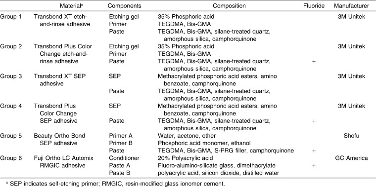
One hundred eight noncarious human premolars, obtained by extraction from patients who were to undergo orthodontic treatment, were allocated into six groups of 18 each. This in vitro study received approval by an ethics committee at the Health Sciences University of Hokkaido. The buccal surfaces of all teeth were cleaned using nonfluoridated pumice. The teeth were subsequently polished using a rubber cup, and thoroughly washed and dried using a moisture-free air source. Maxillary or mandibular stainless steel brackets (Victory Series, 3M Unitek, Monrovia, Calif) were bonded to the enamel samples with each of the six bracket bonding adhesive systems listed in Table 1. Groups 1 and 2 employed conventional etch-and-rinse composite resin adhesive systems. Groups 3, 4, and 5 employed SEP composite resin adhesive systems. Group 6 employed an RMGIC adhesive system. The manufacturers claim that the adhesives employed for groups 2, 4, 5, and 6 have fluoride-releasing ability.
Table 1.
Materials Employed in Present Study
Preparation of Teeth
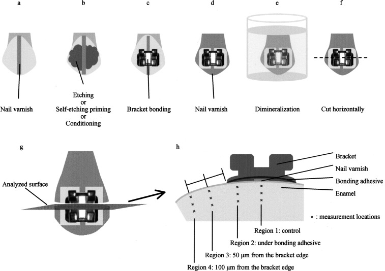
The buccal surfaces of the premolars were coated with a 1.5-mm width of acid-resistant nail varnish from the cusp to the cervix, and these regions under the nail varnish were used as the control (Figure 1a). In groups 1 and 2, the buccal surfaces were etched with conventional 35% phosphoric acid gel for 15 seconds (Figure 1b), washed for 20 seconds, and dried with an oil-free air stream. Transbond XT primer was then applied to the etched surface. In groups 3, 4, and 5, Transbond SEP or Beauty Ortho Bond SEP was applied by rubbing on the buccal enamel surfaces for approximately 3 seconds; afterward an air jet was lightly applied. In group 6, 20% polyacrylic acid Ortho Conditioner was applied for 10 seconds on the buccal enamel surface, which was washed for 20 seconds, and dried with an oil-free air stream. Then, the stainless steel brackets (Victory Series, 3M Unitek) were bonded with the composite resin adhesive pastes or RMGIC paste and light-cured according to the instructions from each manufacturer (Figure 1c). Acid-resistant nail varnish was next applied to each tooth, leaving a 1-mm rim of exposed sound enamel surrounding the bracket (Figure 1d). The specimens were then immersed in a demineralization solution (4% Methocel MC gel [Fluka, Buchs, Switzerland] + 0.1% lactic acid) in a polystyrene vial (Figure 1e). The solution was adjusted to a pH of 4.6. All specimens were immersed in 10 mL of this solution for 96 hours at 37°C, with the solution being changed every 12 hours, and then gently washed with deionized water.
Figure 1.
Schematic illustration of specimen preparation sequence and regions of tooth enamel investigated with the nanoindentation test.
All of the teeth were cut with a slow-speed water-cooled diamond saw (Isomet, Buehler, Lake Bluff, Ill) so that they were divided into occlusal and cervical halves (Figure 1f); one of the sectioned specimens (transverse planes) was then encapsulated in epoxy resin (Epofix, Struers, Copenhagen, Denmark) for the nanoindentation test. All samples were ground (600-grit sandpaper) and polished using diamond suspensions (3-, 1-, and 0.25-µm particle sizes) to obtain a suitable surface for nanoindentation.
Nanoindentation Test
All nanoindentation testing (ENT-1100a, Elionix, Tokyo, Japan) was carried out at 28°C with a peak load of 10 mN using a Berkovich indenter. Each test consisted of three parts: 10 seconds for loading to the peak value, 1 second of holding at the peak load, and 10 seconds for unloading. The indentations were placed at 1–46 µm depth (10 locations spaced 5 µm apart) from the external surface for four regions (region 1: control; region 2: under the bonding adhesive; region 3: 50 µm from the bracket edge; region 4: 100 µm from the bracket edge) (Figure 1h). The measurement points were selected using the optical microscope and charge-coupled device camera coupled to the nanoindentation test apparatus. Linear extrapolation methods (ISO Standard 14577-1)15 were used for the unloading curve between 95% and 70% of the maximum test force to calculate the elastic modulus. The hardness and elastic modulus were calculated by software available with the nanoindentation apparatus.
Statistical Analysis
The experimental results were analyzed using the PASW Statistics software (version 18.0J for Windows, IBM, Armonk, New York). The data for the mean values of hardness and elastic modulus were not normally distributed (Levene test), so the values were compared using the Kruskal-Wallis test and the Mann-Whitney U-test (P < .0125). The purpose of these comparisons was to assess the effect of bracket bonding procedure or immersion in demineralization solution (regions 1–4) for each adhesive.
RESULTS
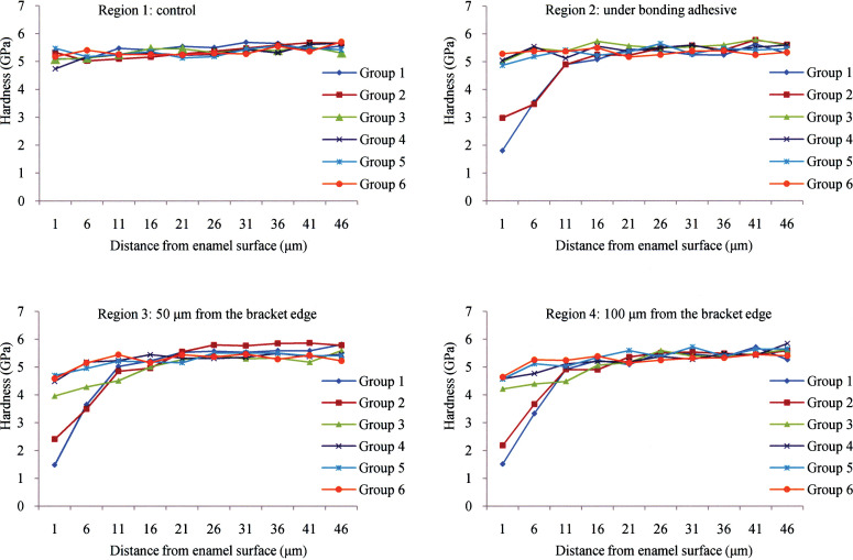
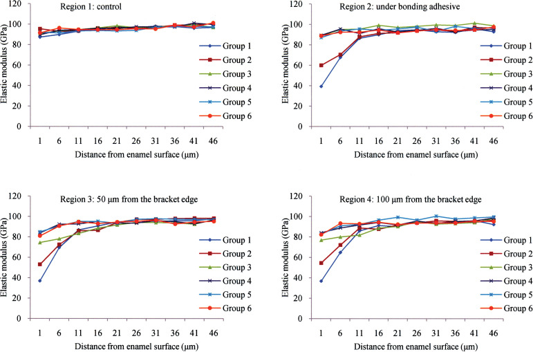
Figures 2 and 3 show mean values for hardness and elastic modulus of the cross-sectioned polished specimens (transverse planes) obtained by the nanoindentation test; the results of statistical comparisons between different regions are summarized in Tables 2 and 3. Locations at 1 µm and 6 µm from the enamel surface for regions 2, 3, and 4 of both conventional etch-and-rinse adhesive systems (groups 1 and 2) had significantly lower hardness and elastic modulus than those for region 1, except for the hardness of region 4 in group 2 at 6 µm depth. For group 3, there was no significant difference in the hardness and elastic modulus between regions 1 and 2. The hardness up to 16 µm in depth and elastic modulus up to 21 µm in depth from the enamel surface for regions 3 and 4 had significantly lower values than those for region 1 and 2 except for elastic modulus at 16 µm depth from the enamel surface for region 4. There was no significant difference in the mean values for hardness and elastic modulus of enamel at each depth for group 4. For group 5, the locations at 1 µm depth from the enamel surface for regions 2, 3, and 4 had significantly lower hardness than region 1, although there were no significant differences in elastic modulus. For group 6, the location at 1 µm below the enamel surface for region 3 had significantly lower hardness than region 2, but region 2 was not significantly different from region 1, and region 3 was not significantly different from region 4. The elastic modulus for group 6 at 1 µm depth was significantly different for regions 1 and 3, but not for regions 1 and 2 nor for regions 3 and 4.
Figure 2.
Mean values of hardness of enamel at different distances from the surface after immersion in demineralization solution. Group 1, Transbond XT etch-and-rinse adhesive system; group 2, Transbond Plus Color Change etch-and-rinse adhesive system; group 3, Transbond XT SEP adhesive system; group 4, Transbond Plus Color Change SEP adhesive system; group 5, Beauty Ortho Bond SEP adhesive system; and group 6, Fuji Ortho LC Automix RMGIC adhesive system.
Figure 3.
Mean values of elastic modulus of enamel at different distances from the surface after immersion in demineralization solution. Group 1, Transbond XT etch-and-rinse adhesive system; group 2, Transbond Plus Color Change etch-and-rinse adhesive system; group 3, Transbond XT SEP adhesive system; group 4, Transbond Plus Color Change SEP adhesive system; group 5, Beauty Ortho Bond SEP adhesive system; and group 6, Fuji Ortho LC Automix RMGIC adhesive system.
Table 2.
Mean Values and Standard Deviation for Cross-sectional Hardness (GPa) of Enamel at Distances From the Surfacea
Table 3.
Mean Values and Standard Deviation for Cross-sectional Elastic Modulus (GPa) of Enamel at Distances From the Surfacea
DISCUSSION
The present in vitro study evaluated the effect of bonding materials on the mechanical properties (hardness and elastic modulus) of enamel around orthodontic brackets. These mechanical properties may change depending upon the bracket bonding procedure and demineralization of enamel, and in this study changes were assessed by cross-sectional nanoindentation analysis. The mechanical properties of enamel are generally linked to its mineral content. Knowledge of the mechanical properties of enamel such as the hardness and elastic modulus may lead to an improved understanding of the bracket bonding and demineralization behavior, along with the effects of new bonding materials. In the nanoindentation test, the minimum distance between indentations should be at least five times the largest indentation diameter.15 This study used a 10 mN peak load, which made indentations having about 2 µm width and length; the distance between the indentations was about 3 µm. Because a greater peak load shows more accurate values and stable data, and because the indentations obtained by our condition did not produce any cracks on the enamel, we used this 10 mN peak load condition. In addition, a distance of less than 5 µm apart between indentations was favorable to analyze the demineralization behavior in this study.
This in vitro study used noncarious human premolars obtained from different patients, and the observed individual variation in acid resistance of the enamel specimens would be expected. However, the hardness and elastic modulus for the control region (region 1) obtained from all adhesive systems (groups 1–6) showed no significant variation for all the locations except that the group 4 location at 1 µm had significantly lower hardness than the other five groups. Mean hardness and elastic modulus for region 1 ranged from 4.7–5.7 GPa and 87.3–99.6 GPa, respectively, and these values were similar to recently published values obtained with the nanoindentation test (3.4–8.3 GPa for hardness; 61–130 GPa for elastic modulus).3,12 The values of hardness and elastic modulus obtained in this study should be affected by the location (depth from enamel surface and analysis region for each adhesive group), along with test load, type of indenter, and direction of the enamel rods.
The locations at 1 µm and 6 µm from the enamel surface for region 2 for both conventional etch-and-rinse adhesive systems (groups 1 and 2) had significantly lower hardness and elastic modulus than those for region 1 (control enamel). The mechanical properties of enamel at least 6 µm from the external surface were influenced by demineralization, and this agrees with the results from previous studies of the effects of conventional etching and self-etching on the mechanical properties (hardness and elastic modulus) of the enamel surface region.3,8 The location at 1 µm from the enamel surface for regions 2, 3, and 4 of groups 1 and 2 had similar values of hardness and elastic modulus, which was also found at the 6 µm location, and additional deterioration of the enamel structure after etching with 35% phosphoric acid was not observed. These results might suggest that Transbond Plus Color Change with fluoride-releasing ability was insufficient for remineralization of etched enamel by 35% phosphoric acid around brackets and that additional application of fluoride-containing products such as toothpaste, bonding materials, and solutions should be needed for the remineralization.
Over the past decade, progress has been made in bracket bonding to enamel with RMGICs and SEPs,2 and their lower etching ability might minimize the potential for iatrogenic damage to enamel. Region 2 for groups 3, 4, 5, and 6 had higher values for hardness and elastic modulus than for groups 1 and 2, suggesting that the surface treatment of SEP and RMGIC adhesive systems causes minimal enamel demineralization; this agrees with the observation from a previous study.3 A previous study16 found that the etching patterns of aprismatic enamel were dependent on the aggressiveness of the acid, but there was no correlation between the aggressiveness and bond strength to intact enamel. Another study17 measured pH values of phosphoric acid and orthodontic SEPs, and the values were 1.39 for 35% phosphoric acid, 1.85 for Transbond SEP, and 2.20 for Beauty Ortho Bond SEP; both SEPs with relatively less acidic pH values had a mild etching effect for intact enamel. In this study, greater demineralization by 35% phosphoric acid compared to both SEPs was due to lower pH value and longer application time.
For group 3, the hardness up to 16 µm in depth and elastic modulus up to 21 µm in depth from enamel surface for regions 3 and 4 generally had significantly lower values than those for regions 1 and 2 (although there were no significant differences for region 1 at 16 µm depth in group 3), indicating that additional deterioration of the enamel structure by immersion in demineralization solution occurred after priming with SEPs. On the other hand, noteworthy deterioration of the enamel structure by immersion in the demineralization solution did not occur for groups 4 and 5. These results may confirm that the fluoride-releasing ability for groups 4 and 5 prevent enamel demineralization around orthodontic brackets.
Glass ionomer cements have high levels of fluoride-releasing ability, although they show poor bracket bond strength and greater bond failure rates than composite resin adhesives.6,18 To obtain adequate bond strength and provide greater fluoride release, RMGICs have been developed. The recently introduced RMGIC, Fuji Ortho LC Automix (GC America), in easy-to-use Automix paste packages, was selected for the present study. While Fuji Ortho LC Automix is considered to be an improved product based on Fuji Ortho LC adhesive (GC America), neither the fluoride release behavior nor the bracket bond strength have been reported for this new adhesive. Previous studies have reported that RMGICs showed more resistance to demineralization of enamel than other bracket bonding materials.1,6,7 However, SEP composite resin adhesive systems (groups 4 and 5) with fluoride-releasing ability showed an equivalent effect for preventive enamel demineralization in the present study.
Further materials-oriented studies are needed to provide insight about the relationships between bonding materials and enamel structures during orthodontic treatment. Such investigations may also provide new ideas for novel design of adhesive materials.
CONCLUSIONS
Under the conditions of this study, the following conclusions can be drawn:
The hardness and elastic modulus of the enamel surface region is decreased by enamel etching with 35% phosphoric acid or priming with SEPs. The influence is minimal for SEP adhesive, and SEP adhesives or RMGIC adhesive systems with fluoride-releasing ability may increase the hardness and elastic modulus of the enamel surface region by remineralization of the enamel.
The fluoride-releasing ability of the composite resin adhesive and RMGIC adhesive prevents enamel demineralization and the deterioration in mechanical properties of enamel around brackets.
Nanoindentation is a useful method for investigating mechanical properties in small regions of enamel, and these properties should be relevant to demineralization.
REFERENCES
- 1.Sudjalim T. R, Woods M. G, Manton D. J, Reynolds E. C. Prevention of demineralization around orthodontic brackets in vitro. Am J Orthod Dentofacial Orthop. 2007;131:705.e1–e9. doi: 10.1016/j.ajodo.2006.09.043. [DOI] [PubMed] [Google Scholar]
- 2.Fjeld M, Øgaard B. Scanning electron microscopic evaluation of enamel surfaces exposed to 3 orthodontic bonding systems. Am J Orthod Dentofacial Orthop. 2006;130:575–581. doi: 10.1016/j.ajodo.2006.07.002. [DOI] [PubMed] [Google Scholar]
- 3.Iijima M, Muguruma T, Brantley W. A, Ito S, Yuasa T, Saito T, Mizoguchi I. Effect of bracket bonding on nanomechanical properties of enamel. Am J Orthod Dentofacial Orthop. 2010;138:735–740. doi: 10.1016/j.ajodo.2009.01.028. [DOI] [PubMed] [Google Scholar]
- 4.Hu W, Zhou Y. H, Wang Q, Fu M. K, Volpe A, Devizio W, Petrone M, Zhang Y. P. Effects of fluoride toothpaste on etched enamel of orthodontic patients. Chin J Dent Res. 1999;2:79–83. [PubMed] [Google Scholar]
- 5.Hu W, Featherstone J. D. Prevention of enamel demineralization: an in-vitro study using light-cured filled sealant. Am J Orthod Dentofacial Orthop. 2005;128:592–600. doi: 10.1016/j.ajodo.2004.07.046. [DOI] [PubMed] [Google Scholar]
- 6.Paschos E, Kleinschrodt T, Clementino-Luedemann T, Huth K. C, Hickel R, Kunzelmann K. H, Rudzki-Janson I. Effect of different bonding agents on prevention of enamel demineralization around orthodontic brackets. Am J Orthod Dentofacial Orthop. 2009;135:603–612. doi: 10.1016/j.ajodo.2007.11.028. [DOI] [PubMed] [Google Scholar]
- 7.Gorton J, Featherstone J. D. In vivo inhibition of demineralization around orthodontic brackets. Am J Orthod Dentofacial Orthop. 2003;123:10–14. doi: 10.1067/mod.2003.47. [DOI] [PubMed] [Google Scholar]
- 8.Pascotto R. C, Navarro M. F, Capelozza Filho L, Cury J. A. In vivo effect of a resin-modified glass ionomer cement on enamel demineralization around orthodontic brackets. Am J Orthod Dentofacial Orthop. 2004;125:36–41. doi: 10.1016/s0889-5406(03)00571-7. [DOI] [PubMed] [Google Scholar]
- 9.Ghiz M. A, Ngan P, Kao E, Martin C, Gunel E. Effects of sealant and self-etching primer on enamel decalcification. Part II: an in-vivo study. Am J Orthod Dentofacial Orthop. 2009;135:206–213. doi: 10.1016/j.ajodo.2007.02.060. [DOI] [PubMed] [Google Scholar]
- 10.Featherstone J. D, ten Cate J. M, Shariati M, Arends J. Comparison of artificial caries-like lesions by quantitative microradiography and microhardness profiles. Caries Res. 1983;17:385–391. doi: 10.1159/000260692. [DOI] [PubMed] [Google Scholar]
- 11.Oliver W. C, Pharr G. M. An improved technique for determining hardness and elastic modulus using load and displacement sensing indentation experiments. J Mater Res. 1992;7:1564–1583. [Google Scholar]
- 12.He L. H, Swain M. V. Influence of environment on the mechanical behaviour of mature human enamel. Biomaterials. 2007;28:4512–4520. doi: 10.1016/j.biomaterials.2007.06.020. [DOI] [PubMed] [Google Scholar]
- 13.Alcock J. P, Barbour M. E, Sandy J. R, Ireland A. J. Nanoindentation of orthodontic archwires: the effect of decontamination and clinical use on hardness, elastic modulus and surface roughness. Dent Mater. 2009;25:1039–1043. doi: 10.1016/j.dental.2009.03.003. [DOI] [PubMed] [Google Scholar]
- 14.Iijima M, Muguruma T, Brantley W. A, Yuasa T, Uechi J, Mizoguchi I. The effect of filler on the grindability of composite resin adhesive. Am J Orthod Dentofacial Orthop. 2010;138:420–426. doi: 10.1016/j.ajodo.2008.08.039. [DOI] [PubMed] [Google Scholar]
- 15.ISO 14577-1 Metallic materials—instrumented indentation test for hardness and materials parameters— Part 1 test method 1st ed. International Organization for Standardization; 2002. [Google Scholar]
- 16.Pashley D. H, Tay F. R. Aggressiveness of contemporary self-etching adhesives. Part II: etching effects on unground enamel. Dent Mater. 2001;17:430–444. doi: 10.1016/s0109-5641(00)00104-4. [DOI] [PubMed] [Google Scholar]
- 17.Iijima M, Ito S, Yuasa T, Muguruma T, Saito T, Mizoguchi I. Bond strength comparison and scanning microscopic evaluation of three orthodontic bonding systems. Dent Mater J. 2008;27:392–399. doi: 10.4012/dmj.27.392. [DOI] [PubMed] [Google Scholar]
- 18.Basdra E. K, Huber H, Komposch G. Fluoride released from orthodontic bonding agents the enamel demineralization in vitro. Am J Orthod Dentofacial Orthop. 1996;109:466–472. doi: 10.1016/s0889-5406(96)70130-0. [DOI] [PubMed] [Google Scholar]


