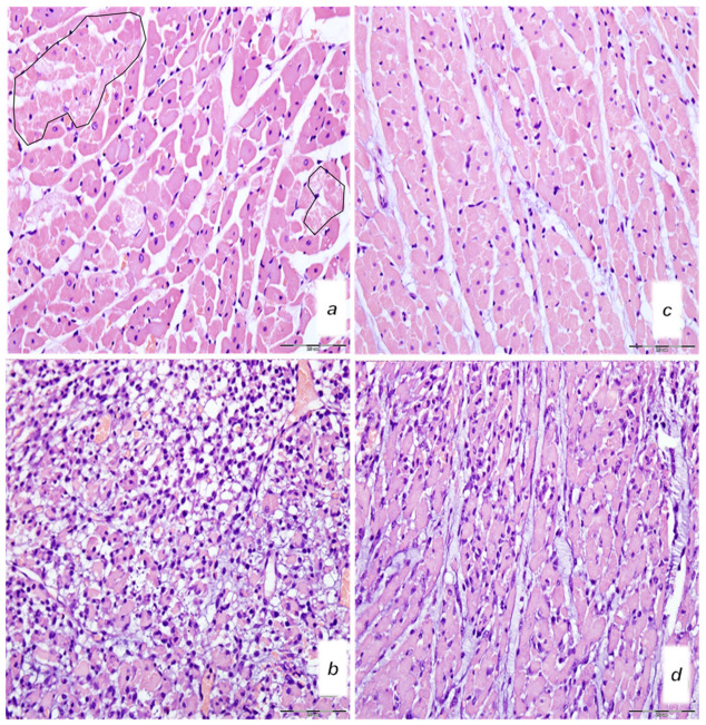
Figure 15.
Microscopy presentation of heart injury. Rats challenged once with isoprenaline 150 mg/kg s.c., mediation at 30 min before isoprenaline (a–d). Upper 24 h. (a,c) Single and small groups of necrotic myocites without inflammation (irregular circles) (magnification 400×, scale bar 20 μm) (saline (5 mL/kg i.p, left)) (a); single and small groups of necrotic myocites without inflammation (magnification 400x, scale bar 20 μm) (BPC 157 (10 μg/kg i.p., right)) (c). Low (b,d). Pronounced inflammatory infiltrate with necrotic myocites (magnification 400×, scale bar 20 μm) (saline (5 mL/kg i.p, left)) (b). Moderate inflammatory infiltrate affecting up to half of the septal thickness (magnification 400×, scale bar 20 μm) (BPC 157 (10 μg/kg i.p., right)). (d). (HE staining).

