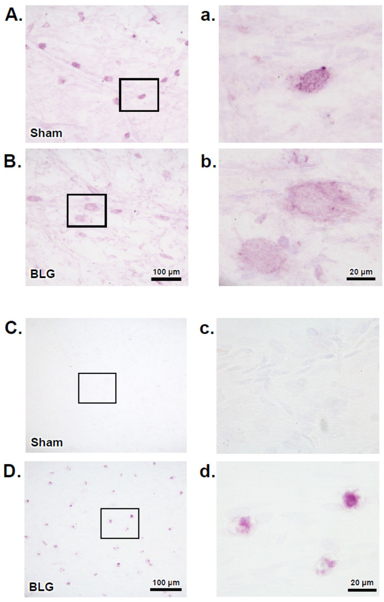
Figure 8.
Immunohistochemical detection of FcεRI and IgE in dural MCs. Dural tissues were collected from the sham and BLG-sensitized mice with prolonged whey-containing diet and stained using antibodies against FcεRI (A,B) and IgE (C,D). The rectangles in panels A–D denote the areas where the corresponding high-magnification photomicrographs in panels a-d were taken. The immunoreactive cells were photographed with 40× (A–D) and 100× objectives (a–d). Scale bars: 100 µm (for panels (A–D)); 20 µm (for panels (a–d)).

