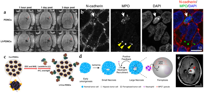
Figure 2.
The iron-based nanoprobes in glioblastoma MRI imaging. (a) T2-weighted MRI images with contrast accumulation in GBM [25]. (b) The confocal images demonstrate that the accumulation of MPO was located in the cytosol (with FITC tracking dye) [26]. (c) Modified with lactoferrin on the surface of iron nanoparticles. (d) Illustration of neutrophil recruitment crossing with targeted magnetic iron nanoparticles (MPO) and (e) in brain tissue GBM MRI imaging. Adapted with permission from Refs. [25,26], Copyright 2016 Wiley and 2020 Springer.

