Abstract
Introduction
Online MR-guided radiotherapy (MRgRT) is a relatively novel advancement in the field of radiation oncology, ensuring superior soft-tissue visualisation, allowing for online plan adaptation to anatomical and functional interfractional changes and improved motion management. Platinum-based chemoradiation followed by durvalumab is the recommended treatment for stage IIB(N1)/III NSCLC. However, this is only the case for patients with favourable risk factors and sufficient pulmonary function and reserve.
Methods
Herein, we present a technical report on tumour motion and breathing curve analyses of the first patient with node-positive stage IIB NSCLC and severely compromised pulmonary function and reserve [total lung capacity (TLC) 8.78L/132% predicted, residual volume (RV) 6.35L/271% predicted, vital capacity (VC) max 2.43L/58% predicted, FEV1 1.19L/38% predicted, DLCO-SB corrected for hemoglobin 2.76 mmol/min/kPa/30% predicted] treated in a prospective observational study with moderately hypofractionated MRgRT to a total dose of 48.0 Gy/16 daily fractions on the MRIdian system (Viewray Inc, Oakwood, USA).
Results
Radiotherapy was well tolerated with no relevant toxicity. First follow-up imaging at 3 months post-radiotherapy showed a partial remission. The distinctive features of this case are the patient’s severely compromised pulmonary function and the first online MR-guided accelerated hypofractionated radiotherapy treatment for primary node-positive NSCLC.
Conclusions
This technical report describes the first patient treated in a prospective observational study evaluating the feasibility of this relatively novel technology in stage IIB(N1)/III disease, proposing a clinical pathway for the management of high-risk patients.
Supplementary Information
The online version contains supplementary material available at 10.1186/s13014-022-02011-8.
Keywords: Hypofractionation, Image-guided radiotherapy, NSCLC, Pulmonary function, Thoracic radiotherapy
Introduction
Chemoradiation (CRT) followed by consolidation with durvalumab is the recommended treatment strategy for the management of stage IIB (N1)/III non-small cell lung cancer (NSCLC) [1]. However, in patients with poor prognostic factors, there is no consensus on the optimal treatment regimen, and patients are often referred to palliative radiotherapy alone or best supportive care. Our group has previously demonstrated the feasibility of PET-based accelerated hypofractionated radiotherapy (AHRT) on a conventional linear accelerator (LINAC) in this distinct patient cohort with encouraging outcome [2, 3].
Online MR-guided radiotherapy is a promising novel advancement in the field of radiation oncology allowing for superior real-time tumour visualisation, plan adaptation to anatomical changes and improved motion management [4–6]. Based on our previous experience, the Ludwig Maximilian University of Munich ethics committee approved a protocol (reference number: 20-793) for MRgRT in high-risk patients. In theory, due to online soft-tissue visualisation and gating capabilities, there is less uncertainty when delivering radiotherapy which subsequently could support dose-escalation strategies. Herein, we present a comprehensive technical report of the first patient treated with this protocol.
Case presentation
A 60-year-old male cachectic patient (172 cm, 45 kg; BMI: 15.2 kg/m2) presented to a specialised clinic in the treatment of respiratory diseases in 11/2020 following detection of a suspicious lung mass in SII of the right lung and an adjacent enlarged 15 mm lymph node (LN) in position 11R on follow-up CT for previous oropharyngeal cancer. The patient had an Eastern Cooperative Oncology Group performance status (ECOG-PS) of 2 and was an active smoker (40 pack years). He presented with chronic shortness of breath and persistent cough. His medical history included previous oropharyngeal cancer with first diagnosis in 11/2016. He underwent transoral laser surgery (pT2 pN0 cM0 R1) and adjuvant CRT to a total dose of 66.0 Gy ex domo. Other secondary diagnoses included COPD GOLD 3; group B, insulin-dependent diabetes mellitus, recurrent acute pancreatitis. A pulmonary function test (PFT) on admission, revealed the following results: total lung capacity (TLC) 8.78L/132% predicted, residual volume (RV) 6.35L/271% predicted, vital capacity (VC) max 2.43L/58% predicted, forced expiratory volume in 1 s (FEV1) 1.19L/38% predicted, single breath diffusing capacity of lung for carbon monoxide corrected for haemoglobin (DLCOcSB) 2.76 mmol/min/kPa/30% predicted. In addition, a blood gas analysis in room air was performed: pH: 7.40, pO2: 67 mmHg, pCO2: 44 mmHg, BE: 2.40 mmol/l, STB: 26.1 mmol/l, SaO2: 95%.
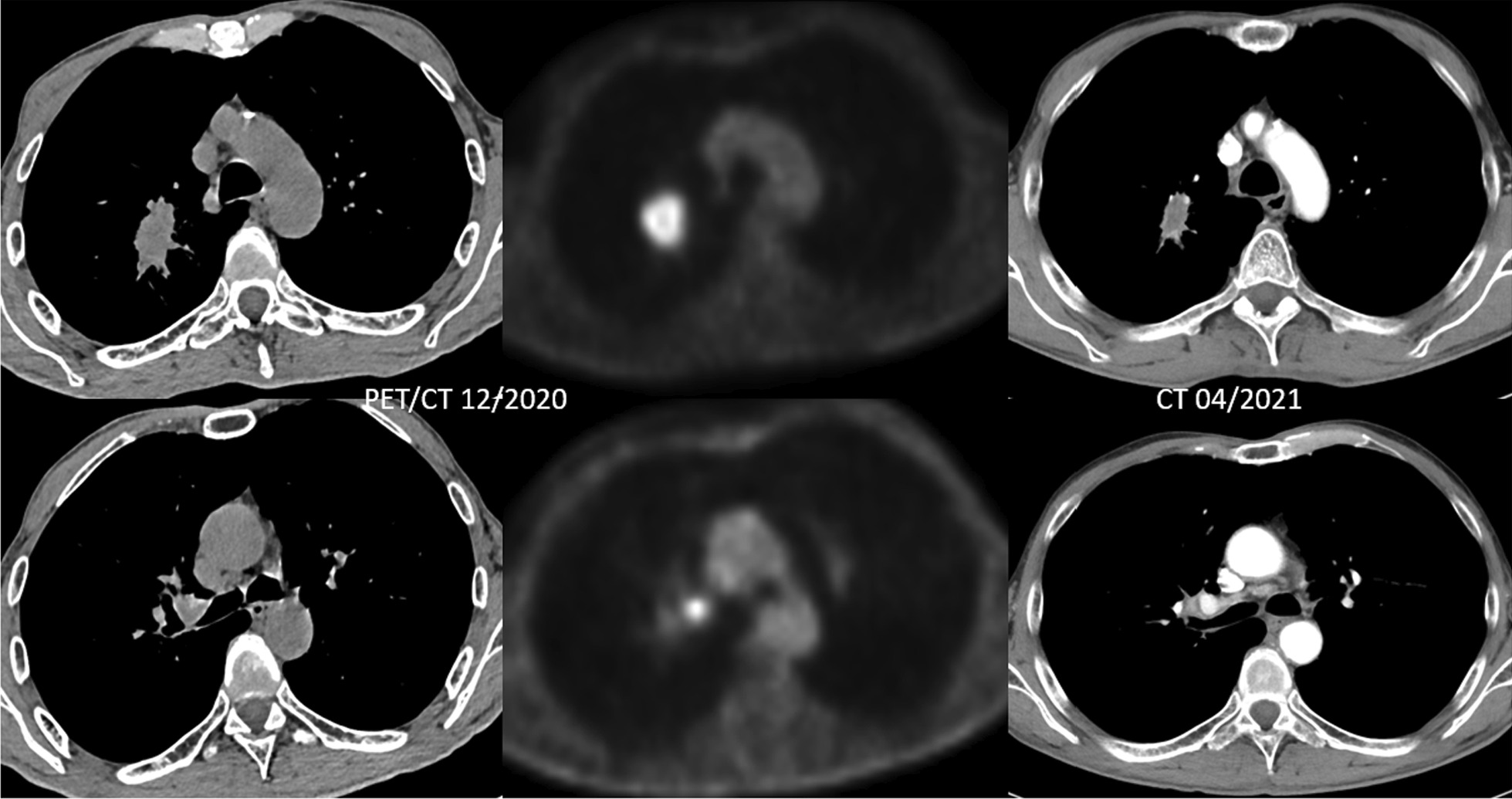
Bronchoscopy and transbronchial biopsy (TBB) with endobronchial ultrasound-guided transbronchial needle aspiration (EBUS-TBNA) 7/11R was performed with the histopathological diagnosis of CK7, Napsin A pos.; CDX2, TTF-1, p40 neg. primary adenocarcinoma of the lung; PD-L1 negative/Cologne Score: 0 (E1L3N). His insurance company did not cover further molecular pathology testing in the primary setting. PET/CT confirmed a 3.2 × 2.5 × 3.6 cm FDG-avid tumour (SUVmax 7) in the right upper lobe with moderate FDG-uptake in a LN at position 11R (SUVmax 4) (Fig. 1). MRI of the brain was inconspicuous, hence stage IIB-cT2a cN1 cM0 (UICC 8th edition) disease. Following discussion at the multidisciplinary tumour board (MTB), the patient was referred for moderately hypofractionated radiotherapy 48.0 Gy in 16 fractions.
Fig. 1.

PET/CT at initial diagnosis 12/2020; note enhanced FDG-uptake in the primary tumour (SUVmax 7) and LN pos. 11R (SUVmax 4) and Follow-up CT 3 months post-MRgRT in 04/2021 demonstrating a partial remission
Treatment planning and delivery
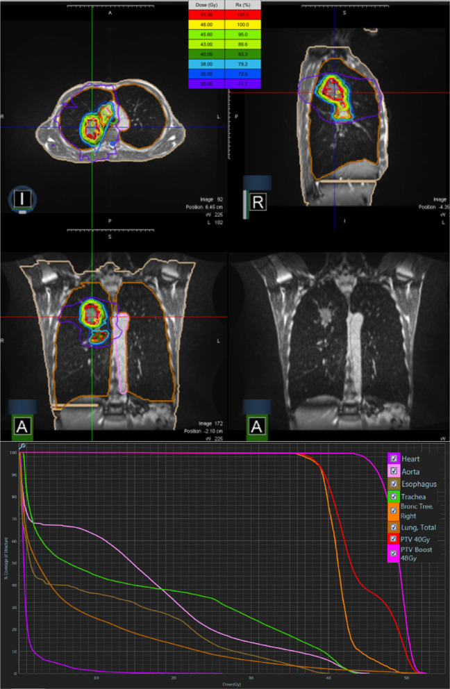
Briefly, the treatment was delivered on one of two commercially available MR-guided linear accelerator systems, the MRIdian system (Viewray Inc, Oakwood, USA) [7, 8]. The patient underwent MRI simulation in inspiration breath-hold (BH) and supine position with arms above the head using a dedicated positioning device (WingSTEP, IT-V, Innsbruck, Austria). Thereafter, a standard planning CT using the same patient positioning and BH level was conducted to obtain electron density information. Image datasets were then co-registered using the deformable registration algorithm of the integrated MRIdian treatment planning system. The target volume and OARs were contoured on the 3D MR simulation scan. An isotropic gross tumour volume (GTV) expansion of 5 mm was used to generate the planning target volume (PTV) primary tumour + LN. Due to multiple suspicious mediastinal LNs in pos. 2R, regional nodal irradiation of stations 10R/4R/2R was performed. GTV primary tumour + LN was 35.12 cc and PTV primary tumour + LN was 80.41 cc. Radiotherapy was delivered in a non-adaptive manner with the baseline plan, due to limited interfractional anatomical changes. This was also confirmed by the minimal dosimetric changes of the GTV/PTV dose coverage over the course of the treatment (Fig. 2). The relevant dosimetric parameters of organs at risk (OARs) for the baseline plan are provided in Table 1. During dose delivery, continuous, real-time 2D cine MRI in a single sagittal plane was used to monitor the target volume. For this purpose, a gating boundary contour was defined by adding an isotropic margin of 3 mm to the GTV and a pre-defined threshold region of interest percentage (ROI%) was set at 5%, allowing a percentage of the GTV to be outside the gating boundary before a beam-hold is triggered. The beam was gated automatically by the gating functionality of the system through a deformable registration-based tracking algorithm. The total delivery time for all fractions was 2.35 h and total “beam-on” time for all fractions was 27.78 min (Table 2). The dose covering 98% (D98), 50% (D50) of the GTV and PTV primary tumour was 47.6 Gy, 49.70 Gy and 44.6 Gy, 49.1 Gy, respectively. The dose to 2% (D2) of the GTV and PTV primary tumour was 51.5 Gy and 51.3 Gy. The isodose distribution for representative slices in all planes and the corresponding dose-volume histogram (DVH) is depicted in Fig. 3. In addition, a cine movie extract of the first fraction is provided in the Additional file 1: Video S1.
Fig. 2.
Box-plot of percentage change in D2, D50, D95 and D98 for all 16 fractions compared to the baseline plan
Table 1.
Relevant organs at risk dosimetric parameters of the baseline plan; MLD mean lung dose, relative organ volume receiving ≥ 5 Gy (V5), ≥ 10 Gy (V10), ≥ 20 Gy (V20)
| Lung, total | |||
| V5 (%) | V10 (%) | V20 (%) | MLD (Gy) |
| 36.0 | 24.9 | 13.3 | 7.4 |
| Lung, right | |||
| V5 (%) | V10 (%) | V20 (%) | MLD (Gy) |
| 45.6 | 40.3 | 24.9 | 11.0 |
| Lung, left | |||
| V5 (%) | V10 (%) | V20 (%) | MLD (Gy) |
| 25.4 | 8.2 | 0.6 | 3.6 |
| Esophagus | |||
| Mean (Gy) | V5 (%) | V10 (%) | V20 (%) |
| 9.5 | 40.2 | 36.2 | 25.5 |
| Heart | |||
| Mean (Gy) | V5 (%) | V10 (%) | V20 (%) |
| 1.2 | 4.6 | 1.1 | 0.1 |
Table 2.
Treatment delivery times and resulting duty cycle efficiencies for each fraction
| Delivery time (mins) | Beam-on time (mins) | Duty cycle efficiency (%) | |
|---|---|---|---|
| Fr 1 | 8.48 | 1.71 | 20.20 |
| Fr 2 | 9.68 | 1.73 | 17.86 |
| Fr 3 | 8.86 | 1.71 | 19.32 |
| Fr 4 | 8.49 | 1.69 | 19.88 |
| Fr 5 | 9.04 | 1.72 | 18.99 |
| Fr 6 | 8.86 | 1.73 | 19.46 |
| Fr 7 | 9.10 | 1.71 | 18.83 |
| Fr 8 | 8.84 | 1.72 | 19.46 |
| Fr 9 | 8.40 | 1.72 | 20.45 |
| Fr 10 | 9.86 | 1.73 | 17.58 |
| Fr 11 | 8.50 | 1.74 | 20.45 |
| Fr 12 | 9.18 | 1.79 | 19.48 |
| Fr 13 | 8.47 | 1.77 | 20.96 |
| Fr 14 | 8.45 | 1.8 | 21.24 |
| Fr 15 | 8.38 | 1.76 | 20.98 |
| Fr 16 | 8.43 | 1.75 | 20.76 |
Fig. 3.
Isodose distribution for representative slices in all main planes and the corresponding dose-volume histogram with the green colour depicting 95% (45.6 Gy) of the prescribed dose to the PTV Boost and the cyan colour depicting 95% (38.0 Gy) of the prescribed dose to the PTV 40 Gy
Tumour motion and gating analysis
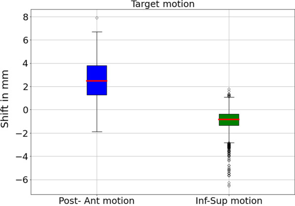
Tumour centroid positions were extracted from the cine videos using an in-house developed code. Using the centroid positions, we then analysed the primary tumour motion in the posterior-anterior and inferior-superior directions for all fractions as shown exemplarily for the first fraction (Fig. 4) and all fractions (Additional file 2: S2-1 to S2-15).
Fig. 4.

Box-plot of the primary tumour motion for fraction 1. We calculated the median and inter-quartile range (IQR) amplitudes of the extracted target centroid positions relative to the centroid position of the boundary contour. For the first fraction, the target posterior-anterior motion was 2.48/2.53 mm (median/IQR) and -0.85/0.98 mm (median/IQR) for the inferior-superior motion. Box-plots for every fraction are provided in the Additional file 2
In total, 2.35 h of real-time cine images were analysed. Using the beam-on status displayed in the cine videos, we computed the total beam-on time for all fractions, which amounted to 27.78 min. Additionally, we calculated the duty cycle efficiency for each fraction, defined as the number of “beam-on” frames divided by the total number of MR cine frames acquired during that fraction, including frames acquired during multileaf collimator motion and gantry rotation according to Finazzi et al. [9]. Information about the imaging pauses was also extracted from the cine videos. The data for all fractions is provided in Table 2.
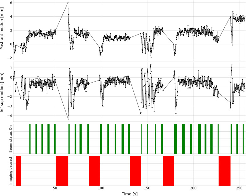
The Primary tumour motion in posterior-anterior and inferior-superior directions for the first few minutes of fraction 1 is depicted in Fig. 5; “Beam-on” time intervals are denoted by the green bars while time intervals where the cine imaging was paused are denoted by the red bars. During gantry rotation, acquisition of cine imaging was paused hence the patient breathes freely.
Fig. 5.
Primary tumour motion in posterior-anterior and inferior-superior directions for the first few minutes of fraction 1; “Beam-on” time intervals are denoted by the green bars while time intervals where the cine imaging was paused are denoted by the red bars. During gantry rotation, acquisition of cine imaging was paused hence the patient breathes freely
Patient outcome
Treatment was well tolerated with no relevant (grade ≥ 2) toxicity. On the first follow-up CT scan 3 months post-MRgRT (04/2021), a decrease in the target lesion dimensions from 3.2 × 2.5 × 3.6 cm to 2.7 × 1.2 × 2.3 cm was noted, hence partial remission per RECIST 1.1 criteria (Fig. 1). The patient remained an active smoker and last visited with his pulmonologist early October 2021 in reduced general condition (age-adjusted Charlson Comorbidity Index: 7; BMI: 14 kg/m2) and died late October 2021 (8.8 months post-MRgRT and 11.2 months from initial diagnosis) with the cause of death unclear but possibly related to pulmonary disease.
Discussion
To the best of our knowledge, the present case is the first report on MR-guided radiotherapy for node-positive NSCLC. Importantly, this patient had reduced PS and limited pulmonary function and reserve, which precluded concurrent CRT. MRgRT was feasible with no significant overlap with the previous radiotherapy in the head-and-neck region. On first follow-up post-RT, the patient presented in stable condition with a partial remission (Fig. 1).
Regarding the concept of moderate hypofractionation, Amini et al. [10] reported comparable oncologic outcome in 300 stage III patients treated with 45.0 Gy/15 vs. ≥ 60.0 Gy/2.0 Gy fractions, although patients in the hypofractionation arm initially had worse prognostic factors. Similarly, we previously published encouraging survival data of prospectively enrolled patients with poor prognostic factors and limited pulmonary reserve (FEV1 ≤ 1.0 L and/or DLCO-SB ≤ 40% predicted and/or on long-term oxygen therapy) treated on a conventional linear accelerator [3]. Hence, due to the severely compromised lung function, a moderate hypofractionation schedule was chosen to account for normal lung tissue dose exposure. Furthermore, the application of MRgRT in this setting has its inherent advantages. Due to the incorporation of a 0.35 T MR scanner, superior soft tissue contrast aids in more accurate delineation of central tumours in close proximity to mediastinal structures [4]. Online MRgRT ensures daily plan adaptation in particular in challenging cases e.g. with accompanying atelectasis and subsequent resolution during treatment thereby mitigating an overdose to organs at risk [11]. In the case of favourable tumour response, online adaptation to morphological changes to the tumour are readily feasible. Importantly, online MRgRT enables real-time tumour monitoring during treatment and motion management by means of gating/tracking strategies thus eliminating the need for larger margins or internal target volume (ITV) concepts to account for tumour mobility. To further unlock new capabilities of this novel technology, functional imaging including diffusion-weighted imaging facilitates response assessment during and after primary treatment [12]. Therefore, promoting adaptive and dose-escalation strategies based on biological response similar to concepts employed with PET imaging [13, 14]. All these aspects in comparison to treatments on a conventional LINAC ensure that there is less uncertainty when delivering radiotherapy and support isotoxic dose escalation strategies. In conjunction with our previous experience, the present analysis provides a clinical pathway for the management of these high-risk patients.
Currently, an ongoing phase II study is exploring the feasibility of MR-guided hypofractionated adaptive radiation therapy of 60.0 Gy/15 fractions with concurrent chemotherapy and consolidation durvalumab in unresectable stages IIB-IIIC NSCLC (NCT03916419). Furthermore, the combination of radiotherapy and immune-checkpoint inhibitors can potentiate anti-tumour response and improve patient outcome [15]. A number of trials are exploring conventional/hypofractionated radiotherapy in node-positive NSCLC with PD-L1 inhibition in (borderline) patients [SWOG S1933 (NCT04310020), ARCHON-1 study (NCT03801902), TRADE-hypo trial (NCT04351256) and the SPIRAL-RT study (JMA-IIA00434)].
Conclusion
We report the first case of a patient with stage IIB(N1) NSCLC and severely limited pulmonary function and reserve treated with MRgRT, hereby proposing a clinical pathway for the management of high-risk patients. We are currently recruiting patients to our MRgRT protocol.
Supplementary Information
Additional file 1: Extract of gated MRgRT delivery for the first fraction of treatment. The gating target (GTV; green contour) and the gating boundary (red contour) are displayed.
Additional file 2: Box-plot of the primary tumour motion in posterior-anterior and inferior-superior directions for fractions 2-16.
Acknowledgements
Not applicable.
Authors' contributions
CE: conceptualisation; data curation; formal analysis; funding acquisition; investigation; methodology; validation; visualisation; roles/writing–original draft; writing–review and editing. EL: data curation; formal analysis; investigation; methodology; validation; visualisation; writing–review and editing. LN: Data curation; formal analysis; investigation; methodology; validation; writing–review and editing. YX: data curation; formal analysis; investigation; methodology; validation; writing–review and editing. MN: conceptualisation; investigation; methodology; supervision; writing–review and editing. CB: conceptualisation; funding acquisition; investigation; methodology; supervision; writing–review and editing. FM: conceptualisation; investigation; methodology; supervision; roles/writing–original draft; writing–review and editing. SC: conceptualisation; data curation; formal analysis; investigation; methodology; supervision; roles/writing–original draft; writing–review and editing. All authors read and approved the final manuscript.
Funding
Chukwuka Eze received funding in the form of a research grant from the German Cancer Aid [Mildred Scheel-Stipendienprogramm für Krebsforschung, 2018 (57468956)].
Availability of data and materials
Substantial data generated or analysed during this study are included in this published article [and its supplementary information files]. Further datasets during and/or analysed during the current study are available from the corresponding author on reasonable request.
Declarations
Ethics approval and consent to participate
The Ludwig Maximilian University of Munich Ethics Committee approved this study (reference number: 20-793). Furthermore, the study is also registered in the German Clinical Trials Register (DRKS00026665).
Consent for publication
The individual described in this analysis provided consent for publication of this data.
Competing interests
SC and CB have received research grants and speaker fees/travel support from Viewray Inc (Oakwood Village, Ohio). MN has received research grants from Viewray (Oakwood Village, Ohio). All other authors declare no conflict of interest related to this work.
Footnotes
Publisher's Note
Springer Nature remains neutral with regard to jurisdictional claims in published maps and institutional affiliations.
References
- 1.Ettinger DS, Wood DE, Aisner DL, et al. NCCN guidelines insights: non-small cell lung cancer, version 2.2021: featured updates to the NCCN guidelines. J Natl Compr Cancer Netw. 2021;19(3):254–266. doi: 10.6004/JNCCN.2021.0013. [DOI] [PubMed] [Google Scholar]
- 2.Eze C, Taugner J, Roengvoraphoj O, et al. Initial report on feasibility of PET/CT-based image-guided moderate hypofractionated thoracic irradiation in node-positive non-small cell lung Cancer patients with poor prognostic factors and strongly diminished lung function: a retrospective analysis. Radiat Oncol. 2019;14(1):1–7. doi: 10.1186/s13014-019-1304-2. [DOI] [PMC free article] [PubMed] [Google Scholar]
- 3.Eze C, Taugner J, Schmidt-Hegemann NS, et al. Feasibility of hypofractionated radiotherapy in inoperable node-positive NSCLC patients with poor prognostic factors and limited pulmonary reserve: a prospective observational study. Acta Oncol (Madr) 2021 doi: 10.1080/0284186X.2021.1941244. [DOI] [PubMed] [Google Scholar]
- 4.Crockett CB, Samson P, Chuter R, et al. Initial clinical experience of MR-guided radiotherapy for non-small cell lung cancer. Front Oncol. 2021 doi: 10.3389/fonc.2021.617681. [DOI] [PMC free article] [PubMed] [Google Scholar]
- 5.Corradini S, Alongi F, Andratschke N, et al. MR-guidance in clinical reality: current treatment challenges and future perspectives. Radiat Oncol. 2019;14(1):1–12. doi: 10.1186/s13014-019-1308-y. [DOI] [PMC free article] [PubMed] [Google Scholar]
- 6.Corradini S, Alongi F, Andratschke N, et al. ESTRO-ACROP recommendations on the clinical implementation of hybrid MR-linac systems in radiation oncology. Radiother Oncol. 2021;159:146–154. doi: 10.1016/j.radonc.2021.03.025. [DOI] [PubMed] [Google Scholar]
- 7.Mutic S, Dempsey JF. The viewray system: magnetic resonance-guided and controlled radiotherapy. Semin Radiat Oncol. 2014;24(3):196–199. doi: 10.1016/j.semradonc.2014.02.008. [DOI] [PubMed] [Google Scholar]
- 8.Klüter S. Technical design and concept of a 0.35 T MR-Linac. Clin Transl Radiat Oncol. 2019;18:98–101. doi: 10.1016/j.ctro.2019.04.007. [DOI] [PMC free article] [PubMed] [Google Scholar]
- 9.Finazzi T, van Sörnsen de Koste JR, Palacios MA, et al. Delivery of magnetic resonance-guided single-fraction stereotactic lung radiotherapy. Phys Imaging Radiat Oncol. 2020;14:17–23. doi: 10.1016/j.phro.2020.05.002. [DOI] [PMC free article] [PubMed] [Google Scholar]
- 10.Amini A, Lin SH, Wei C, Allen P, Cox JD, Komaki R. Accelerated hypofractionated radiation therapy compared to conventionally fractionated radiation therapy for the treatment of inoperable non-small cell lung cancer. Radiat Oncol. 2012;7(1):1–7. doi: 10.1186/1748-717X-7-33. [DOI] [PMC free article] [PubMed] [Google Scholar]
- 11.Chin S, Eccles CL, McWilliam A, et al. Magnetic resonance-guided radiation therapy: a review. J Med Imaging Radiat Oncol. 2020;64(1):163–177. doi: 10.1111/1754-9485.12968. [DOI] [PubMed] [Google Scholar]
- 12.Jagoda P, Fleckenstein J, Sonnhoff M, et al. Diffusion-weighted MRI improves response assessment after definitive radiotherapy in patients with NSCLC. Cancer Imaging. 2021;21(1):1–11. doi: 10.1186/S40644-021-00384-9. [DOI] [PMC free article] [PubMed] [Google Scholar]
- 13.Unterrainer M, Eze C, Ilhan H, et al. Recent advances of PET imaging in clinical radiation oncology. Radiat Oncol. 2020;15(1):1–15. doi: 10.1186/s13014-020-01519-1. [DOI] [PMC free article] [PubMed] [Google Scholar]
- 14.Eze C, Schmidt-Hegemann NS, Sawicki LM, et al. PET/CT imaging for evaluation of multimodal treatment efficacy and toxicity in advanced NSCLC—current state and future directions. Eur J Nucl Med Mol Imaging. 2021;48:3975–3989. doi: 10.1007/s00259-021-05211-8. [DOI] [PMC free article] [PubMed] [Google Scholar]
- 15.Käsmann L, Eze C, Taugner J, et al. Chemoradioimmunotherapy of inoperable stage III non-small cell lung cancer: immunological rationale and current clinical trials establishing a novel multimodal strategy. Radiat Oncol. 2020;15(1):167. doi: 10.1186/s13014-020-01595-3. [DOI] [PMC free article] [PubMed] [Google Scholar]
Associated Data
This section collects any data citations, data availability statements, or supplementary materials included in this article.
Supplementary Materials
Additional file 1: Extract of gated MRgRT delivery for the first fraction of treatment. The gating target (GTV; green contour) and the gating boundary (red contour) are displayed.
Additional file 2: Box-plot of the primary tumour motion in posterior-anterior and inferior-superior directions for fractions 2-16.
Data Availability Statement
Substantial data generated or analysed during this study are included in this published article [and its supplementary information files]. Further datasets during and/or analysed during the current study are available from the corresponding author on reasonable request.


