Abstract
Endogenous retroviruses (ERVs) are the remnants of past retroviral infections that once invaded the host’s germline and were vertically transmitted. ERV sequences have been reported in mammals, but their distribution and diversity in cervids are unclear. Using next-generation sequencing, we identified a nearly complete genome of an endogenous betaretrovirus in fallow deer (Dama dama). Further genomic analysis showed that this provirus, tentatively named cervid endogenous betaretrovirus 1 (CERV β1), has typical betaretroviral genome features (gag-pro-pol-env) and the betaretrovirus-specific dUTPase domain. In addition, CERV β1 pol sequences were detected by PCR in the six non-native deer species with wild populations in Australia. Phylogenetic analyses demonstrated that CERV β1 sequences from subfamily Cervinae clustered as sister taxa to ERV-like sequences in species of subfamily Muntiacinae. These findings, therefore, suggest that CERV β1 endogenisation occurred after the split of these two subfamilies (between 3.3 and 5 million years ago). Our results provide important insights into the evolution of betaretroviruses in cervids.
Keywords: betaretrovirus, class II retroviruses, deer, endogenous retroviruses, genetic characterisation
1. Introduction
Endogenous retroviruses (ERVs) represent remnants of past retrovirus infections, which became integrated into the host germline and were passed to progeny cells; and can comprise up to 10% of vertebrate genomes [1,2]. Most ERVs have accumulated genetic defects that render them unable to express infectious viruses or proteins. However, some ERVs are transcriptionally active and have maintained intact open reading frames for some of their genes [3], suggesting that these elements may benefit their hosts, possibly protecting against exogenous retrovirus infection [4]. Based on phylogenetic relatedness of reverse transcriptase sequences, ERVs are grouped into class I (gamma- and epsilon-RVs), class II (lentiviruses, alpha-, beta-, and delta-RVs), and class III (spumaviruses) [5].
In contrast to classes I and III, the class II ERVs have a more restricted host range, comprised mainly of mammals and birds; however, they have also recently been detected in amphibians [6,7,8]. Among class II ERVs, full-length endogenous betaretroviruses have been detected in the genomes of livestock [9,10,11,12], rodents [9,13], primates [9,14,15], bats [16,17], and the common brushtail possum (Trichosurus vulpecula) [18]. Betaretrovirus-related elements have been previously detected in wild cervid genomes such as caribou (Rangifer tarandus) and white-tailed deer (Odocoileus virginianus) utilising PCR assays targeting conserved regions of the retroviral pro and/or pol genes; however, these elements still remain uncharacterised [6].
The family Cervidae comprises 40 deer species within four subfamilies (Cervinae, Muntiacinae, Hydropotinae, and Odocoileinae), and deer are distributed throughout the northern hemisphere, South America, and Southeast Asia [19,20]. Deer were introduced to Australia in the 19th century, and six non-native deer species (chital, fallow, hog, red, rusa, and sambar) have expanded considerably in numbers and distribution in recent decades [21]. These six species belong to the subfamily Cervinae [19,20]. We herein characterise an endogenous betaretrovirus in fallow deer (Dama dama) and rusa deer (Rusa timorensis) detected by next-generation sequencing. Subsequent PCR-based screening of the six non-native Australian deer species determined the distribution of this betaretrovirus.
2. Materials and Methods
2.1. Sampling
Deer blood samples were collected in eastern Australia as previously described [22,23]. Briefly, blood was drawn from the jugular vein, the heart or thoracic cavity of dead deer immediately after killing via shooting during operational culling programs and collected in tubes with and without anticoagulant (EDTA). Collection tubes were immediately refrigerated and transported to the laboratory. Tubes were centrifuged for 10 min at 2000 g, and aliquots of blood pellet and serum/plasma were stored at −20 °C and −80 °C, respectively, until required.
2.2. Nucleic Acid Extraction and Next-Generation Sequencing (NGS)
Genomic DNA (gDNA) was extracted from blood samples using the MAX™CORE Nucleic Acid Purification Kit (Applied Biosystems, Foster City, CA, USA) as previously described [23]. Sequencing libraries were constructed using the Nextera XT Flexi DNA Library kit (Illumina, San Diego, CA, USA) according to the manufacturer’s instructions. The prepared 2 nM final pooled library sample was sequenced using the NextSeq 500 Sequencing System with the Illumina NextSeq 500/550 HighOutput Kit v2.5 (300 cycles). Additionally, NGS data obtained previously from deer serum and plasma samples [24] were included in the present study.
2.3. Bioinformatic Analysis
Sequencing reads were analysed using the previously described computational workflow [24]. Briefly, raw data was demultiplexed, trimmed, and filtered out against the deer RefSeq genome (GenBank GCF_002102435.1) using Trim_Galore v0.4.5, bwa v0.7.17, samtools v1.6, and bedtools v2.26. The cleaned reads were de novo assembled and resulting contigs were compared against the nonredundant nucleotide database on GenBank using BLASTn. Gene prediction was performed using ORFfinder (https://www.ncbi.nlm.nih.gov/orffinder/, accessed on 12 November 2021). Protein domains were predicted using Pfam conserved domain search (http://pfam.xfam.org/, accessed on 15 November 2021) and NCBI conserved domain search (https://www.ncbi.nlm.nih.gov/Structure/cdd/wrpsb.cgi, accessed on 15 November 2021).
2.4. PCR Detection and Sanger Sequencing
Standard precautions to avoid product contamination were taken for all PCR assays, including filter pipette tips and physically separated rooms for PCR setup. A non-template (negative control) was interspersed with actual samples. The following primer set: 5′-CCTCGGGACTTGGAAGAAATAA-3′ and 5′-GCAAGATGTAGGTAGGGTCTAATC-3′, were designed on the identified retrovirus-like sequences to amplify an approximately 900 bp fragment of the pol gene, covering the entire reverse transcriptase region. PCR amplification was performed using 0.2 μM of both forward and reverse primers, GoTaq G2 DNA polymerase (Promega, Madison, WI, USA), and 1 μL of cDNA. Moreover, PCR conditions were 95 °C for 2 min, 40 cycles of 95 °C for 30 s, 57 °C for 30 s, 72 °C for 60 s, with a final extension at 72 °C for 5 min. Nucleotide sequencing was performed by Sanger sequencing at the Australian Genome Research Facility, Melbourne, Australia.
2.5. Phylogenetic Analysis
Multiple alignments of amino acid or nucleotide sequences were conducted using ClustalW implemented in Geneious software (Biomatters Ltd., Auckland, New Zealand, version 11.1.4). The best-fitting substitution model was determined based on the lowest BIC scores in MEGA 7 [25]. Phylogenetic trees were also constructed with this software, using the maximum likelihood method. Statistical support for the trees was evaluated by bootstrapping based on 1000 repetitions.
3. Results
3.1. Identification of Betaretrovirus in Australian Deer Genome
As part of a pathogen search in Australian wild deer [23], gDNA extracted from blood specimens collected in four fallow deer were subjected to next-generation sequencing. Illumina sequencing of these samples generated a total of 107,542,698 paired-end (PE) reads, ranging from 16,837,618 to 33,656,032 PE reads. After trimming and host-genome removing, a total of 22,814,671 PE reads (range 653,018–10,289,520 PE reads) were retained. These datasets were searched for possible pathogen-derived sequences, and here we focus on reporting the presence of apparent endogenous retrovirus-like sequences, displaying the closest relatedness with betaretroviruses identified in goats (Table 1).
Table 1.
Percentage identities of the assembled retroviral contigs.
| Deer ID | Deer Species | Source | Contig Length (nt) | Accession Number | Best hit (Accession Number) | Subject Cover (%) | Identity (%) |
|---|---|---|---|---|---|---|---|
| NSW48 | Fallow | gDNA from blood | 7288 | OL547611 | ENTV-2CHN11 (KU258877) | 76 | 68 |
| NSW96 | Fallow | gDNA from blood | 7277 | OL547612 | ENTV/CH/GT/2015 (MK210250) | 76 | 68 |
| VIC43 | Fallow | gDNA from blood | 2020 | OL547609 | ENTV-2CHN6 (KU258875) | 73 | 74 |
| VIC44 | Fallow | gDNA from blood | 1975 | OL547610 | ENTV-2CHN6 (KU258875) | 73 | 74 |
| NSW164 | Rusa | RNA from plasma | 1922 | OL547613 | ENTV-2CHN10 (KU258879) | 73 | 73 |
Additionally, bioinformatic analysis of NGS data obtained previously by our team [24] revealed the presence of one betaretrovirus-like contig in RNA extracted from one rusa deer plasma sample. Alignments of these collective betaretrovirus-like contigs showed near-identical sequences (99.7–100% nucleotide similarities), suggesting the presence of the same provirus in the fallow deer genome, which we provisionally named cervid endogenous betaretrovirus 1 (CERV β1).
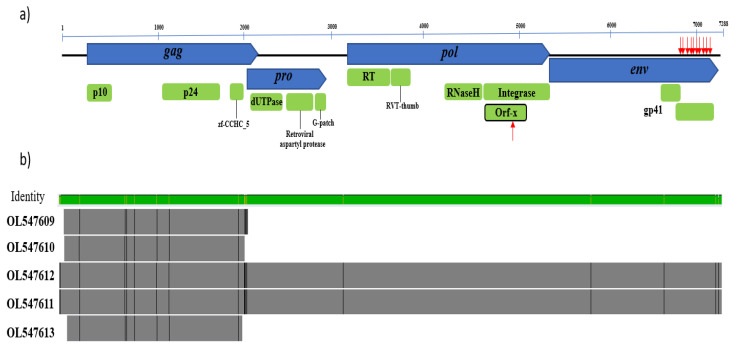
We detected five CERV β1 sequences, with variants NSW48 and NSW96 being near full-length proviruses with high homology (Figure 1). Sequence analysis of the longest contig (OL547611) revealed four open reading frames (ORFs) characteristic of betaretroviruses encoding gag, pol, pro, and env proteins. Several stop codons were found in the env protein at its 3′ end; however, the gag, pro, and pol proteins are intact, suggesting they might code for functional proteins (Figure 1a and Figure S1).
Figure 1.
Genomic structure of CERV β1. (a) The core retroviral genes gag, pro, pol, and env are blue, while coding regions and predicted conserved domains are green. The position of aberrant stop codons is denoted with red arrows. (b) Alignment of the four detected retroviral contigs. Sequences OL547611 and OL547612 are nearly complete proviruses, while sequences OL547609, OL0547610, and OL0547613 matched the gag gene. Genome alignments are represented by the outlined bars in grey, with divergent sites highlighted in black. The green bar above indicates the percentage identity among the sequences (green is the highest identity).
Analysis of the genomic structure of CERV β1 revealed that gag (1890 nt/630 aa) contains the structural betaretrovirus proteins, p10 or matrix (83 aa) and p24 or core nucleocapsid (192aa), along with a predicted zinc finger domain (zf-CCHC_5; pfam14787). In addition, the major homology region (MHR; nucleotide coordinates 1517–1570) conserved among retroviruses [26] was also identified in CERV β1 gag protein. This MHR showed the conserved motif QGxxExxxxFxxRLxxxx as was previously identified in other retroviruses [27]. Pro (870 nt/290 aa) was shown to overlap 115 nt with the gag gene. In general, pro-encoded proteins have two domains, a pseudoprotease (protease-like) domain that has deoxyuridine triphosphatase (dUTPase) activity and an active protease (PR) [28,29]. We found that the CERV β1 pro contains a dUTPase domain at its 5′ end (Figure 1), and the protease activity was confirmed since this sequence bears the core amino acid sequence of a retroviral aspartyl protease Leu-Asp-Thr-Gly (nt 2536–2547) [28,30]. Interestingly, such as other betaretroviruses [6], CERV β1 pro encodes a glycine-rich region called the G-patch domain [31].
The pol gene (2241 nt/747 aa) encodes a reverse transcriptase (RT), an RT thumb domain, the RNase H transcriptase, and an integrase. The integrase is composed of three subdomains, namely, integrase zinc-binding (pfam02022), integrase core domain (pfam00665), and integrase DNA binding domain (pfam00552). In addition, it was interesting to note that CERV β1 pol includes an additional ORF, defined as orf-X, of 159 aa in reading frame -1 within the integrase domain. Orf-x was previously identified in Jaagsiekte sheep retrovirus [32] and in endogenous retroviruses of bat (Desmodus rotundus) [17] and armadillo (Dasypus novemcinctus) [33]. However, the CERV β1 orf-X is shorter, shows stop codons and revealed low amino acid similarity (<50%) with orf-X previously reported (Figure S2). Conserved retroviral active site motifs were present in the protease (DxG), reverse transcriptase (YXDD), and integrase (DDE) domains. A truncated env protein was found with ten stop codons (positions 473, 475, 477, 492, 507, 511, 522, 534, 562, 579 and 590); thus, the transmembrane domain gp41 (pfam00517) is truncated and divided into two portions with different read frames: one of 49 aa (read frame 1) and the other of 146 aa (read frame 3).
3.2. Detection of CERV β1-Related Sequences in Deer Genomes
Translated CERV β1 gag, pro, pol, and env ORFs were first compared to deer genomes deposited in NCBI GenBank databases. This analysis showed homology with protein sequences from five deer species, sharing similarities of 83.7–98.6% (Table 2).
Table 2.
Comparison of CERV β1 ORFs and proteins from other deer species deposited in GenBank.
| Deer Species | Pairwise Amino Acid Identity % (Accession Number) | |||
|---|---|---|---|---|
| Gag | Pro | Pol | Env | |
| Cervus hanglu yarhandensis | 97.6 (KAF4008627) | 98.6 (KAF4023393) | N.D | 83.7 (KAF4008560) |
| Cervus canadensis | 97.5 (XP_043296825) | N.D | N.D | 91.3 (XP_043339709) |
| Cervus elaphus | 97.3 (XP_043768122) | N.D | N.D | 95.2 (XP_043772720) |
| Muntiacus muntjak | 93.3 (KAB0338462) | 96.2 (KAB338400) | 94.1 (KAB0338155) | 93.3 (KAB0337844) |
| Muntiacus reevesi | 92.5 (KAB0339023) | 96.6 (KAB0386166) | 94 (KAB0374272) | 93.5 (KAB0338018) |
N.D: no data.
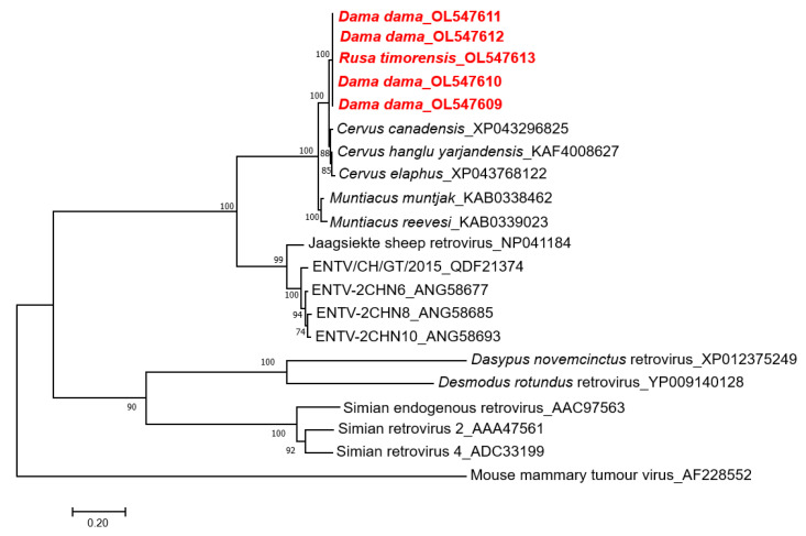
To examine the phylogenetic relationship, the CERV β1 gag amino acid sequences were aligned with known endogenous and exogenous betaretroviruses and related proteins found in deer genomes (Figure 2). This phylogenetic tree showed that CERV β1 gag sequences from fallow and rusa deer formed a monophyletic clade. Moreover, these sequences do not group with other previously described betaretroviruses but cluster with sequences identified in Cervus sp. and Muntiacus sp. genomes forming a well-supported clade. CERV β1 sequences are grouped within this clade with Cervus sp. sequences and form a sister taxon with Muntiacus sp. sequences (Figure 2).
Figure 2.
Phylogenetic analysis of CERV β1 sequences obtained by next-generation sequencing (in red), related sequences in deer genomes, and representative members of the genus Betaretrovirus. The tree was generated based on the gag protein by the maximum-likelihood method, and JTT + G + I substitution model with 1000 bootstrap replicates. Bootstrap values >70% are displayed at the tree branches. The scale bar indicates amino acid substitutions per site. ENTV: Enzootic nasal tumour virus.
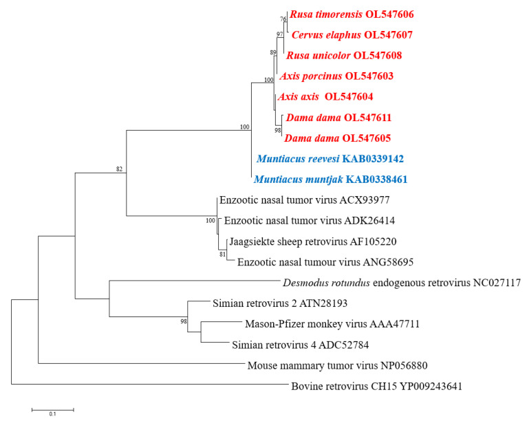
CERV β1 pol-like sequences in gDNA of the six Australian non-native deer species belonging to the subfamily Cervinae were amplified and analysed. Sequences were deposited in GenBank under accession numbers OL547603-OL547608. One blood sample per deer species was screened using the designed primers, which amplified an 828 nt (276 aa) fragment of the pol gene. This fragment covers the entire reverse transcriptase region (RT Rtv; Pfam CL0027). The accuracy and specificity of the designed primers were demonstrated by the close correlation between the amplified sequence and the contig used for primer design, as they created a unique cluster (Figure 3). Furthermore, the identity matrix of the non-native Australian deer species sequences derived from the alignment showed more than 94% homology in nucleotide and amino acid identity. Of the 276 aa examined, only 16 (5.8%) positions were variable. Surprisingly, the RT in rusa deer, red deer, and sambar deer is truncated with a single stop codon in position 90 (Figure S3). The translated partial pol sequence converged into a well-supported clade and clustered separately from members of the subfamily Muntiacinae (Figure 3).
Figure 3.
Phylogenetic analysis of partial pol aa sequences. Translated sequences obtained by PCR in members of subfamily Cervinae are in red: chital deer (Axis axis), fallow deer (Dama dama), hog deer (Axis porcinus), red deer (Cervus elaphus), rusa deer (Rusa timorensis), and sambar deer (Rusa unicolor). Sequences of members of subfamily Muntiacinae are in blue. The tree was generated by the maximum-likelihood method based on JTT + G + I substitution model with 1000 bootstrap replicates. Bootstrap values >70% are displayed at the tree branches. The scale bar indicates amino acid substitutions per site.
4. Discussion
This study reports the identification and genome characterisation of an endogenous betaretrovirus in deer, which we have provisionally named cervid endogenous betaretrovirus 1 (CERV β1). To our knowledge, the only prior retrovirus reported and characterised in the deer genome is CrERVγ (cervid endogenous gammaretrovirus) [34]. Therefore, CERV β1 represents the first cervid endogenous betaretrovirus described in detail. The CERV β1 sequence was initially detected in blood and plasma samples by viral metagenomics. Further analysis shows that CERV β1 displays typical betaretroviral genome features and the betaretrovirus-specific dUTPase domain [16]. However, we were unable to identify the long terminal repeats (LTRs). Moreover, CERV β1 conserves almost total coding capacity in its genes excepting the env gene, which showed several single stop codons.
Phylogenetic analysis of the CERV β1 gag gene detected in fallow deer and rusa deer revealed a relationship with Cervus and Muntiacus species, the latter forming a sister clade. Additionally, CERV β1 pol sequences were detected by PCR in blood samples of the six deer species established in the wild in Australia, all members of the subfamily Cervinae. These sequences formed a high, supported cluster and were phylogenetically distinct with Muntiacus sp. sequences. These results suggest that CERV β1 endogenised before the radiation of the subfamily Cervinae, between 3.3 and 5 million years ago [19,20].
Three modes of proliferation (reinfection, retrotransposition and complementation in trans) were described in ERVs, directly related to the integrity of their genes [35]. Thus, ERV lineages with functional genes are reinfecting; those with a non-functional env gene proliferate by retrotransposition, whereas complementation in trans is observed in provirus with several inactivated genes [35,36]. Furthermore, retrotransposition has been suggested as one of the traits that lead ERVs to become genomic superspreaders [36]. Our analysis revealed that CERV β1 ORFs are largely intact except for the env gene, which is interrupted in the transmembrane subunit, indicating that CERV β1 may proliferate by retrotransposition. Similarly, most EVRs in mammals appear to be retrotransposed due to the non-functional env gene or lack of this gene [36].
Attempts at identifying the integration sites, as well as the LTR from sequencing reads, were unsuccessful and should form the basis of future studies. ERVs in wildlife often reflect unknown retrovirus variants from many millions of years ago that have ceased to circulate in animal populations [37,38]. These ERVs are not known to be infectious; however, their discovery provides historical knowledge about what viruses may have circulated at the time of endogenisation and how host-virus interactions might have influenced their coevolution. Further genomic examination of wildlife will elucidate the relationship and genetic history of endogenous and exogenous retroviruses.
5. Conclusions
This study identifies and describes a cervid endogenous betaretrovirus for the first time. Our study revealed this provirus has circulated in species of subfamily Cervinae for most of their evolutionary history, suggesting CERV β1 was integrated into these deer species genomes recently and may have infectious members today. Finally, our results provide important insights into the evolution of betaretroviruses in cervids.
Acknowledgments
We would like to thank Jake Haddad (VPAC), Andrew Bengsen, Troy Crittle, Quentin Hart (all New South Wales Department of Primary Industries), and the staff from Parks Victoria for assisting with sample collection. In addition, we thank Gowtan Chowdary and Shannon Hedtke for laboratory support in library preparation and Erin Hill for donating hog deer gDNA.
Supplementary Materials
The following supporting information can be downloaded at: https://www.mdpi.com/article/10.3390/v14020252/s1, Figure S1: The complete nucleotide sequence of the CERV β1 genome with deduced amino acid sequences of the ORFs for the gag, pro, pol and env genes. The stop codons are indicated as *. The major homology region is marked with a red box. Figure S2: Orf-x alignment. Alignment of the predicted amino acid sequence of orf-x from betaretrovirus detected in armadillo, bat, deer, and sheep. Gaps in alignment are shown by dashes; * indicates stop codons and letter highlighted in red represents identity. Figure S3: Alignment of pol gene aa sequences obtained in this study. Numbers indicate nucleotide positions. ‘*’ denotes a stop codon. Variable regions are highlighted in red. # indicates sequence OL547611 obtained by next-generation sequencing.
Author Contributions
Conceptualization, C.P., T.G.C. and K.J.H.; methodology, J.L.H., C.P. and K.J.H.; formal analysis, J.L.H.; investigation, J.L.H., C.P. and K.J.H.; resources, C.P., D.M.F., A.P., J.O.H. and K.J.H.; data curation, J.L.H.; writing—original draft preparation, J.L.H. and K.J.H.; writing—review and editing, C.P., D.M.F., A.P., J.O.H., T.G.C. and K.J.H.; visualization, J.L.H. and K.J.H.; supervision, C.P., T.G.C. and K.J.H.; project administration, C.P., D.M.F., A.P., T.G.C. and K.J.H.; funding acquisition, C.P., D.M.F., A.P., T.G.C. and K.J.H. All authors have read and agreed to the published version of the manuscript.
Funding
This study was funded by the Centre for Invasive Species Solutions (PO1-L-002).
Institutional Review Board Statement
The work presented in this manuscript required no specific ethical approval due to the use of opportunistic samples.
Informed Consent Statement
Not applicable.
Data Availability Statement
All data can be obtained from the authors on request.
Conflicts of Interest
The authors declare no conflict of interest.
Footnotes
Publisher’s Note: MDPI stays neutral with regard to jurisdictional claims in published maps and institutional affiliations.
References
- 1.Saxena S.K. Molecular Biology and Pathogenesis of Retroviruses. In: Saxena S.K., Chitti S.V., editors. Advances in Molecular Retrovirology. IntechOpen; London, UK: 2016. [Google Scholar]
- 2.Johnson W.E. Endogenous retroviruses in the genomics era. Annu. Rev. Virol. 2015;2:135–159. doi: 10.1146/annurev-virology-100114-054945. [DOI] [PubMed] [Google Scholar]
- 3.Stoye J.P. Endogenous retroviruses: Still active after all these years? Curr. Biol. CB. 2001;11:R914–R916. doi: 10.1016/S0960-9822(01)00553-X. [DOI] [PubMed] [Google Scholar]
- 4.Best S., Le Tissier P.R., Stoye J.P. Endogenous retroviruses and the evolution of resistance to retroviral infection. Trends Microbiol. 1997;5:313–318. doi: 10.1016/S0966-842X(97)01086-X. [DOI] [PubMed] [Google Scholar]
- 5.Gifford R., Tristem M. The evolution, distribution and diversity of endogenous retroviruses. Virus Genes. 2003;26:291–315. doi: 10.1023/A:1024455415443. [DOI] [PubMed] [Google Scholar]
- 6.Gifford R., Kabat P., Martin J., Lynch C., Tristem M. Evolution and distribution of class II-related endogenous retroviruses. J. Virol. 2005;79:6478–6486. doi: 10.1128/JVI.79.10.6478-6486.2005. [DOI] [PMC free article] [PubMed] [Google Scholar]
- 7.Herniou E., Martin J., Miller K., Cook J., Wilkinson M., Tristem M. Retroviral diversity and distribution in vertebrates. J. Virol. 1998;72:5955–5966. doi: 10.1128/JVI.72.7.5955-5966.1998. [DOI] [PMC free article] [PubMed] [Google Scholar]
- 8.Chen M., Guo X., Zhang L. Unexpected discovery and expression of amphibian class II endogenous retroviruses. J. Virol. 2021;95:e01806-20. doi: 10.1128/JVI.01806-20. [DOI] [PMC free article] [PubMed] [Google Scholar]
- 9.Baillie G.J., van de Lagemaat L.N., Baust C., Mager D.L. Multiple groups of endogenous betaretroviruses in mice, rats, and other mammals. J. Virol. 2004;78:5784–5798. doi: 10.1128/JVI.78.11.5784-5798.2004. [DOI] [PMC free article] [PubMed] [Google Scholar]
- 10.van der Kuyl A.C. Characterization of a full-length endogenous beta-retrovirus, EqERV-beta1, in the genome of the horse (Equus caballus) Viruses. 2011;3:620–628. doi: 10.3390/v3060620. [DOI] [PMC free article] [PubMed] [Google Scholar]
- 11.Torresi C., Casciari C., Giammarioli M., Feliziani F., De Mia G.M. Characterization of a novel full-length bovine endogenous retrovirus, BERV-beta1. Arch. Virol. 2015;160:3105–3114. doi: 10.1007/s00705-015-2603-z. [DOI] [PubMed] [Google Scholar]
- 12.Arnaud F., Varela M., Spencer T.E., Palmarini M. Coevolution of endogenous betaretroviruses of sheep and their host. Cell Mol. Life Sci. 2008;65:3422–3432. doi: 10.1007/s00018-008-8500-9. [DOI] [PMC free article] [PubMed] [Google Scholar]
- 13.Horvath K.B., Boros A., Kalman E., Pankovics P., Delwart E., Reuter G. Characterization of an integrated, endogenous mouse mammary tumor virus-like (MMTV) betaretrovirus genome in a black Syrian hamster (Mesocricetus auratus) Infect. Genet. Evol. 2019;75:103995. doi: 10.1016/j.meegid.2019.103995. [DOI] [PubMed] [Google Scholar]
- 14.Apakupakul K., Deem S.L., Maqsood R., Sithiyopasakul P., Wang D., Lim E.S. Endogenization of a prosimian retrovirus during lemur evolution. Viruses. 2021;13:383. doi: 10.3390/v13030383. [DOI] [PMC free article] [PubMed] [Google Scholar]
- 15.Montiel N.A. An updated review of simian betaretrovirus (SRV) in macaque hosts. J. Med. Primatol. 2010;39:303–314. doi: 10.1111/j.1600-0684.2010.00412.x. [DOI] [PubMed] [Google Scholar]
- 16.Hayward J.A., Tachedjian M., Cui J., Field H., Holmes E.C., Wang L.F., Tachedjian G. Identification of diverse full-length endogenous betaretroviruses in megabats and microbats. Retrovirology. 2013;10:35. doi: 10.1186/1742-4690-10-35. [DOI] [PMC free article] [PubMed] [Google Scholar]
- 17.Escalera-Zamudio M., Mendoza M.L., Heeger F., Loza-Rubio E., Rojas-Anaya E., Mendez-Ojeda M.L., Taboada B., Mazzoni C.J., Arias C.F., Greenwood A.D. A novel endogenous betaretrovirus in the common vampire bat (Desmodus rotundus) suggests multiple independent infection and cross-species transmission events. J. Virol. 2015;89:5180–5184. doi: 10.1128/JVI.03452-14. [DOI] [PMC free article] [PubMed] [Google Scholar]
- 18.Baillie G.J., Wilkins R.J. Endogenous type D retrovirus in a marsupial, the common brushtail possum (Trichosurus vulpecula) J. Virol. 2001;75:2499–2507. doi: 10.1128/JVI.75.5.2499-2507.2001. [DOI] [PMC free article] [PubMed] [Google Scholar]
- 19.Gilbert C., Ropiquet A., Hassanin A. Mitochondrial and nuclear phylogenies of Cervidae (Mammalia, Ruminantia): Systematics, morphology, and biogeography. Mol. Phylogenet Evol. 2006;40:101–117. doi: 10.1016/j.ympev.2006.02.017. [DOI] [PubMed] [Google Scholar]
- 20.Zhang W.Q., Zhang M.H. Phylogeny and evolution of Cervidae based on complete mitochondrial genomes. Genet. Mol. Res. 2012;11:628–635. doi: 10.4238/2012.March.14.6. [DOI] [PubMed] [Google Scholar]
- 21.Davis N.E., Bennett A., Forsyth D.M., Bowman D.M.J.S., Lefroy E.C., Wood S.W., Woolnough A.P., West P., Hampton J.O., Johnson C.N. A systematic review of the impacts and management of introduced deer (family Cervidae) in Australia. Wildl Res. 2016;43:515–532. doi: 10.1071/WR16148. [DOI] [Google Scholar]
- 22.Huaman J.L., Pacioni C., Forsyth D.M., Pople A., Hampton J.O., Carvalho T.G., Helbig K.J. Serosurveillance and molecular investigation of wild deer in Australia reveals seroprevalence of Pestivirus infection. Viruses. 2020;12:752. doi: 10.3390/v12070752. [DOI] [PMC free article] [PubMed] [Google Scholar]
- 23.Huaman J.L., Pacioni C., Forsyth D.M., Pople A., Hampton J.O., Helbig K.J., Carvalho T.G. Evaluation of haemoparasite and Sarcocystis infections in Australian wild deer. Int. J. Parasitol. Parasites Wildl. 2021;15:262–269. doi: 10.1016/j.ijppaw.2021.06.006. [DOI] [PMC free article] [PubMed] [Google Scholar]
- 24.Huaman J.L., Pacioni C., Sarker S., Doyle M., Forsyth D.M., Pople A., Hampton J.O., Carvalho T.G., Helbig K.J. Molecular epidemiology and characterization of Picobirnavirus in wild deer and cattle from Australia: Evidence of genogroup I and II in the upper respiratory tract. Viruses. 2021;13:1492. doi: 10.3390/v13081492. [DOI] [PMC free article] [PubMed] [Google Scholar]
- 25.Kumar S., Stecher G., Tamura K. MEGA7: Molecular Evolutionary Genetics Analysis Version 7.0 for bigger datasets. Mol. Biol. Evol. 2016;33:1870–1874. doi: 10.1093/molbev/msw054. [DOI] [PMC free article] [PubMed] [Google Scholar]
- 26.George M., Schwecke T., Beimforde N., Hohn O., Chudak C., Zimmermann A., Kurth R., Naumann D., Bannert N. Identification of the protease cleavage sites in a reconstituted Gag polyprotein of an HERV-K(HML-2) element. Retrovirology. 2011;8:30. doi: 10.1186/1742-4690-8-30. [DOI] [PMC free article] [PubMed] [Google Scholar]
- 27.Tanaka M., Robinson B.A., Chutiraka K., Geary C.D., Reed J.C., Lingappa J.R., Sundquist W.I. Mutations of conserved residues in the major homology region arrest assembling HIV-1 gag as a membrane-targeted intermediate containing genomic RNA and cellular proteins. J. Virol. 2016;90:1944–1963. doi: 10.1128/JVI.02698-15. [DOI] [PMC free article] [PubMed] [Google Scholar]
- 28.Vogt V.M. Proteolytic processing and particle maturation. Curr Top. Microbiol. Immunol. 1996;214:95–131. doi: 10.1007/978-3-642-80145-7_4. [DOI] [PubMed] [Google Scholar]
- 29.Elder J.H., Lerner D.L., Hasselkus-Light C.S., Fontenot D.J., Hunter E., Luciw P.A., Montelaro R.C., Phillips T.R. Distinct subsets of retroviruses encode dUTPase. J. Virol. 1992;66:1791–1794. doi: 10.1128/jvi.66.3.1791-1794.1992. [DOI] [PMC free article] [PubMed] [Google Scholar]
- 30.Turnbull M.G., Douville R.N. Related endogenous retrovirus-K elements harbor distinct protease active site motifs. Front. Microbiol. 2018;9:1577. doi: 10.3389/fmicb.2018.01577. [DOI] [PMC free article] [PubMed] [Google Scholar]
- 31.Aravind L., Koonin E.V. G-patch: A new conserved domain in eukaryotic RNA-processing proteins and type D retroviral polyproteins. Trends Biochem. Sci. 1999;24:342–344. doi: 10.1016/S0968-0004(99)01437-1. [DOI] [PubMed] [Google Scholar]
- 32.Rosati S., Pittau M., Alberti A., Pozzi S., York D.F., Sharp J.M., Palmarini M. An accessory open reading frame (orf-x) of jaagsiekte sheep retrovirus is conserved between different virus isolates. Virus Res. 2000;66:109–116. doi: 10.1016/S0168-1702(99)00118-5. [DOI] [PubMed] [Google Scholar]
- 33.Malicorne S., Vernochet C., Cornelis G., Mulot B., Delsuc F., Heidmann O., Heidmann T., Dupressoir A. Genome-wide screening of retroviral envelope genes in the nine-banded armadillo (Dasypus novemcinctus, Xenarthra) reveals an unfixed chimeric endogenous betaretrovirus using the ASCT2 receptor. J. Virol. 2016;90:8132–8149. doi: 10.1128/JVI.00483-16. [DOI] [PMC free article] [PubMed] [Google Scholar]
- 34.Elleder D., Kim O., Padhi A., Bankert J.G., Simeonov I., Schuster S.C., Wittekindt N.E., Motameny S., Poss M. Polymorphic integrations of an endogenous gammaretrovirus in the mule deer genome. J. Virol. 2012;86:2787–2796. doi: 10.1128/JVI.06859-11. [DOI] [PMC free article] [PubMed] [Google Scholar]
- 35.Bannert N., Kurth R. The evolutionary dynamics of human endogenous retroviral families. Annu Rev. Genomics Hum. Genet. 2006;7:149–173. doi: 10.1146/annurev.genom.7.080505.115700. [DOI] [PubMed] [Google Scholar]
- 36.Magiorkinis G., Gifford R.J., Katzourakis A., De Ranter J., Belshaw R. Env-less endogenous retroviruses are genomic superspreaders. Proc. Natl. Acad. Sci. USA. 2012;109:7385–7390. doi: 10.1073/pnas.1200913109. [DOI] [PMC free article] [PubMed] [Google Scholar]
- 37.Han G.Z., Worobey M. Endogenous lentiviral elements in the weasel family (Mustelidae) Mol. Biol. Evol. 2012;29:2905–2908. doi: 10.1093/molbev/mss126. [DOI] [PMC free article] [PubMed] [Google Scholar]
- 38.Lamere S.A., St Leger J.A., Schrenzel M.D., Anthony S.J., Rideout B.A., Salomon D.R. Molecular characterization of a novel gammaretrovirus in killer whales (Orcinus orca) J. Virol. 2009;83:12956–12967. doi: 10.1128/JVI.01354-09. [DOI] [PMC free article] [PubMed] [Google Scholar]
Associated Data
This section collects any data citations, data availability statements, or supplementary materials included in this article.
Supplementary Materials
Data Availability Statement
All data can be obtained from the authors on request.


