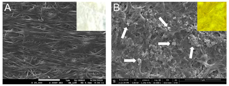
Figure 1.
Scanning electron microscopy (SEM) show (A) the pristine BCM (at 25,000×) and (B) the AgNMS@BCM (16,000×). The white arrows in (B) indicate the AgNMS fragments impregnated into the networks of membrane fibers. The insets in (A,B) demonstrate that upon impregnation with the AgNMS, the bacterial cellulose membrane (originally white, inset (A)) acquires the characteristic yellow color of the AgNMS compound (inset (B)).

