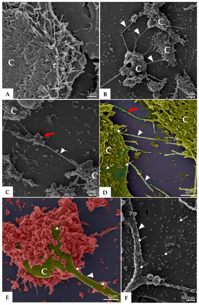
Figure 2.
Vero-E6 cells 72 h post-infection with SARS-CoV-2 (HIM images). (A) Uninfected Vero-E6 cell at 72 h of cultivation (cell control). (B–E) Infected Vero-E6 monolayer, connection (red arrow) between cells mediated by filopodia was observed (B–D). Virus particles (thick arrow, blue structures) was detected attached to cell filopodia (green, image (D)) and with cell membrane (green or red, image (D–F). Cell (C), filopodia (arrowhead). The images (D,E) were colored in Adobe photoshop.

