PLOS ONE logoLink to PLOS ONE
. 2022 Feb 25;17(2):e0264387. doi: 10.1371/journal.pone.0264387

Prognostic value of 11C-methionine volume-based PET parameters in IDH wild type glioblastoma

Bart R J van Dijken 1,*, Alfred O Ankrah 2, Gilles N Stormezand 2, Rudi A J O Dierckx 1,2, Peter Jan van Laar 1,3, Anouk van der Hoorn 1
Editor: Kevin Camphausen4
PMCID: PMC8880430  PMID: 35213602

Abstract

Purpose

11C-Methionine (11C-MET) PET prognostication of isocitrate dehydrogenase (IDH) wild type glioblastomas is inadequate as conventional parameters such as standardized uptake value (SUV) do not adequately reflect tumor heterogeneity. We retrospectively evaluated whether volume-based parameters such as metabolic tumor volume (MTV) and total lesion methionine metabolism (TLMM) outperformed SUV for survival correlation in patients with IDH wild type glioblastomas.

Methods

Thirteen IDH wild type glioblastoma patients underwent preoperative 11C-MET PET. Both SUV-based parameters and volume-based parameters were calculated for each lesion. Kaplan-Meier curves with log-rank testing and Cox regression analysis were used for correlation between PET parameters and overall survival.

Results

Median overall survival for the entire cohort was 393 days. MTV (HR 1.136, p = 0.007) and TLMM (HR 1.022, p = 0.030) were inversely correlated with overall survival. SUV-based 11C-MET PET parameters did not show a correlation with survival. In a paired analysis with other clinical parameters including age and radiotherapy dose, MTV and TLMM were found to be independent factors.

Conclusions

MTV and TLMM, and not SUV, significantly correlate with overall survival in patients with IDH wild type glioblastomas. The incorporation of volume-based 11C-MET PET parameters may lead to a better outcome prediction for this heterogeneous patient population.

Introduction

The use of amino acid PET such as L-methyl-11C-methionine (11C-MET) for prognostication of gliomas has recently been recommended [1]. 11C-MET PET has an excellent tumor-to-background discrimination and provides supplementary metabolic information in addition to MRI [2].

Survival and treatment strategies differ significantly among glioma patients, necessitating reliable preoperative prognostication [3]. Gliomas without isocitrate dehydrogenase (IDH) mutation, including the WHO grade 4 glioblastoma, are now recognized as the most malignant subtype [4]. These IDH wild type gliomas are heterogeneous tumors characterized by a more aggressive and infiltrative nature than IDH mutant gliomas [5]. Glioblastomas IDH wild type WHO grade 4 are defined due to either aggressive histological features such as microvascular proliferation and necrosis, or by specific molecular markers such as TERT promoter mutation, EGFR amplification, and chromosome 7 gain/chromosome 10 loss [4]. Outcome in glioblastomas is generally poor.

The prognostic use of 11C-MET PET in gliomas has previously been explored but showed inconclusive results [2]. Studies were usually limited to conventional parameters such as standardized uptake value (SUV), which do not reliably detect the heterogeneity of IDH wild type glioblastomas [6]. Recently, volume-based parameters have been introduced, possibly better reflecting the heterogeneity of IDH wild type glioblastomas [7, 8]. Volume-based parameters such as metabolic tumor volume (MTV) and total lesion methionine metabolism (TLMM), a combination of MTV and SUVmean, provide additional information about the tumor extent. The aim of this study was to investigate the value of conventional and volume-based 11C-MET PET parameters for prognostication in IDH wild type glioblastomas.

Methods

We performed a retrospective search among all 11C-MET PET examinations in our tertiary university hospital between 2011–2018. Inclusion criteria were glioblastoma, IDH wild type, WHO grade 4 according to the 2021 WHO guidelines, confirmed by biopsy or surgical resection ≤3 months after PET examination [4]. Exclusion criteria were previous cranial surgery or cerebral irradiation, known other primary tumor, and pediatric patients (<18 years). The study was approved by the institutional review board and the need for written informed consent was waived.

The tracer was prepared as previously published [9] and imaging was performed in accordance with the 2006 procedure guidelines of the European Association of Nuclear Medicine for brain tumor imaging using labelled amino acid analogues [10]. Patients underwent static PET imaging 20 minutes after intravenous injection of 200 MBq (range: 180–220 MBq) 11C-MET in one bed position of five minutes. Images were acquired on a Biograph mCT PET/CT system (Siemens/CTI, Knoxville, TN, USA) with 2 mm spatial resolution. The images were reconstructed using Truex + TOF with three iterations and 21 subsets in a 400 x 400 matrix size (zoom 1.0) and a 2 mm Gaussian filter.

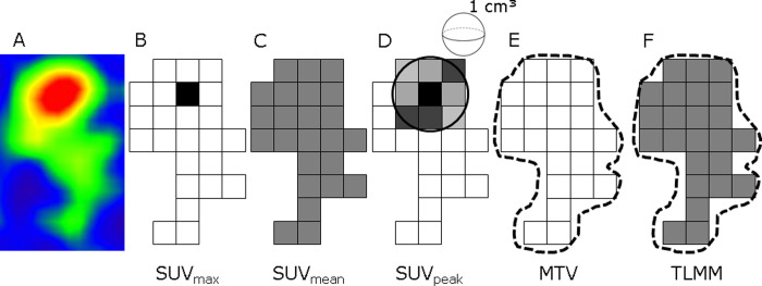
Volumes of interest (VOI) were defined using Syngo.via (Siemens Medical Solutions Inc., Knoxville, TN, USA). A circle large enough to include all visual uptake was manually drawn by one researcher (BD) and checked by a nuclear physician with 3 years of experience (GS). The VOI were semi-automatically defined using a 40% threshold of SUVmax [6], which is standard value for delineation used in our research group and leads to good tumor definition when compared to visual inspection. Syngo.via allowed for extraction of the following PET parameters within the VOI: SUVmax, SUVpeak, SUVmean, MTV and TLMM (Fig 1). TLMM was automatically calculated in Syngo.via by multiplying SUVmean and MTV within the lesion border, in an equal manner as the Total Lesion Glycolysis (TLG) in FDG imaging.

Fig 1. Schematic overview of metabolic and volume-based 11C-MET PET parameters.

Fig 1

(A) Snapshot of lesion with positive uptake of 11C-MET. (B) SUVmax is defined as voxel with highest uptake within the lesion. (C) SUVmean is an average of all SUV values from included voxels within the lesion. (D) SUVpeak is defined as the average value within a 1cm3 sphere surrounding the SUVmax. Volume-based parameters are shown in E-F. (E) MTV is the volume of increased uptake within the lesion borders. (F) For TLMM the SUVmean and MTV within lesion borders are combined and multiplied to provide both metabolic and volume-based information. TLMM is automatically calculated and extracted within the Syngo.via software. Abbreviations: MTV = metabolic tumor volume, SUV = standardized uptake value, TLMM = total lesion methionine metabolism.

Statistics were done in SPSS (IBM corp, version 25.0, Armonk, NY). Log-rank testing after dichotomization based on median values and Cox regression analysis were used for univariate survival analysis. The influence of various clinical variables (age, tumor grade, extent of resection and radiation dose) was also determined since the sample size was insufficient for multivariate analysis.

Results

A total of 13 patients with a median age of 57 years (range 28–70) and of whom 62% were male were included in the study. Adjuvant treatment consisted of radiotherapy plus or minus chemotherapy. One patient refused to undergo additional treatment after biopsy and one patient had to discontinue radiotherapy due to adverse events after a dose of 32.4 Gy. General characteristics are shown in Table 1.

Table 1. General characteristics of included patients.

Patient Gender Age WHO Grade EOR Treatment RT dose Survival
1 F 70 GBM, gr 4* GTR RT 57.6 1325
2 F 63 GBM, gr 4 STR C+RT 60 195
3 M 28 GBM, gr 4* B C+RT 60 32
4 F 64 GBM, gr 4* B C+RT 32.4 221
5 F 57 GBM, gr 4 B C+RT 60 418
6 M 65 GBM, gr 4 B None - 266
7 M 59 GBM, gr 4 B C+RT 60 200
8 M 44 GBM, gr 4 GTR C+RT 60 277
10 M 47 GBM, gr 4 B C+RT 60 171
11 M 61 GBM, gr 4 B C+RT 60 618
12 M 52 GBM, gr 4 B C+RT 60 469
13 M 67 GBM, gr 4* B RT 59.4 688
15 F 60 GBM, gr 4* B C+RT 59.4 237

* defined by EGFR amplification

discontinued radiotherapy due to adverse events.

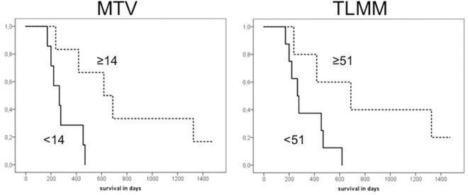
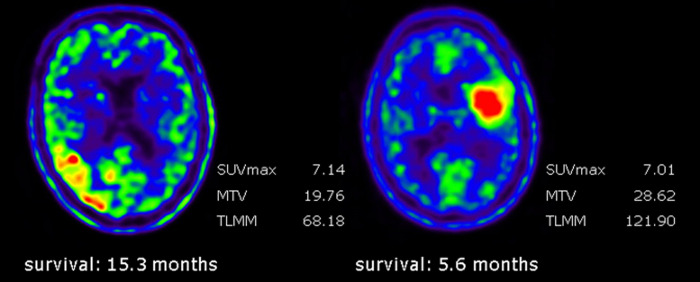
Median survival was 393 days. The results of univariate analysis for the different PET parameters are shown in Table 2. MTV and TLMM were the only parameters that significantly correlated with overall survival (hazard ratio [HR] 1.136 (95%CI 1.035–1.246), p = 0.007 and HR 1.022 (95%CI 1.002–1.043), p = 0.030, respectively). Kaplan-Meier curves for MTV and TLMM are shown in Fig 2. Paired analysis with other clinical parameters demonstrated that MTV and TLMM were correlated with survival, independent of age, extent of resection or radiation dose. SUV based parameters were not correlated with survival (Fig 3).

Table 2. Univariate analysis of 11C-MET PET parameters and survival.

Parameter HR (95%CI) p value
SUVmax 0.818 (0.583–1.148) 0.246
SUVpeak 0.833 (0.549–1.263) 0.389
SUVmean 0.669 (0.366–1.221) 0.190
MTV 1.136 (1.035–1.246) 0.007
TLMM 1.022 (1.002–1.043) 0.030

Abbreviations: HR = hazard ratio, MTV = metabolic tumor volume, SUV = standard uptake value, TLMM = total lesion methionine metabolism.

Fig 2. Kaplan-Meier curves for MTV and TLMM.

Fig 2

Survival curves for MTV and TLMM. Dichotomisation of the data based on the median (≥14 for MTV and ≥51 for TLMM). The group with higher values is displayed with the dotted line while the group with lower values is displayed with the bold line. The survival difference between patients with higher and lower values was statistically significant for both MTV (p = 0.014) and TLMM (p = 0.041). Abbreviations: MTV = metabolic tumor volume, TLMM = total lesion methionine metabolism.

Fig 3. Differences in metabolic and volumetric parameters in two glioblastoma patients.

Fig 3

Left image: 52-year-old male with right temporoparietal glioblastoma. Right image: 47-year-old male with left frontal glioblastoma. Despite relatively similar SUVmax values, there was a significant difference in survival between the two patients. The tumor of the patient on the right side demonstrated higher MTV and TLMM values than the tumor on the left side, corresponding to the lower survival of this patient.

Discussion

Volume-based 11C-MET PET parameters were significantly correlated with survival in this small retrospective study among IDH wild type glioblastoma patients, while SUV based parameters were not. TLMM, combining both volume-based and metabolic information, can thus be potentially employed for prognostication in IDH wild type glioblastomas.

Conventional SUV measures are the most commonly used quantitative PET parameters. Several studies have investigated the use of 11C-MET SUV measures for glioma prognostication, with inconclusive results [2, 1114]. SUV measures, however, do not provide information about tumor heterogeneity, nor do they visualize the extent of the lesion. In the current study we did not find a correlation between SUV measures and survival in IDH wild type glioblastomas, while volume-based parameters did demonstrate this correlation.

Our findings are in accordance with another 11C-MET PET study which also demonstrated that a larger MTV and TLMM were correlated with lower survival [7]. However, important limitations of that study were the inclusion of oligodendrogliomas and lack of correcting for IDH mutation status, whilst it is known that both survival and 11C-MET uptake values significantly differ between IDH wild type and IDH-1 gliomas [15, 16]. Furthermore, IDH wild type glioblastomas, are currently seen as separate entity and have a poorer prognosis than IDH-1 gliomas [4]. The prognostic role of volume-based parameters in gliomas have also been demonstrated for other amino acid PET tracers, such as O-(2-[18F]fluoroethyl)-l-tyrosine (18F-FET) [1719]. for oncology prognostication has also been demonstrated in other populations [6, 2023].

Volume-based PET parameters are strongly dependent on the used delineation method [6]. This was recently demonstrated for 18F-FDG PET in non-small cell lung cancer patients, where MTV significantly differed with chosen delineation method [6]. According to the authors of that study, the approach that reached best agreement was 40% of SUVmax, corresponding to the method used in the present study [6]. However, these results have not yet been validated for 11C-MET PET. Moreover, it is not known if these results can be translated to gliomas. More research should therefore be aimed at establishing a robust delineation method for 11C-MET PET in gliomas.

The most important limitation of this study is the small sample size. Due to this, a comprehensive multivariate analysis was not possible. Nevertheless, in paired analysis with other clinical parameters TLMM demonstrated to be independently correlated to survival. Another limitation is the retrospective nature of this study. Due to this, molecular markers, such as TERT promoter mutation, EGFR amplification, and gain of chromosome 7 with loss of chromosome 10 status was not always known, contributing to the smaller sample size. Our results should thus be seen as an encouragement for larger prospective studies focusing on the prognostic role of volume-based 11C-MET PET parameters. At present, a prospective study on 11C-MET PET for treatment evaluation in glioma patients is ongoing at out tertiary center (Netherlands Trial Register number NL6536) which will also address the utility of volume-based parameters for prognostication.

Conclusion

Volume-based 11C-MET PET parameters MTV and TLMM were significantly correlated with overall survival in patients with IDH wild type glioblastomas. Volume-based parameters could play a future role in tailoring treatment decisions in patients with IDH wild type glioblastoma.

Supporting information

S1 File

(PDF)

S2 File

(PDF)

S1 Data

(XLSX)

Data Availability

We have uploaded the minimal (anonymized) data set as supporting information.

Funding Statement

The author(s) received no specific funding for this work.

References

  • 1.Albert NL, Weller M, Suchorska B, et al. Response Assessment in Neuro-Oncology working group and European Association for Neuro-Oncology recommendations for the clinical use of PET imaging in gliomas. Neuro Oncol. 2016;18:1199–108. doi: 10.1093/neuonc/now058 [DOI] [PMC free article] [PubMed] [Google Scholar]
  • 2.Glaudemans AWJM, Enting RH, Heesters MAAM, et al. Value of 11C-methionine PET in imaging brain tumours and metastases. Eur J Nucl Med Mol Imaging. 2013;40:615–635. doi: 10.1007/s00259-012-2295-5 [DOI] [PubMed] [Google Scholar]
  • 3.van den Bent MJ, Baumert B, Erridge SC, et al. Interim results from the CATNON trial (EORTC study 26053–22054) of treatment with concurrent and adjuvant temozolomide for 1p/19q non-co-deleted anaplastic glioma: a phase 3, randomised, open-label intergroup study. Lancet. 2017;390:1645–1653. doi: 10.1016/S0140-6736(17)31442-3 [DOI] [PMC free article] [PubMed] [Google Scholar]
  • 4.Louis DN, Perry A, Reifenberger G, et al. The 2016 World Health Organization Classification of Tumors of the Central Nervous System: a summary. Acta Neuropathol. 2016;131:803–820. doi: 10.1007/s00401-016-1545-1 [DOI] [PubMed] [Google Scholar]
  • 5.Eckel-Passow JE, Lachance DH, Molinaro AM, et al. Glioma Groups Based on 1p/19q, IDH, and TERT Promoter Mutations in Tumors. N Engl J Med. 2015;372:2499–2508. doi: 10.1056/NEJMoa1407279 [DOI] [PMC free article] [PubMed] [Google Scholar]
  • 6.Guilherme KD, Vállez Garcia D, Kramer GM, et al. Repeatability of [18 F]FDG PET/CT Total Metabolic Active Tumour Volume and Total Tumour Burden in NSCLC Patients. EJNMMI Res. 2019;9:14. doi: 10.1186/s13550-019-0481-1 [DOI] [PMC free article] [PubMed] [Google Scholar]
  • 7.Kobayashi K, Hirata K, Yamaguchi S, et al. Prognostic value of volume-based measurements on (11)C-methionine PET in glioma patients. Eur J Nucl Med Mol Imaging. 2015;42:1071–1080. doi: 10.1007/s00259-015-3046-1 [DOI] [PubMed] [Google Scholar]
  • 8.Yoo MY, Paeng JC, Cheon GJ, et al. Prognostic Value of Metabolic Tumor Volume on (11)C-Methionine PET in Predicting Progression-Free Survival in High-Grade Glioma. 2015;49:291–297. [DOI] [PMC free article] [PubMed] [Google Scholar]
  • 9.Langstrom B, Antoni G, Gullberg P, et al. Synthesis of L- and D-[methyl-11C]methionine. J Nucl Med. 1987;28:1037–1040. [PubMed] [Google Scholar]
  • 10.Vander Borght T, Asembaum S, Bartenstein P, et al. EANM procedure guidelines for brain tumour imaging using labelled amino acid analogues. Eur J Nucl Med Mol Imaging. 2006;33:1374–1380. doi: 10.1007/s00259-006-0206-3 [DOI] [PubMed] [Google Scholar]
  • 11.Ceyssens S, van Laere K, de Groot T, et al. [11C]methionine PET, Histopathology, and Survival in Primary Brain Tumors and Recurrence. AJNR Am J Neuroradiol. 2006;27:1432–1437. [PMC free article] [PubMed] [Google Scholar]
  • 12.Cicuendez M, Lorenzo-Bosquet C, Cuberas-Borrós G, et al. Role of [(11)C] Methionine Positron Emission Tomography in the Diagnosis and Prediction of Survival in Brain Tumours. Clin Neurol Neurosurg. 2015;139:328–333. doi: 10.1016/j.clineuro.2015.10.035 [DOI] [PubMed] [Google Scholar]
  • 13.Lopci E, Riva M, Olivari L, et al. Prognostic Value of Molecular and Imaging Biomarkers in Patients With Supratentorial Glioma. Eur J Nucl Med Mol Imaging. 2017;44:1155–1164. doi: 10.1007/s00259-017-3618-3 [DOI] [PubMed] [Google Scholar]
  • 14.Poetsch N, Woehrer A, Gesperger J, et al. Visual and Semiquantitative 11C-methionine PET: An Independent Prognostic Factor for Survival of Newly Diagnosed and Treatment-Naïve Gliomas. Neuro Oncol. 2018;20:411–419. doi: 10.1093/neuonc/nox177 [DOI] [PMC free article] [PubMed] [Google Scholar]
  • 15.Takei H, Shinoda J, Ikuta S, et al. Usefulness of Positron Emission Tomography for Differentiating Gliomas According to the 2016 World Health Organization Classification of Tumors of the Central Nervous System. J Neurosurg. 2019;16:1–10. [DOI] [PubMed] [Google Scholar]
  • 16.Ogawa T, Kawai N, Miyake K, et al. Diagnostic Value of PET/CT With 11C-methionine (MET) and 18 F-fluorothymidine (FLT) in Newly Diagnosed Glioma Based on the 2016 WHO Classification. EJNMMI Res. 2020;10:44. doi: 10.1186/s13550-020-00633-1 [DOI] [PMC free article] [PubMed] [Google Scholar]
  • 17.Suchorska B, Jansen NL, Linn J, et al. Biological tumor volume in 18FET-PET before radiochemotherapy correlates with survival in GBM. Neurology. 2015;84:710–719. doi: 10.1212/WNL.0000000000001262 [DOI] [PubMed] [Google Scholar]
  • 18.Bauer EK, Stoffels G, Blau T, et al. Prediction of survival in patients with IDH-wildtype astrocytic gliomas using dynamic O-(2- [18 F]-fluoroethyl)-L-tyrosine PET. Eur J Nucl Med Mol Imaging. 2020;47:1486–1495. doi: 10.1007/s00259-020-04695-0 [DOI] [PMC free article] [PubMed] [Google Scholar]
  • 19.Ceccon G, Lohman P, Werner JM, et al. Early Treatment Response Assessment Using 18 F-FET PET Compared with Contrast-Enhanced MRI in Glioma Patients After Adjuvant Temozolomide Chemotherapy. J Nucl Med. 2021;62:918–925. doi: 10.2967/jnumed.120.254243 [DOI] [PubMed] [Google Scholar]
  • 20.Morales-Lozano MIM, Viering O, Samnick S, et al. 18F-FDG and 11C-Methionine PET/CT in Newly Diagnosed Multiple Myeloma Patients: Comparison of Volume-Based PET Biomarkers. Cancers (Basel). 2020;12:1042. doi: 10.3390/cancers12041042 [DOI] [PMC free article] [PubMed] [Google Scholar]
  • 21.Cegla P, Burchardt E, Roszak A, et al. Influence of Biological Parameters Assessed in [18F]FDG PET/CT on Overall Survival in Cervical Cancer Patients. Clin Nucl Med. 2019;44:860–863. doi: 10.1097/RLU.0000000000002733 [DOI] [PubMed] [Google Scholar]
  • 22.Carretta A, Bandiera A, Muriana P, et al. Prognostic Role of Positron Emission Tomography and Computed Tomography Parameters in Stage I Lung Adenocarcinoma. Radiol Oncol. 2020. Online ahead of print. doi: 10.2478/raon-2020-0034 [DOI] [PMC free article] [PubMed] [Google Scholar]
  • 23.Creff G, Devillers A, Depeursinge A, et al. Evaluation of the Prognostic Value of FDG PET/CT Parameters for Patients With Surgically Treated Head and Neck Cancer: A Systematic Review. JAMA Otolaryngol Head Neck Surg. 2020;146:471–479. doi: 10.1001/jamaoto.2020.0014 [DOI] [PubMed] [Google Scholar]

Decision Letter 0

Kevin Camphausen

1 Dec 2021

PONE-D-21-28633Prognostic value of 11C-methionine volume-based PET parameters in IDH wild type gliomasPLOS ONE

Dear Dr. Van Dijken,

Thank you for submitting your manuscript to PLOS ONE. After careful consideration, we feel that it has merit but does not fully meet PLOS ONE’s publication criteria as it currently stands. Therefore, we invite you to submit a revised version of the manuscript that addresses the points raised during the review process. Please submit your revised manuscript by Jan 15 2022 11:59PM. If you will need more time than this to complete your revisions, please reply to this message or contact the journal office at plosone@plos.org. When you're ready to submit your revision, log on to https://www.editorialmanager.com/pone/ and select the 'Submissions Needing Revision' folder to locate your manuscript file.

Please include the following items when submitting your revised manuscript:

  • A rebuttal letter that responds to each point raised by the academic editor and reviewer(s). You should upload this letter as a separate file labeled 'Response to Reviewers'.

  • A marked-up copy of your manuscript that highlights changes made to the original version. You should upload this as a separate file labeled 'Revised Manuscript with Track Changes'.

  • An unmarked version of your revised paper without tracked changes. You should upload this as a separate file labeled 'Manuscript'.

If you would like to make changes to your financial disclosure, please include your updated statement in your cover letter. Guidelines for resubmitting your figure files are available below the reviewer comments at the end of this letter.

If applicable, we recommend that you deposit your laboratory protocols in protocols.io to enhance the reproducibility of your results. Protocols.io assigns your protocol its own identifier (DOI) so that it can be cited independently in the future. For instructions see: https://journals.plos.org/plosone/s/submission-guidelines#loc-laboratory-protocols. Additionally, PLOS ONE offers an option for publishing peer-reviewed Lab Protocol articles, which describe protocols hosted on protocols.io. Read more information on sharing protocols at https://plos.org/protocols?utm_medium=editorial-email&utm_source=authorletters&utm_campaign=protocols.

We look forward to receiving your revised manuscript.

Kind regards,

Kevin Camphausen

Academic Editor

PLOS ONE

Journal Requirements:

When submitting your revision, we need you to address these additional requirements.

1. Please ensure that your manuscript meets PLOS ONE's style requirements, including those for file naming. The PLOS ONE style templates can be found at 

https://journals.plos.org/plosone/s/file?id=wjVg/PLOSOne_formatting_sample_main_body.pdf and 

https://journals.plos.org/plosone/s/file?id=ba62/PLOSOne_formatting_sample_title_authors_affiliations.pdf

2. We note that you have indicated that data from this study are available upon request. PLOS only allows data to be available upon request if there are legal or ethical restrictions on sharing data publicly. For more information on unacceptable data access restrictions, please see http://journals.plos.org/plosone/s/data-availability#loc-unacceptable-data-access-restrictions. 

In your revised cover letter, please address the following prompts:

a) If there are ethical or legal restrictions on sharing a de-identified data set, please explain them in detail (e.g., data contain potentially sensitive information, data are owned by a third-party organization, etc.) and who has imposed them (e.g., an ethics committee). Please also provide contact information for a data access committee, ethics committee, or other institutional body to which data requests may be sent.

b) If there are no restrictions, please upload the minimal anonymized data set necessary to replicate your study findings as either Supporting Information files or to a stable, public repository and provide us with the relevant URLs, DOIs, or accession numbers. For a list of acceptable repositories, please see http://journals.plos.org/plosone/s/data-availability#loc-recommended-repositories.

We will update your Data Availability statement on your behalf to reflect the information you provide

[Note: HTML markup is below. Please do not edit.]

Reviewers' comments:

Reviewer's Responses to Questions

Comments to the Author

1. Is the manuscript technically sound, and do the data support the conclusions?

The manuscript must describe a technically sound piece of scientific research with data that supports the conclusions. Experiments must have been conducted rigorously, with appropriate controls, replication, and sample sizes. The conclusions must be drawn appropriately based on the data presented.

Reviewer #1: Yes

Reviewer #2: No

**********

2. Has the statistical analysis been performed appropriately and rigorously?

Reviewer #1: Yes

Reviewer #2: N/A

**********

3. Have the authors made all data underlying the findings in their manuscript fully available?

The PLOS Data policy requires authors to make all data underlying the findings described in their manuscript fully available without restriction, with rare exception (please refer to the Data Availability Statement in the manuscript PDF file). The data should be provided as part of the manuscript or its supporting information, or deposited to a public repository. For example, in addition to summary statistics, the data points behind means, medians and variance measures should be available. If there are restrictions on publicly sharing data—e.g. participant privacy or use of data from a third party—those must be specified.

Reviewer #1: Yes

Reviewer #2: No

**********

4. Is the manuscript presented in an intelligible fashion and written in standard English?

PLOS ONE does not copyedit accepted manuscripts, so the language in submitted articles must be clear, correct, and unambiguous. Any typographical or grammatical errors should be corrected at revision, so please note any specific errors here.

Reviewer #1: Yes

Reviewer #2: Yes

**********

5. Review Comments to the Author

Please use the space provided to explain your answers to the questions above. You may also include additional comments for the author, including concerns about dual publication, research ethics, or publication ethics. (Please upload your review as an attachment if it exceeds 20,000 characters)

Reviewer #1: This paper on the prognostic value of 11C-methionine volume-based PET parameters in IDH wild type

gliomas is timely and deals with an important imaging niche (11C-methionine PET) and specifically the need for acquired parameters to become increasingly clinically meaningful (SUV vs. volume based ). It is to be commended on not simply going after the SUV and in its emphasis on focusing on wild type IDH mutant gliomas. However it deals with only 15 patients (which the authors acknowledge as a limitation) and a larger cohort as well as validation with larger scale data perhaps from another institution and/or in conjunction with large scale data bases (TCGA/BRATS) would add weight to the findings and advance the field.

Reviewer #2: Prognostic value of 11C-methionine volume-based PET parameters in IDH wild type gliomas.

This article aims to demonstrate the prognostic value of 11C-methionine (11C-met) PET in the survival of IDH wild type glioma. While previous trials of 11C-met trials have proven inconclusive, the authors of this paper employ volume-based parameters such as metabolic tumor volume (MTV) and total lesion methionine metabolism (TLMM) in an attempt to improve prognostic accuracy. This is an important question, as better non-invasive methods of diagnosing glioma are desperately needed. Unfortunately, this paper has several structural issues that cannot be fixed and prevent its publication:

1) The sample size of this study is too small and heterogenous to make any conclusions. While it is true that IDH wild type gliomas do generally carry worse prognosis that IDH mutants, grade IV, grade III and grade II gliomas represent different pathologies with different behaviors and responses to treatment.

2) The definition of MTV and TLMM are unclear. In the legend of Figure 1, you state that SUVmean and MTV are combined and multiplied to obtain TLMM however the diagram accompanied does not clearly demonstrate how this is applied in practice.

3) The Kaplan Meier curves from figure 2 is unclear. Are the values based on volume or metabolic activity?

4) While table 1 provides individual data on extent of resection, age and radiation dose, graphical data may better delineate how these factors impact survival

**********

6. PLOS authors have the option to publish the peer review history of their article (what does this mean?). If published, this will include your full peer review and any attached files.

If you choose “no”, your identity will remain anonymous but your review may still be made public.

Do you want your identity to be public for this peer review? For information about this choice, including consent withdrawal, please see our Privacy Policy.

Reviewer #1: No

Reviewer #2: No

[NOTE: If reviewer comments were submitted as an attachment file, they will be attached to this email and accessible via the submission site. Please log into your account, locate the manuscript record, and check for the action link "View Attachments". If this link does not appear, there are no attachment files.]

While revising your submission, please upload your figure files to the Preflight Analysis and Conversion Engine (PACE) digital diagnostic tool, https://pacev2.apexcovantage.com/. PACE helps ensure that figures meet PLOS requirements. To use PACE, you must first register as a user. Registration is free. Then, login and navigate to the UPLOAD tab, where you will find detailed instructions on how to use the tool. If you encounter any issues or have any questions when using PACE, please email PLOS at figures@plos.org. Please note that Supporting Information files do not need this step.

PLoS One. 2022 Feb 25;17(2):e0264387. doi: 10.1371/journal.pone.0264387.r002

Author response to Decision Letter 0


26 Jan 2022

Response to the reviewers

Reviewer #1:

Comment:

This paper on the prognostic value of 11C-methionine volume-based PET parameters in IDH wild type gliomas is timely and deals with an important imaging niche (11C-methionine PET) and specifically the need for acquired parameters to become increasingly clinically meaningful (SUV vs. volume based ). It is to be commended on not simply going after the SUV and in its emphasis on focusing on wild type IDH mutant gliomas. However, it deals with only 15 patients (which the authors acknowledge as a limitation) and a larger cohort as well as validation with larger scale data perhaps from another institution and/or in conjunction with large scale data bases (TCGA/BRATS) would add weight to the findings and advance the field.

Answer:

We would like to thank the reviewer for critically reading our manuscript and commending us on the scope of this study. The small sample size indeed is one of the most important limitations of our study, which we also acknowledge in the discussion section. A larger study cohort would definitely strengthen the study. However, due to the retrospective nature of our study and the study niche (11C-methionine PET in IDH wild type gliomas) this was not possible. We thank the reviewer for the suggestion of using large scale databases to increase the number of subjects. Unfortunately, the TCGA and BRATS databases do not include preoperative 11C-methionine PET data. Therefore, producing a larger cohort for our study at this stage was not deemed feasible.

Despite small sample size, the found correlation of the volume-based parameters MTV and TLMM with survival was statistically significant and TLMM was even suggested to be an independent factor in paired analysis with other clinical parameters. We feel that our results should thus be seen as an encouragement for larger prospective studies focusing on the prognostic role of volume-based 11C-MET PET parameters to confirm our results. At a present, a prospective study on 11C-MET PET treatment evaluation in glioma patients is undertaken at our tertiary hospital (Netherlands Trial Register number NL6536; https://www.trialregister.nl/trial/6536), which includes preoperative imaging as well. The value of volume-based parameters for prognostication (and treatment response assessment) will also be a scope of this study. A sentence to highlight this was added to the final paragraph of the discussion.

Reviewer #2:

Comment:

Prognostic value of 11C-methionine volume-based PET parameters in IDH wild type gliomas. This article aims to demonstrate the prognostic value of 11C-methionine (11C-met) PET in the survival of IDH wild type glioma. While previous trials of 11C-met trials have proven inconclusive, the authors of this paper employ volume-based parameters such as metabolic tumor volume (MTV) and total lesion methionine metabolism (TLMM) in an attempt to improve prognostic accuracy. This is an important question, as better non-invasive methods of diagnosing glioma are desperately needed. Unfortunately, this paper has several structural issues that cannot be fixed and prevent its publication.

Answer:

We thank the reviewer for the thorough assessment of our manuscript. We agree with the reviewer that establishing more accurate non-invasive methods for the diagnosis and prognostication of IDH wild type gliomas is crucial. This study investigated the value of volume-based 11C-MET PET parameters in addition to SUV, as it is known that SUV does not reliably detect the heterogeneity of IDH wild type gliomas.

Comment:

The sample size of this study is too small and heterogenous to make any conclusions. While it is true that IDH wild type gliomas do generally carry worse prognosis that IDH mutants, grade IV, grade III and grade II gliomas represent different pathologies with different behaviors and responses to treatment.

Answer:

We understand the concern of the reviewer about the small sample size of this study. We also acknowledge this limitation in the last paragraph of the discussion section. Nevertheless, our results were statistically significant and showed that volume-based parameters were correlated with overall survival while conventional parameters were not. Volume-based parameters could play a future role in tailoring treatment decisions in patients with IDH wild type glioblastomas, but we agree that future studies are necessary to confirm our results. We also refer to our answer to reviewer #1.

Historically, the pathological grade was one of the most important predictors of survival in glioma patients. However, with recent guideline updates, molecular markers now play a prominent role. Due to the poor prognosis, IDH wild type tumors are seen as a different entity than IDH mutated gliomas. In addition, certain molecular markers (TERT promoter mutation, EGFR gene amplification, +7/-10 chromosome changes) now define glioblastoma IDH wild type WHO grade 4, even if histologically the tumor is of lower grade. We agree with the reviewer that the heterogeneity was a point of concern. The reviewer’s comment prompted us to critically re-review our dataset according to the most recent guidelines (2021 WHO Classification of Tumors of the Central Nervous System, Louis et al, Neuro-Oncology, 2021). EGFR gene amplification was seen in a subset of patients in absence of glioblastoma grade 4 defining features such as microvascular proliferation or necrosis. We updated the grades of these patients in table 1. Two patients did not demonstrate any glioblastoma-defining molecular markers, and were therefore deleted from the analysis. In agreement with the newest guidelines, we have also changed the terminology throughout the manuscript to IDH wild type glioblastoma, WHO grade 4.

Comment:

The definition of MTV and TLMM are unclear. In the legend of Figure 1, you state that SUVmean and MTV are combined and multiplied to obtain TLMM however the diagram accompanied does not clearly demonstrate how this is applied in practice.

Answer:

We agree that the legend of figure 1 was somewhat brief. TLMM is the product of SUVmean and MTV within the lesion border. This parameter can be of course be calculated by hand, but most software allow for automatic calculation and extraction of this TLMM; equal to Total Lesion Glycolysis (TLG) in FDG PET imaging. To clarify the definitions of MTV and TLMM to the reader, we have made the following changes to the text and the legend of figure 1:

Method section, final sentence of paragraph 3: TLMM was automatically calculated in Syngo.via by multiplying SUVmean and MTV within the lesion border, in an equal manner as the Total Lesion Glycolysis (TLG) in FDG imaging.

Legend of figure 1: (…) (E) MTV is the volume of increased uptake within the lesion borders. (F) For TLMM the SUVmean and MTV within lesion borders are combined and multiplied to provide both metabolic and volume-based information. TLMM is automatically calculated and extracted within the Syngo.via software.

Comment:

The Kaplan Meier curves from figure 2 is unclear. Are the values based on volume or metabolic activity?

Answer:

We agree with the reviewer that the Kaplan Meier curves from figure 2 are unclear. Therefore, to increase clarity, we included the thresholds (median values) of both MTV (�14) and TLMM (�51) for which the data were dichotomized. The group with higher values is displayed with the dotted line while the group with lower values is displayed with the bold line as can be read in the legend. The values for MTV are based on volume and TLMM is a dimensionless parameter which combines the volumetric and metabolic information and is thus not expressed in a particular unit.

Comment:

4) While table 1 provides individual data on extent of resection, age and radiation dose, graphical data may better delineate how these factors impact survival.

Answer:

In accordance with the reviewer’s suggestion we have included the paired analyses between MTV/TLMM and clinical parameters, as well as graphical data (Kaplan Meier curves) for the impact of clinical parameters on survival in supplementary data 1 and 2, respectively.

Decision Letter 1

Kevin Camphausen

10 Feb 2022

Prognostic value of 11C-methionine volume-based PET parameters in IDH wild type glioblastoma

PONE-D-21-28633R1

Dear Dr. Van Dijken,

We’re pleased to inform you that your manuscript has been judged scientifically suitable for publication and will be formally accepted for publication once it meets all outstanding technical requirements.

Within one week, you’ll receive an e-mail detailing the required amendments. When these have been addressed, you’ll receive a formal acceptance letter and your manuscript will be scheduled for publication.

An invoice for payment will follow shortly after the formal acceptance. To ensure an efficient process, please log into Editorial Manager at http://www.editorialmanager.com/pone/, click the 'Update My Information' link at the top of the page, and double check that your user information is up-to-date. If you have any billing related questions, please contact our Author Billing department directly at authorbilling@plos.org.

If your institution or institutions have a press office, please notify them about your upcoming paper to help maximize its impact. If they’ll be preparing press materials, please inform our press team as soon as possible -- no later than 48 hours after receiving the formal acceptance. Your manuscript will remain under strict press embargo until 2 pm Eastern Time on the date of publication. For more information, please contact onepress@plos.org.

Kind regards,

Kevin Camphausen

Academic Editor

PLOS ONE

Additional Editor Comments (optional):

Reviewers' comments:

Acceptance letter

Kevin Camphausen

14 Feb 2022

PONE-D-21-28633R1

Prognostic value of 11C-methionine volume-based PET parameters in IDH wild type glioblastoma

Dear Dr. Van Dijken:

I'm pleased to inform you that your manuscript has been deemed suitable for publication in PLOS ONE. Congratulations! Your manuscript is now with our production department.

If your institution or institutions have a press office, please let them know about your upcoming paper now to help maximize its impact. If they'll be preparing press materials, please inform our press team within the next 48 hours. Your manuscript will remain under strict press embargo until 2 pm Eastern Time on the date of publication. For more information please contact onepress@plos.org.

If we can help with anything else, please email us at plosone@plos.org.

Thank you for submitting your work to PLOS ONE and supporting open access.

Kind regards,

PLOS ONE Editorial Office Staff

on behalf of

Dr. Kevin Camphausen

Academic Editor

PLOS ONE


Articles from PLoS ONE are provided here courtesy of PLOS

RESOURCES