Figure 2.
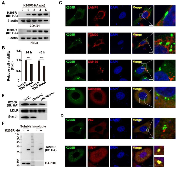
Expression and subcellular localization of K205R. (A) 3D4/21 and HeLa cells were transfected with K205R-HA plasmid as indicated for 24 h. The expression of K205R-HA was detected with immunoblotting analysis. (B) HeLa cells were transfected with empty vector or K205R-HA plasmid for 24 or 48 h. Cell viability was assessed with CCK-8 assays. *** p < 0.001. (C) HeLa cells were transfected with K205R-GFP plasmid for 24 h. Colocalization of K205R with LAMP1 (lysosome), TOM20 (mitochondria), GM130 (Golgi), and calnexin (ER) was analyzed with immunofluorescence analysis. Scale bar: 10 μm. (D) HeLa cells were transfected with K205R-GFP plasmid for 24 h. Colocalization of K205R with P62 (aggrephagy marker) and TIA-1 (SG marker) was determined with immunofluorescence analysis. Scale bar: 10 μm. (E) HeLa cells were transfected with K205R-HA plasmid for 24 h. The distribution of K205R in the cytosolic and membrane fractions was detected with immunoblotting analysis. (F) HeLa cells were transfected with empty vector or K205R-HA plasmid for 24 h. The distribution of K205R in soluble and insoluble fractions was detected with immunoblotting analysis.

