Abstract
Most bacterial genomes contain horizontally acquired and transmissible mobile genetic elements, including temperate bacteriophages and integrative and conjugative elements. Little is known about how these elements interact and co-evolved as parts of their host genomes. In many cases, it is not known what advantages, if any, these elements provide to their bacterial hosts. Most strains of Bacillus subtilis contain the temperate phage SPß and the integrative and conjugative element ICEBs1. Here we show that the presence of ICEBs1 in cells protects populations of B. subtilis from predation by SPß, likely providing selective pressure for the maintenance of ICEBs1 in B. subtilis. A single gene in ICEBs1 (yddK, now called spbK for SPß killing) was both necessary and sufficient for this protection. spbK inhibited production of SPß, during both activation of a lysogen and following de novo infection. We found that expression spbK, together with the SPß gene yonE constitutes an abortive infection system that leads to cell death. spbK encodes a TIR (Toll-interleukin-1 receptor)-domain protein with similarity to some plant antiviral proteins and animal innate immune signaling proteins. We postulate that many uncharacterized cargo genes in ICEs may confer selective advantage to cells by protecting against other mobile elements.
Author summary
Chromosomes from virtually all organisms contain genes that were horizontally acquired. In bacteria, many of the horizontally acquired genes are located in mobile genetic elements, elements that promote their own transfer from one cell to another. These elements include viruses and conjugative elements that are parts of the host genome and they can contain genes involved in metabolism, pathogenesis, symbiosis, and antibiotic resistances. Interactions between these elements are poorly understood. Furthermore, the majority of these elements confer no obvious benefit to host cells. We found that the presence of an integrative and conjugative element (ICE) in a bacterial genome protects host cells from predation by a bacteriophage (virus). There is a single gene in the integrative and conjugative element that confers this protection, and one gene in the bacteriophage that likely works together with the ICE gene. When expressed at the same time, these two genes cause cell death, before functional viruses can be made and released to kill other cells. We postulate that other ICEs may confer selective advantage to their host cells by protecting against other mobile elements.
Introduction
Mobile genetic elements can move between host genomes or within a host’s genome. The genomes of many bacterial species contain multiple functional and defective mobile elements, including insertion sequences, transposons, temperate phages, genomic islands, and integrative and conjugative elements (ICEs; also called conjugative transposons). In some cases, these elements constitute a substantial portion of the host genome [1–4]. Multiple elements within a given host have the potential to interact with each other, and likely co-evolve.
ICEs are mobile genetic elements that reside integrated in a host chromosome and are replicated and segregated to daughter cells along with the host genome [5–7]. Under certain conditions, or stochastically, an ICE can excise from the chromosome and be transferred to a recipient cell via the element-encoded conjugation machinery, typically a type IV secretion system.
ICEs frequently carry cargo genes that are not essential for their own lifecycle, but instead benefit the host. Most well-studied ICEs were discovered because of such phenotypes [6]. For example, the ICE Tn916 was discovered because it confers tetracycline resistance to host cells and can move between cells via conjugation [8,9]. Likewise, many other ICEs were identified because they carry genes that confer specific phenotypes including: antibiotic resistance [10–15], pathogenesis [16], symbiosis [17], and metabolic functions [18–21].
Many ICEs have been identified by means other than the phenotype conferred by their cargo genes. In these cases, the functions of the cargo genes are largely unknown. We suspect that many of these cargo genes encode functions that are beneficial to the host under certain conditions, but that the appropriate conditions have not been identified.
Many strains of Bacillus subtilis contain at least two functional mobile genetic elements, the integrative and conjugative element ICEBs1 [22,23] and the temperate phage SPß [24]. B. subtilis strains also contain several defective mobile genetic elements [25–27].
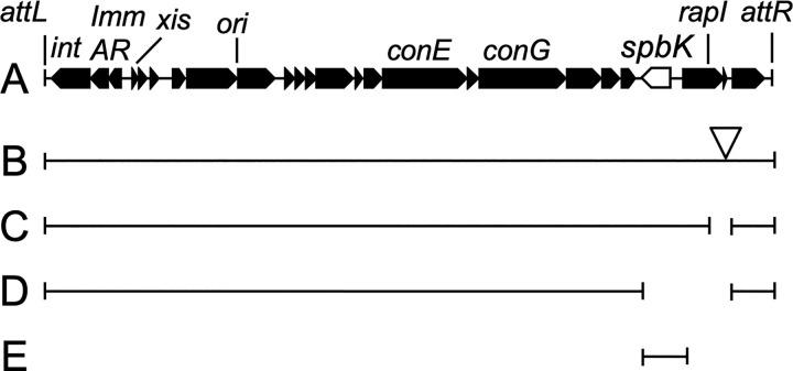
ICEBs1 (Fig 1) is found integrated in the B. subtilis genome in trnS-leu2, the gene for a leucine-tRNA. While integrated, most ICEBs1 genes are repressed [22,28]. ICEBs1 is activated during the recA-dependent SOS response to DNA damage or in the presence of B. subtilis cells that do not have the element [22]. Under these conditions, ICEBs1 gene expression is derepressed, the element excises from the chromosome and can transfer to an available recipient via the element-encoded conjugation machinery. ICEBs1 was identified because of homology to other ICEs [29] and because it is regulated by cell-cell signaling [22]. At the time of its discovery, it was not known if ICEBs1 conferred a beneficial phenotype to its host.
Fig 1. Map of ICEBs1 and some mutants.
A. A linear map of ICEBs1 is shown. Genes are indicated as filled boxes with arrowheads at the ends indicating the direction of transcription. spbK is shown as an open arrow. The attachment sites attL and attR mark the junctions between ICEBs1 and chromosomal sequences. Below the map are shown the ICEBs1 mutants used in this work. The regions of ICEBs1 that are present in each construct are shown as bars beneath the map. B. ICEBs1::kan. The open triangle indicates that this construct contains a kan gene inserted in the intergenic region between rapI and yddM. C. ICEBs1 Δ(rapI-phrI). D. ICEBs1 Δ(spbK-phrI). E. ICEBs10 spbK+. Many of these derivatives of ICEBs1 are used in multiple strains as indicated in Table 1.
SPß is a temperate phage found in the chromosome of many isolates of B. subtilis. Historically, SPß was thought to be a defective phage {reviewed in [30]}. When strains cured of SPß (SPß0) were isolated, it became clear that it is functional [31], and cured strains are used to grow the phage. A widely used strain missing SPß is also missing ICEBs1 [32,33].
In strains lysogenic for SPß, the phage is integrated in spsM, near the terminus of replication [32,34,35]. SPß contains genes needed for production of and resistance to the peptide antibiotic sublancin [35,36], providing a growth advantage to the host in the presence of cells sensitive to sublancin. Most phage genes are repressed in the lysogen, but during the recA-dependent SOS response to DNA damage, SPß gene expression is induced and the phage excises from the host chromosome. In cells capable of producing phage particles, the activated phage enters the lytic cycle, produces progeny phage, and causes cell lysis and release of phage particles.
We found that the presence of ICEBs1 in B. subtilis inhibited production of active SPß, both when the phage was activated from the lysogenic state and during de novo infection. The ICEBs1 gene spbK, although dispensable for conjugation, was necessary and sufficient for the inhibition of SPß. The spbK gene product contains a Toll/Interleukin-1 Receptor (TIR) domain that was needed for function. The anti-SPß phenotype (abortive infection) caused by spbK was dependent on the SPß gene yonE. We found that yonE was essential for SPß lytic growth, but not for establishing a lysogen. Co-expression of spbK and yonE inhibited host cell growth and caused a drop in cell viability, even in the absence of any other ICEBs1 or SPß genes. The presence of ICEBs1 in cells prevented the spread of SPß, thereby protecting nearby B. subtilis cells from infection and allowing the population to continue growing. This phenotype likely provides strong selective pressure to maintain ICEBs1 in B. subtilis. We postulate that other ICEs might encode abortive infection, or other anti-phage systems, providing selective pressure for host cells to maintain these ICEs.
Results
ICEBs1 prevents SPß from forming plaques
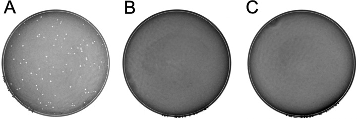
ICEBs1 was not known to confer phenotypes to B. subtilis, aside from those directly related to conjugation. However, the left end of ICEBs1 (Fig 1) encodes a phage-like repressor ImmR [28], anti-repressor ImmA [37], and recombinase Int [38]. In addition, the strain CU1050, that is cured of and often used to grow the temperate subtilis phage SPß [30,31], still contains the defective prophage PBSX and skin but is cured of ICEBs1 [33]. This information led us to wonder if there might be some interaction between ICEBs1 and SPß. We tested if the presence of ICEBs1 in B. subtilis altered the ability of SPß to make plaques. We mixed SPß with two B. subtilis strains, one that was missing ICEBs1 (ICEBs10) and that has been used as an indicator strain for SPß strain (CU1050) [32,33] and an isogenic derivative that contained ICEBs1 (CMJ81; ICEBs1+). SPß formed plaques on a lawn of the ICEBs10 strain (Fig 2A), but not on a lawn of the isogenic ICEBs1+ strain (Fig 2B), even when 100-fold more plaque forming units (PFUs) were mixed with cells (Fig 2C). Based on these results, we conclude that the presence of ICEBs1 inhibited plaque formation by SPß.
Fig 2. ICEBs1 prevents plaque formation by SPß.
Various numbers of PFUs of SPß were mixed with the indicated strains, plated, incubated overnight and checked for the presence of plaques (methods). A. Approximately 100 PFUs of SPß were mixed with the indicator strain CU1050. B. Approximately 100 PFUs of SPß mixed with strain CMJ81 (the indicator strain CU1050 carrying ICEBs1). C. Approximately 104 PFU of SPß mixed with strain CMJ81 (the indicator strain CU1050 carrying ICEBs1).
ICEBs1 reduces phage production during infection
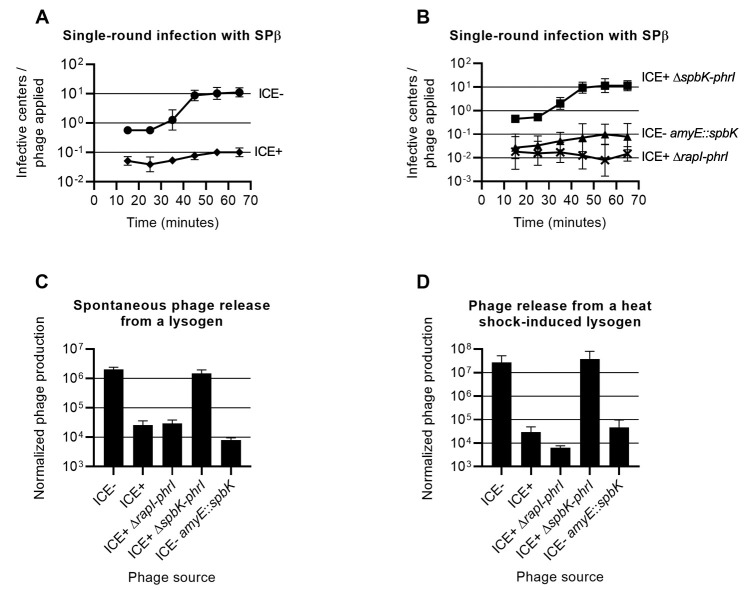
To quantify the effects of ICEBs1 on the production of SPß, we measured the kinetics of phage production during a single round of infection (Fig 3A and 3B). We mixed ~105 PFUs of SPß with ~107 cells (MOI ~0.01) for 5 min at 37°C, pelleted the cells by centrifugation, washed the cells to remove unattached phage, and resuspended the cells in LB medium at 37°C to allow for phage growth. The initial number of infective cells in the medium was determined by measuring the number of infective centers (PFUs) following the initial adsorption, and new phage production was monitored by tracking the subsequent increase in infective centers. For a strain without ICEBs1, the initial number of infective centers in the culture was about 90% of the initial number of phage used to inoculate the culture (Fig 3A). The number of infective centers in the culture began to increase about 25 minutes after initial infection, and plateaued about 45 minutes after initial infection. This indicated that SPß had an eclipse period of about 25 minutes (Fig 3A). The burst size (number of phage produced per infective cell) was 20 ± 7, somewhat less than the previously reported burst size of about 30 phage [24].
Fig 3. Production of SPß during the lytic cycle is reduced in cells containing ICEBs1 or spbK.
A. Effect of ICEBs1 on single-round infection of B. subtilis cultures. SPß null strains were grown in rich medium, infected with phage (MOI = 0.01) and then diluted in fresh medium. The number of infective centers in the culture was tracked over time using strain CU1050 as the indicator (methods). circles, ICEBs10 (CU1050); diamonds, ICEBs1+ (CMJ81). B. Effect of spbK on single-round infection of B. subtilis cultures. SPß null strains were grown and infected with SPß as in 2A. crosses, ICEBs1+ ΔrapI-phrI (CMJ913); squares, ICEBs1+ Δ(spbK-rapI-phrI) (CMJ914); triangles, ICEBs10 amyE::spbK (CMJ82). C. Effect of ICEBs1 and spbK on spontaneous phage production. Strains carrying wild type SPß lysogens and different ICEBs1 variants were grown in rich medium; ICEBs10 (JMA222), ICEBs1+ (AG174), ICEBs1+ ΔrapI-phrI (IRN342), ICEBs1+ Δ(spbK-rapI-phrI) (CAL1500), ICEBs10 amyE::spbK (CMJ74). Supernatant was collected from each culture during exponential growth and used as a phage source in a plaque assay (methods). D. Effect of ICEBs1 and spbK on phage production after induction of a lysogen. Strains carrying ts SPß lysogens and different ICEBs1 variants were grown in rich medium; ICEBs10 (CMJ114), ICEBs1+ (CMJ826), ICEBs1+ ΔrapI-phrI (CMJ917), ICEBs1+ Δ(spbK-rapI-phrI) (CMJ918), ICEBs10 amyE::spbK (CMJ116). SPß lysogens were induced by a heat shock during exponential growth and supernatants were collected and used as a phage source in a plaque assay (methods). For C and D the Y axis shows the number of PFU/ml of culture divided by the OD600 of the culture.
Cells with ICEBs1 that were exposed to SPß were less likely to become infective centers, and produced fewer phage per initial infective center. At an MOI of 0.01, the number of cells that produced any phage was reduced at least 10-fold relative to cells without ICEBs1 (Fig 3A). Furthermore, the number of phage produced per initial infective center was ~2.2 ± 0.4 (Fig 3A). Based on these results, we conclude that the presence of ICEBs1 in cells reduced the total number of phage released from the infected culture by at least 100-fold, or to about 0.1 progeny phage per infecting phage. This reduction did not support propagation of phage in the lytic cycle.
ICEBs1 has little or no effect on entry of phage into cells
The ICEBs1-dependent reduction in plaque formation and phage production could be due to reduced entry of phage into cells. Alternatively, a step in the phage lifecycle after entry could be inhibited. If the presence of ICEBs1 was causing a block in phage entry, then there should be a corresponding reduction in the frequency of lysogen formation. We used SPß that contained spc, conferring resistance to spectinomycin, to measure the frequency of lysogenization. Cells with or without ICEBs1 were mixed with SPß::spc98 (MOI ~ 0.001), unbound phage were washed off, and cells were spread on plates containing spectinomycin to select for lysogens. The lysogenization frequency of cells without ICEBs1 (CMJ472) was ~1% (1.1x10⁻² ± 0.46x10⁻²), or approximately one lysogen per 100 initial phage. Similarly, the lysogenization frequency of ICEBs1+ cells (CMJ827) was ~0.4% (4.3x10⁻³±1.3x10⁻³), or about 40% of that of the ICEBs10 cells. These results indicate that ICEBs1 has a relatively minor (if any) effect on lysogenization frequency and that the anti-SPß phenotype conferred by ICEBs1 was not due to a block in adhesion or entry of the phage.
ICEBs1 reduces the number of phage released by SPß lysogens
We found that the presence of ICEBs1 in an SPß lysogen inhibited phage production. We grew lysogens in liquid medium and measured the number of PFUs present in the supernatant. We found that cultures of a lysogen without ICEBs1 had approximately 100-fold more PFUs/ml than cultures of an ICE+ lysogen (Fig 3C). Together our results demonstrate that ICEBs1 acts primarily by blocking production of phage by infected cells, rather than by preventing infection of cells in the first place.
We also found that the presence of ICEBs1 prevented production of SPß following induction of a temperature sensitive lysogen. We grew strains with a temperature sensitive SPß lysogen (SPßc2) in rich medium, induced the lysogen by heat shock, and measured phage release. Phage production was reduced by ~1,000-fold in cells with ICEBs1 compared to cells without (Fig 3D). Although production of functional phage particles was reduced, the cells were still killed following phage induction. Cell viability, as measured by colony forming units (CFUs), was reduced to ~0.1% after phage induction compared to right before phage induction for strains with (CMJ826) and without ICEBs1 (CMJ114). Based on these results we conclude that ICEBs1 was probably not preventing induction of SPß but rather was inhibiting production of active phage particles post-induction.
The ICEBs1 gene spbK is necessary and sufficient to inhibit SPß
We were interested in determining which ICEBs1 gene was responsible for the inhibition of SPß. Most ICEBs1 genes are repressed when ICEBs1 is integrated in the host genome. Because the inhibition of SPß did not appear to depend on activation of ICEBs1, we focused on the handful of ICEBs1 genes that are constitutively expressed, including genes toward the left and right ends of the element (Fig 1). Preliminary experiments led us to focus on spbK (formerly yddK). These experiments included testing for the presence of SPß in the culture supernatant from lysogens, essentially as described above, with various regions of ICEBs1 deleted. Most deletion mutants tested had been described previously [22,39–41]. The preliminary results indicated that strains in which spbK was intact, including ΔcwlT and ΔrapI-yddM, inhibited phage release. In contrast, strains in which spbK had been deleted, including ΔconG-yddM, ΔydcB-yddM, ΔnicK-yddM, and ΔydcQ-yddM [39], did not inhibit phage production. Based on these results, we inferred that spbK was likely needed for ICEBs1-mediated inhibition of spontaneous release of SPß from a lysogen and tested this directly. We used three different assays to test the effects of spbK on SPß. In all three assays, we compared three B. subtilis strains: an ICEBs1+ strain with spbK (Δ(rapI-phrI)::kan, Fig 1C), an ICEBs1+ strain lacking spbK (Δ(spbK-phrI)::kan, Fig 1D), and an ICEBs10 strain expressing spbK from its own promoter at an ectopic locus (ICEBs10 amyE::{spbK kan}, Fig 1E). We measured: 1) the appearance of infective centers following a single round of infection with SPß (Fig 3A and 3B); 2) the number of phage spontaneously released from an SPß lysogen (Fig 3C); and 3) the number of phage produced after induction of a temperature sensitive SPß lysogen (Fig 3D). In all cases, we found that spbK was necessary for ICEBs1 to inhibit the formation of infective centers and the production of phage, and that ICEBs1+ ΔspbK strains were indistinguishable from strains entirely lacking ICEBs1. Furthermore, ectopic expression of spbK was sufficient to inhibit phage production in the absence of ICEBs1 in all three assays.
Expression of the SPß gene yonE inhibits acquisition of ICEBs1 and this inhibition is dependent on the ICEBs1 gene spbK
Based on the results described above, we thought that there might be at least one gene in SPß that was needed for the spbK-mediated inhibition of phage production. Results described below indicate that yonE is this SPß gene.
In previous work [42], we used Tn-seq to identify genes in recipients that affected the efficiency of stable acquisition of ICEBs1 in conjugation. Briefly, a library of random transposon insertions in a strain that is an SPß lysogen and cured of ICEBs1 was used as the recipient in conjugation. We selected for transconjugants that had acquired ICEBs1. Insertion mutations that cause a decrease in acquisition of ICEBs1 were underrepresented in transconjugants relative to controls. We found that insertions in some position in the SPß gene yonF were underrepresented, indicating that these insertion mutations reduced the ability of would-be recipients to stably acquire ICEBs1 from donors. Because the frequency of insertions in other positions in yonF was unaltered in transconjugants [42], and because neither yonF nor yonE is normally expressed in SPß lysogens, the phenotype could not be due to loss of yonF. We hypothesized that the reduction in acquisition of ICEBs1 might be due to inappropriate expression of yonE, the gene immediately downstream of yonF, likely transcribed from the promoter for the antibiotic resistance gene (spc) in the transposon. We therefore tested directly the effects of inappropriate expression of yonE on acquisition of ICEBs1.
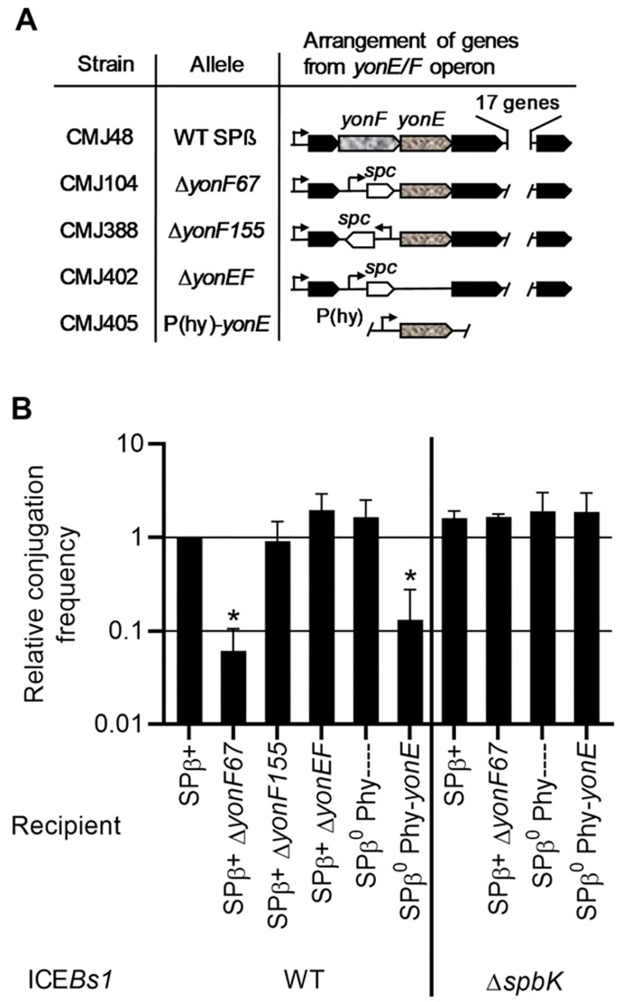
We found that inappropriate expression of yonE reduced the ability of cells to stably acquire a copy of ICEBs1. We made a series of mutations in SPß (Fig 4A) and tested these for effects on the ability of cells to act as ICEBs1 recipients during conjugation. We found that an insertion of spc into a deletion of yonF (ΔyonF::spc) reduced acquisition of ICEBs1 only when spc was co-directional with yonE. Furthermore, deletion of yonE in this context eliminated the defect in acquisition of ICEBs1 (Fig 4B). In the absence of all other SPß genes, expression of yonE from the IPTG-inducible promoter Pspank(hy) was sufficient to inhibit acquisition of ICEBs1 (Fig 4B). We conclude that yonE in SPß is both necessary and sufficient to cause the decrease in stable acquisition of ICEBs1. Results presented below demonstrate that when ICEBs1 is transferred to cells expressing yonE, those nascent transconjugants die. It is for this reason that there are no stable transconjugants recovered.
Fig 4. Expression of yonE in recipients reduces acquisition of ICEBs1 via conjugation.
A. Map of the operon in SPß that contains yonF and yonE, and relevant mutations. Genes are shown as arrows. yonF and yonE are indicated by arrows filled with a mottled pattern. spc is shown as an open arrow. Promoters are shown as bent arrows. The allele and the recipient strain carrying that allele are indicated. B. The relative conjugation frequencies are shown, normalized to the conjugation frequency between a donor carrying a wild type ICEBs1 (KM250) and a recipient with a wild type SPß (CMJ48) within the same experiment, (average 9.7 x 10⁻⁴ ± 1.3 x 10⁻³ transconjugants/donor). The ΔspbK donor (CMJ431) carries an ICEBs1 in which spbK-rapI-phrI have been deleted. The recipient with the promoter Pspank(hy) and no yonE allele is CMJ405. Other recipient strain numbers are indicated in panel A. Each experiment was repeated ≥ 3 times. Asterisks indicate that the conjugation frequency with the given recipient is significantly different than that with the wild type control (p<0.05, t-test).
The decreased acquisition of ICEBs1 by recipients expressing yonE was dependent on the ICEBs1 gene spbK. We tested strains expressing yonE for the ability to acquire ICEBs1 that was missing spbK (ICEBs1 ΔspbK), and found that they all acquired the ΔspbK element at the same frequency as wild type recipients not expressing yonE (Fig 4B, right end of panel). From these results we conclude that expression of yonE caused a defect in the stable acquisition of ICEBs1 and that this defect was dependent on the presence of spbK in the incoming ICEBs1. We note that loss of spbK caused no reduction in conjugation efficiency (Fig 4B), demonstrating that it is dispensable for conjugation. We also note that the presence or absence of wild type SPß had no detectable effect on conjugation efficiency (Fig 4B).
Co-expression of yonE and spbK causes a defect in cell growth and a drop in cell viability
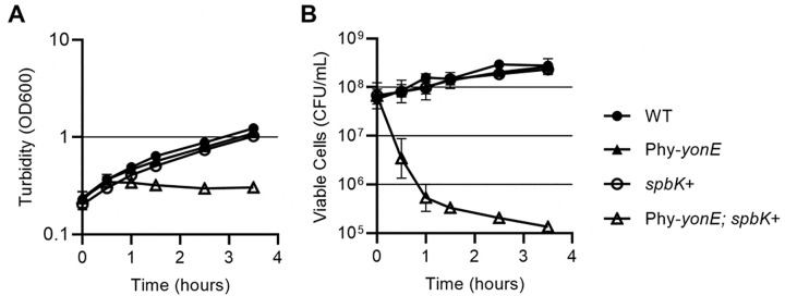
We found that expression of spbK (from its own promoter) and yonE (from Pspank(hy)) together caused a severe growth defect. We grew cells containing both spbK and yonE in defined minimal medium and added IPTG (time = 0) to increase expression of yonE (Fig 5). This caused a rapid growth arrest as measured by optical density (Fig 5A) and an ~1000-fold drop in viability as measured by plating for CFUs on LB plates made with Noble agar (Fig 5B; see below). In contrast, expression of either gene alone, spbK from its own promoter (lacA::spbK), or yonE from an inducible promoter (amyE::Pspank(hy)-yonE), had no obvious effect on growth (Fig 5A and 5B). Together, our results indicate that co-expression of yonE and spbK is detrimental to cell growth. In an SPß lysogen that also contains ICEBs1, spbK, is expressed, but yonE is not. yonE would be expressed only if SPß were activated, or upon infection of non-lysogens.
Fig 5. Co-expression of spbK and yonE kills cells and results in a growth defect.
Strains null for ICEBs1 and SPß (closed circles, PY79), expressing yonE (closed triangles, amyE::{Pspank(hy)-yonE}, CMJ616), expressing spbK (open circles, lacA::spbK, CMJ684), or both yonE and spbK (open triangles, CMJ685) were grown in minimal medium. yonE expression was induced by the addition of 1 mM IPTG and the culture turbidity (A), and cell viability (B) were followed over time. T = 0 samples were collected immediately prior to induction with IPTG. Cell viability was measured as the number of colony forming units per ml, measured by plating for CFUs on LB plates made with Noble agar. Experiment was repeated 3 times.
Despite growing normally in defined liquid medium prior to adding IPTG, cells containing both lacA::spbK and amyE::Pspank(hy)-yonE had a substantial plating defect (~200-fold) when plated on LB plates made with standard bacto-agar (Difco), and had a distinct small colony morphology even in the absence of IPTG. The plating and colony size defects were eliminated when the cells were plated on LB plates made with Noble agar (Difco) (S1 Fig), a purified form of agar that is used to culture some fastidious organisms. We hypothesize that a component of bacto-agar sensitizes cells to the detrimental impact of co-expressing spbK and yonE, such that leaky expression from Pspank(hy)-yonE is sufficient to trigger the growth defect.
yonE is needed for phage production
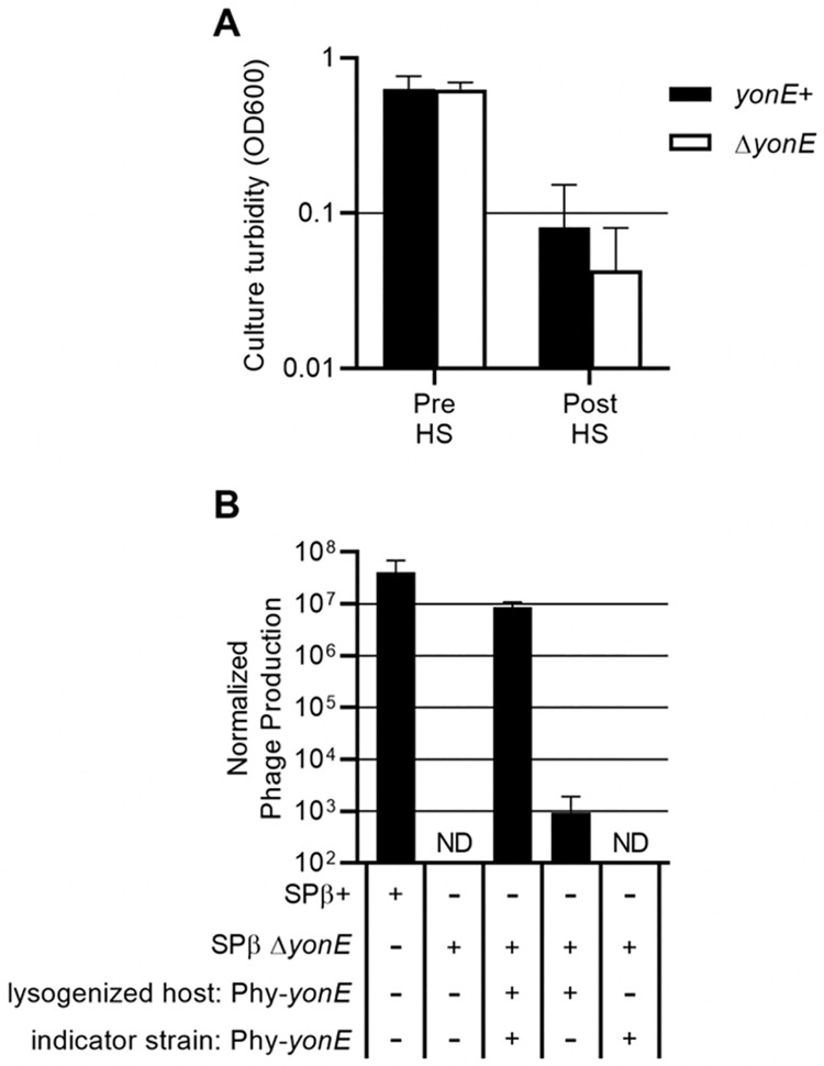
To determine the effect of yonE on phage production, we made an unmarked deletion of yonE (ΔyonE443) in a temperature-sensitive SPß lysogen. We found that cultures of this inducible ΔyonE lysogen cleared comparably to a yonE+ strain following a shift to high temperature (Fig 6A), demonstrating that yonE is not needed for induction of SPß from a lysogenic state, nor is it needed to cause host cell lysis.
Fig 6. yonE is needed for production of infectious phage.
A. Strains carrying a temperature-sensitive SPß lysogen with a wild type yonE allele (black bars, CMJ114) or ΔyonE (white bars, CMJ455) were cultured in rich medium, then heat shocked for 20 minutes (methods). The Y axis shows the average and standard deviation of the OD600 from each culture immediately before and 70 minutes (± 5 minutes) after the heat shock. Each experiment was repeated ≥3 times. B. Phage were prepared by culturing strains with a temperature sensitive SPß lysogen with a wild type yonE allele (CMJ114), ΔyonE (CMJ455), or a ΔyonE allele with yonE complemented from the chromosome (lacA::{Pspank(hy)-yonE}, CMJ457) to an OD600 of approximately 0.4, and then heat shocking the cultures and collecting phage (methods). Lysates were then spread on lawns of the indicator strain CU1050 or an indicator with lacA::{Pspank(hy)-yonE} (CMJ440) and incubated overnight to allow plaque formation. The Y-axis shows the average and standard deviation of the number of infectious phage /ml, normalized by dividing by the OD600 of the culture at the time of heat shock. ND = not detected. Each experiment was repeated ≥3 times.
Despite the fact that the ΔyonE host cells lysed, there were no detectable viable phage (< 10 PFUs/ml) produced by the mutant lysogen (Fig 6B, first two columns). This defect in phage production was partially complemented by expression of yonE from an ectopic chromosomal locus. These ΔyonE phage (recovered from the complemented strain) were capable of forming plaques on an indicator strain that also expressed yonE (Fig 6B, last three columns). Although a small number of phage produced by a ΔyonE lysogen were able to form plaques on a yonE- indicator strain, analysis of lysogenized cells obtained from these plaques revealed that the phage had a wild type copy of yonE, likely obtained through homologous recombination with the yonE allele on the chromosome of the original host strain. Based on these results, we conclude that yonE is essential for production of SPß.
To determine if yonE is needed to form a lysogen, we made a stock of spc-marked ΔyonE phage by growing the ΔyonE mutant on a B. subtilis strain ectopically expressing yonE. The frequency of lysogenization of spc-marked yonE+ and ΔyonE phage were both approximately 1%, indicating that yonE is not needed for lysogen formation.
The function of yonE is not known. However, there are homologs in other phages, including the phage C-ST from Clostridium botulinum. The region of homology between C-ST and SPß extends from yonG to yomZ, indicating that these genes may encode conserved phage functions [43]. The phage E3 from Geobacillus encodes a putative portal protein (accession number AJA41333) that is 25% identical to YonE [44]. Portal proteins are one of three molecular components involved in packaging the phage genome into the capsid during maturation. The other two components are the large and small terminase subunits [45]. Additional homology searches using NCBI BLAST revealed that YonF is a member of the terminase 1 superfamily and encodes a terminase 6 multidomain, typical of large terminase subunit proteins. These results indicate that YonE and YonF may be a part of the SPß head packaging machinery. This notion is consistent with the need for yonE in production of functional phage, but not in host cell lysis or formation of lysogens.
SpbK contains a TIR domain involved in protein-protein interaction
spbK is predicted to encode a 266 amino acid protein. Using the NCBI Delta-BLAST search tool [46] we found that the C-terminal region of SpbK (amino acids 113–266) contains a Toll Interleukin-1 Receptor (TIR) domain in the TIR_2 superfamily (accession: cl23749) (S2A Fig). Proteins containing TIR domains have been found in animals, plants, and bacteria. In animals, such proteins are involved in signaling cascades in development and in immune activation [47]. In plants they mediate disease resistance, often in response to infectious agents [48]. Some pathogenic bacteria encode TIR domain proteins that interact with eukaryotic host TIR domain proteins to modulate the host immune response [49]. Many non-pathogenic bacteria also contain TIR domain proteins and it is thought that the TIR domains mediate protein-protein interaction [50]. Recent work has also implicated some bacterial TIR domain proteins as components of anti-phage defense systems, though the mechanism of defense is not understood [51].
Where they have been studied, TIR domains mediate protein-protein interactions by interacting with other TIR domains. spbK is the only gene in B. subtilis, including all horizontally acquired sequences (e.g, ICEBs1 and SPß), predicted to encode a TIR domain. Using a yeast two-hybrid assay (Methods), we found that full-length SpbK multimerizes in vivo (S2B Fig). Additionally, we found that the TIR domain alone interacted with both full-length SpbK and with the TIR domain, but that deleting the TIR domain abolished all interaction between SbpK proteins (S2B Fig). We also tested for, but were unable to detect, interaction between SpbK and YonE, indicating that if these two proteins interact, that interaction was not detectable with the yeast two-hybrid system that we used (Methods).
ICEBs1 protects B. subtilis populations from attack by SPß
As described above, when SPß undergoes lysogenic to lytic conversion, SPß lysogens that contain ICEBs1 die without significant production of progeny phage. De novo infection of non-lysogens that contain ICEBs1 also die without producing progeny phage. We found that in a population of cells, this abortive infection system in ICEBs1 protected cells from killing by SPß. We grew SPß-cured strains of B. subtilis that either contained or did not contain ICEBs1, infected the cultures with SPß at a low multiplicity of infection (MOI ~0.01), and tracked the growth (optical density) of the culture, the concentration of viable cells (including lysogens), and free phage over time (Fig 7). We suspected that use of SPß that is capable of making lysogens could mask possible effects on cell death and would be measuring possible protection of the population by ICEBs1 and by the formation of lysogens (which are themselves immune to superinfection, see below). Therefore, we first analyzed a clear plaque mutant (incapable of making lysogens; Methods) to eliminate possible effects of lysogeny. We then measured effects of ICEBs1 on phage that could form lysogens.
Fig 7. ICEBs1 protects B. subtilis populations against SPß.
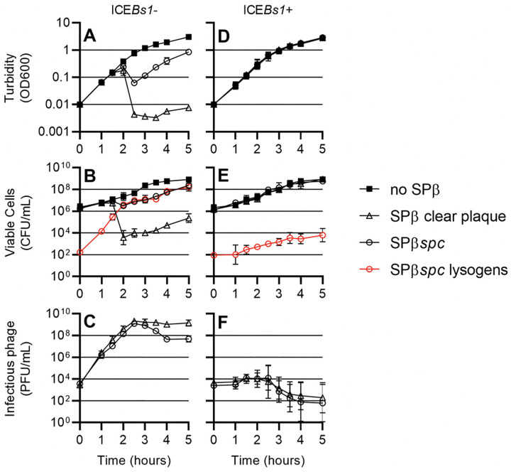
A, B, C, SPß0 ICEBs10 (CU1050) and D, E, F, SPß0 ICEBs1+ (CMJ81) were grown in rich medium, infected with no phage (filled squares), SPß clear plaque (open triangles), or SPß::spc (open circles) at an MOI of approximately 1:100 and diluted in fresh medium to an OD600 of 0.01. Culture turbidity (A, D), CFUs/ml (B, E), and the number of infectious phage per ml of culture (C, D) were tracked over time. Additionally, for cultures infected with SPß::spc, the number of lysogenized cells were tracked over time (red open circles).
When cultures of an ICEBs10 strain were infected with a clear plaque mutant of SPß (MOI ~0.01) the cells continued to grow at the same rate as an uninfected culture for approximately 1.5 hours, then the majority of the cells abruptly died, as evidenced by a decrease in optical density (Fig 7A) and an approximately 5,000-fold decrease in CFUs (Fig 7B). During this time (1.5 hrs) the concentration of phage in the culture (Fig 7C) surpassed the concentration of cells (Fig 7B).
In contrast, when an ICEBs1+ strain was infected with the clear plaque mutant of SPß (MOI ~0.01), cell growth was indistinguishable from an uninfected culture as measured by both the optical density (Fig 7D) and the number of CFUs (Fig 7E). The population of phage in the culture generally decreased to below the initial inoculum (Fig 7F). These results indicate that the presence of ICEBs1 is beneficial to the population of cells even though individual infected cells may not survive.
Experiments described above were done with an SPß mutant that was unable to form lysogens. We repeated these experiments using SPß::spc, that, other than the spc insertion is wild type and able to form lysogens. Lysogens were detected as spectinomycin-resistant colonies.
When cells without ICEBs1 were infected with SPß::spc (MOI ~0.01), there was a 10-fold drop in both the optical density of the culture (Fig 7A) and the number of CFUs (Fig 7B). During the experiment, many cells became lysogenized with SPß. Lysogens are then protected from killing by new SPß infection [24]. These lysogens continued to grow, and after about five hours the population of cells had increased and virtually all cells were SPß lysogens (Fig 7B). Although wt SPß killed only ~90% of the cells, (compared to >99.9% killing by the clear plaque mutant), wt SPß became established in the entire outgrown population.
When cells with ICEBs1 were infected with SPß::spc (MOI ~0.01), cell growth continued and there was no obvious drop in optical density (Fig 7D) nor in the number of CFUs (Fig 7E). Five hours after the initial infection, the number of phage in the culture was below the initial inoculum (Fig 7E) and the number of SPß lysogens remained at approximately 104–105 per ml (Fig 7F), a relatively small fraction of the total number of cells.
Together, these results indicate that the presence of ICEBs1 in cells limits phage production, thereby protecting a population of cells from predation by SPß. The presence of ICEBs1 does not limit initial lysogenization. However, because ICEBs1 limits the production of new phage, the number of lysogens is limited by the initial inoculum of phage. In this way, ICEBs1 protects the population from killing by SPß, and secondarily, prevents SPß from invading all the cells, thereby preventing lysogens taking over the population.
Discussion
Results presented here demonstrate that ICEBs1 encodes an anti-phage system that inhibits production of the phage SPß. This inhibition occurs upon de novo infection by SPß and also upon induction of an SPß lysogen. There is little or no direct effect on the formation of lysogens. The ICEBs1 gene spbK is both necessary and sufficient to inhibit SPß: deleting spbK from ICEBs1 abolishes the phenotype, and expressing spbK in a strain missing ICEBs1 fully inhibits SPß. Expression of yonE apparently triggers this anti-phage response, and co-expression of spbK and yonE in a strain that otherwise lacks ICEBs1 and SPß rapidly kills the cells. There are multiple possibilities for how YonE triggers this killing. It could directly interact with SpbK and the two proteins together, perhaps with other host products, and could disrupt an essential host function. YonE could somehow modify SpbK, perhaps covalently, or by stabilizing it, thereby activating it. Alternatively, YonE could modify an essential host product, making cells susceptible to SpbK. Of course, it is possible that SpbK makes cells susceptible to YonE, and that cells with ICEBs1 are ’primed’ to be killed when yonE is expressed.
Whatever the mechanism, we conclude that ICEBs1 encodes an abortive infection system that protects its host from predation by SPß. Below, we briefly describe the yonE and spbK gene products and TIR-domains, comment generally on abortive infection systems, and conclude with general thoughts about cargo genes in ICEs.
Genes involved in protection against SPß
yonE is essential for the phage lytic cycle, but is not required for lysogen formation. Bioinformatic analysis of yonE and yonF revealed a possible role for these genes as components of the phage head-packing machinery, needed during the final stages of a phage’s lytic cycle.
SpbK contains a TIR domain. Where TIR domains have been studied they generally mediate protein-protein interactions through recognition of other TIR domains. However, examples of heterotypic interactions of TIR domains with non-TIR domain proteins have been described [52]. In some bacterial pathogens, TIR-domain proteins modulate the host immune response [50,53–55]. Recent work has implicated some bacterial TIR-domain proteins as being essential components of a class of anti-phage defense systems (“Thoeris”) [51]. Two of these Thoeris systems (from Bacillus cereus and Bacillus amyloliquefaciens) have been shown to non-specifically confer resistance to some myophages when reconstituted in B. subtilis, although the mechanism of this resistance is not understood.
Although SpbK and the Thoeris systems appear to have a common purpose, SpbK does not appear to be a component of a B. subtilis Thoeris antiphage system. The hallmark of Thoeris systems is a single gene encoding a NAD+ binding protein (ThsA) in proximity to (typically multiple) genes encoding TIR-domain proteins (ThsB) [51]. spbK is the only gene encoding a TIR-domain protein in B. subtilis. Furthermore, of all the genes in ICEBs1, spbK is both necessary and sufficient for protection from SPß, and there is no indication that SpbK contains a nucleotide binding domain. Irrespective of these differences, our analysis of SpbK raises the possibility that Thoeris anti-phage systems might function as abortive infection systems.
Many isolates of B. subtilis have both ICEBs1 and SPß. These elements likely co-evolved and it is possible that the spbK-mediated abortive infection is specific to SPß. However, we suspect that spbK-mediated abortive infection might respond to other phages, perhaps those with yonE orthologs.
Abortive infection systems
The anti-phage phenotype encoded by ICEBs1 resembles abortive infection systems that have been described for Lactococcus, Escherichia coli, and other bacteria. Such systems detect infection of a bacterial cell by phage and respond by inhibiting a host process needed for phage maturation and release [56]. The mechanisms of inhibition vary widely, but often target a critical host process. The cellular process that is inhibited by SpbK is evidently essential for the host, as co-expression of spbK and yonE results in cell death. We have not yet determined what essential process(es) are targeted to cause SpbK-YonE-induced cell death.
Cargo genes in ICEs
The cargo genes of mobile genetic elements, including ICEs, are those genes that are not necessary for the function of the mobile element, but are part of and transferred with the element. Cargo genes on a mobile element can often allow bacteria to rapidly acquire a new phenotype through acquisition of the element. Historically, most well studied ICEs were identified because of the phenotype(s) conferred by the cargo genes [6]. Identification and characterization of the responsible genes revealed that they were in an ICE.
Many ICEs are being identified by bioinformatic analyses of sequenced bacterial genomes [29,57]. In most of these analyses, it is not clear what, if any, phenotype is conferred by the ICE to its host. We suspect that many other ICEs with cargo genes of unknown function likely assist their hosts in mediating interactions with other mobile genetic elements.
Methods
Media and growth conditions
E. coli cells were grown in LB medium and on LB plates containing 1.5% agar at 37°C. S. cereviseae cells were grown in YPAD and on YPAD plates or synthetic dropout (SD) plates containing 2% agar and appropriate supplements to test for the indicated auxotrophies [58,59]. B. subtilis cells were grown in LB or S750 defined minimal medium with 0.1% glutamate [60] with either glucose or arabinose (1% w/v) as a carbon source and on LB plates containing 1.5% bacto-agar or on Noble Agar (1.5%) for strains expressing both spbK and yonE. Antibiotics and other additives were used at the following concentrations for E. coli: carbenicillin (100 μg/ml), B. subtilis: kanamycin (5 μg/ml), spectinomycin (100 μg/ml), chloramphenicol (5 μg/ml). The Pspank(hy) promoter was activated with 1 mM isopropyl-ß-D-thiogalactopyranoside (IPTG), and the Pxyl promoter was activated with 1% (w/v) xylose, typically in cells grown in arabinose.
Strains and alleles
B. subtilis strains and genotypes are listed in Table 1. Specific alleles are described below. All B. subtilis strains are derived from parent AG174 (JH642), unless otherwise indicated.
Table 1. B. subtilis strains used.
| B. subtilis Strain | Genotype (reference) |
|---|---|
| AG174 | trpC2 pheA1 a.k.a., JH642 [27] |
| CAL321 | trpC2 pheA1 Δ(rapI-yddM)318::kan [39] |
| CAL322 | trpC2 pheA1 Δ(yddG-yddM)319::kan [39] |
| CAL323 | trpC2 pheA1 Δ(yddB-yddM)320::kan [39] |
| CAL347 | trpC2 pheA1 Δ(ydcR-yddM)347::kan [39] |
| CAL348 | trpC2 pheA1 Δ(ydcQ-yddM)348::kan [39] |
| CAL1500 | trpC2 pheA1 Δ(spbK-rapI-phrI)1500::kan |
| CMJ48 | PY79 (ICEBs10) SPß+ amyE::{Pspank(hy)-empty lacI spc} |
| CMJ74 | trpC2 pheA1 ICEBs10 amyE::{spbK cat} |
| CMJ81 | CU1050 (SPß0) ICEBs1::kan (non-disruptive); note: made by crossing donor JMA448 with recipient CU1050 |
| CMJ82 | CU1050 (ICEBs10) (SPß0) amyE::{spbK cat} |
| CMJ98 | trpC2 pheA1 ICEBs10 yolBC98::spc |
| CMJ104 | PY79 (ICEBs10) SPß+ ΔyonF67::spc [42] |
| CMJ114 | CU1050 (ICEBs10) SPßc2 yolBC98::spc |
| CMJ116 | CU1050 (ICEBs10) SPßc2 yolBC98::spc amyE::{spbK cat} |
| CMJ388 | PY79 (ICEBs10) SPß+ ΔyonF155::spc (reverse orientation) [42] |
| CMJ402 | PY79 (ICEBs10) SPß+ ΔyonEF396::spc |
| CMJ403 | PY79 (ICEBs10) (SPß0) amyE::{Pspank(hy)-empty lacI spc} lacA::{Pspank(hy)-yonE lacI mls} |
| CMJ405 | PY79 (ICEBs10) (SPß0) amyE::{Pspank(hy)-empty lacI spc} lacA::{Pspank(hy)-empty lacI mls} |
| CMJ431 | trpC2 pheA1 amyE::{Pxyl-rapI xylR cat} Δ(spbK-rapI-phrI)1500::kan |
| CMJ434 | trpC2 pheA1 argA85::Tn917 SPßc2 ΔyonE434::lox-cat |
| CMJ440 | CU1050 (ICEBs10) (SPß0) lacA::{Pspank(hy)-yonE lacI mls} |
| CMJ443 | CU1050 (ICEBs10) SPßc2 ΔyonE443 (unmarked) |
| CMJ455 | CU1050 (ICEBs10) SPßc2 ΔyonE443 (unmarked) yolBC98::spc |
| CMJ457 | CU1050 (ICEBs10) SPßc2 ΔyonE443 (unmarked) yolBC98::spc lacA::{Pspank(hy)-yonE lacI mls} |
| CMJ472 | CU1050 (ICEBs10) (SPß0) comK::cat |
| CMJ616 | PY79 (ICEBs10) (SPß0) amyE::{Pspank(hy)-yonE lacI spc} |
| CMJ684 | PY79 (ICEBs10) (SPß0) lacA::{spbK kan} |
| CMJ685 | PY79 (ICEBs10) (SPß0) amyE::{Pspank(hy)-yonE lacI spc} lacA::{spbK kan} |
| CMJ826 | CU1050 SPßc2 yolBC98::spc ICEBs1::kan (non-disruptive) |
| CMJ827 | CU1050 (SPß0) ICEBs1::kan, comK::cat |
| CMJ913 | CU1050 (SPß0) ICEBs1+ Δ(rapI-phrI)342::kan |
| CMJ914 | CU1050 (SPß0) ICEBs1+ Δ(spbK-rapI-phrI)1500::kan |
| CMJ917 | CU1050 SPßc2 ICEBs1+ Δ(rapI-phrI)342::kan, yolBC98::spc |
| CMJ918 | CU1050 SPßc2 ICEBs1+ Δ(spbK-rapI-phrI)1500::kan, yolBC98::spc |
| CU1050 | ICEBs10 SPß0 metA thrC leu codY sup-3 (trnS-lys) [31,33]; (CMJ28) |
| IRN342 | trpC2 pheA1 Δ(rapI-phrI)342::kan [22] |
| JMA222 | trpC2 pheA1 ICEBs10 [22] |
| JMA448 | trpC2 pheA1 ICEBs1::kan amyE::{Pspank(hy)-rapI spc} [22] note: used as donor with recipient CU1050 to create CMJ81 |
| KM250 | trpC2 pheA1 Δ(rapI-phrI)342::kan amyE::{Pxyl-rapI cat} [61] |
| PY79 | ICEBs10 SPß0 [62] |
SPß::spc
spc (spectinomycin resistance) was inserted between yolB and yolC in SPß. spc was amplified by PCR, and Gibson assembly [63] was used to join this fragment to genomic sequences containing the apparent bidirectional terminator located between the convergently transcribed genes yolB and yolC [64] such that a copy of the terminator is located on each side of spc. This was then used to transform naturally competent B. subtilis cells selecting for resistance to spectinomycin. An antibiotic resistant strain (CMJ98) was identified and the location of the spc gene verified by sequencing. This strategy resulted in duplication of the terminator with spc located between the terminators. The resulting phage is referred to as SPß::spc98, or SPß::spc.
Clear plaque mutant of SPß
SPß typically makes turbid plaques, characteristic of temperate phages. In the course of determining the titre of a stock of SPß::spc, we noticed a plaque that appeared clear. We picked this plaque and designated the phage SPß::spc-clear. We grew the phage and then tested for the ability to form lysogens by infecting cells and selecting for spectinomycin resistance. Cells infected with the SPß::spc phage readily formed lysogens. We did not detect any lysogens (spectinomycin resistance) from cells infected with SPß::spc-clear. In addition, all plaques observed were clear, verifying that this phage was indeed unable to form lysogens.
Δ(spbK-rapI-phrI)1500::kan
The region of ICEBs1 encoding spbK-rapI-phrI was replaced with kan. The deletion replaces all of spbK, rapI, and phrI, and was designed such that the orientation of kan and the phrI deletion boundary would be identical to the Δ(rapI-phrI)342::kan allele of IRN342 [22]. The linkage between ΔspbK and Δ(rapI-phrI)342::kan was used to transfer the ΔspbK allele as needed.
ΔyonEF396::spc
A deletion-insertion that replaces yonE and yonF with a co-directional spc insertion.
ΔyonE443
The unmarked ΔyonE443 allele was made by replacing yonE with the cat gene, flanked by lox sites (CMJ434). The Cre recombinase, expressed from the temperature-sensitive plasmid, pDR244, was then used to remove the lox-flanked cat gene by recombination. Strains were then cured of pDR244 by culturing them on LB + 1.5% agar at 45°C, as previously described [42,65], resulting in strain CMJ443.
Overexpression of yonE
The yonE coding sequence was cloned into a plasmid containing the IPTG-inducible Pspank(hy) promoter [66], lacI, and either spc situated between genomic sequence from amyE, or mls situated between genomic sequence from lacA. The resulting construct was then transformed into competent B. subtilis cells. The following strains carrying a double-crossover of the given construct were identified by antibiotic resistance and PCR: CMJ403, lacA::{Pspank(hy)-yonE lacI mls}; CMJ616, amyE::{Pspank(hy)-yonE lacI spc}. Pspank(hy) is only partly repressed by LacI and was fully derepressed upon addition of 1 mM IPTG. Constructs lacking the yonE insert were also transformed into B. subtilis to generate the control alleles lacA::{Pspank(hy)-empty lacI mls} and amyE::{Pspank(hy)-empty lacI spc}.
Expression of spbK
To study expression of spbK in the absence of other ICEBs1 genes, a fragment containing the spbK coding sequence and 330 bp upstream was amplified by PCR and cloned into a plasmid for double-crossover integration into lacA or amyE. For cloning into lacA, the spbK fragment was cloned by Gibson assembly into a plasmid containing kan and parts of lacA suitable for double crossover. For cloning into amyE, the spbK fragment was cloned into a plasmid containing cat flanked by genomic sequences flanking the amyE locus by Gibson assembly. The resulting constructs were transformed into naturally competent B. subtilis cells and strains carrying a double crossover were identified as above, resulting in strains CMJ74 {amyE:: (spbK cat)} and CMJ684 {lacA:: (spbK kan)}.
Plaque assays
To quantify the number of PFUs, samples with phage were diluted in LB and 100 μl of appropriate dilutions were mixed with 300 μl of an indicator strain at an OD600 of 0.5. Phage and cells were incubated at room temperature for 5 minutes, then mixed with 3 ml of soft agar (soft agar contains 10 g/l tryptone, 5 g/l yeast extract, 10 g/l NaCl, 6.5 g/l agar). The soft agar was spread on warm LB plates and incubated overnight at 37°C, allowing a lawn of cells to form. Plaques in the lawn were then counted. To photograph plaques, bacterial lawns were stained with 2,3,5—triphenyltetrazolium chloride [67] (TTC, Sigma). Briefly, 8 ml of 0.1% TTC in LB was pipetted onto plates and incubated at 37°C for 30 minutes. The TTC solution was then aspirated off and the petri dishes were photographed.
Single-round infection experiments
Cells of the strain to be infected were cultured in rich medium to mid- to late exponential phase. The OD600 was then adjusted to 0.5 and 100 μl of cells was mixed with 10 μl medium containing 105 PFU of SPß (MOI 1:100). Cells and phage were co-incubated for 5 min at 37°C, then washed 3 times by adding 1 ml LB, pelleting the cells in a microcentrifuge and removing the supernatant. The washed pellet was resuspended in 10 ml LB and incubated at 37°C with aeration to allow the phage to develop. Samples of the culture were taken at various time points and used immediately for plaque assays to quantify the concentration of infective centers (free phage + infected cells) in the culture.
Quantification of lysogeny
The frequency of lysogenization was determined using SPß::spc98. 104 PFUs of SPß::spc98 in 10 μl LB were added to 100 μl of an indicator strain at an OD600 of 0.5 (an MOI of approximately 1:1000). Phage and cells were incubated at room temperature for 5 minutes, then cells were washed 3x with 1 ml LB to remove unbound phage. Cells were then spread on LB plates with spectinomycin to select for cells that had become lysongenized with SPß::spc.
Yeast two-hybrid assays
Yeast strains are listed in Table 2 and were derived from PJ69-4A [59]. The yeast two hybrid strains and vectors used in this study have been previously described [59]. Briefly, the coding sequence for spbK from amino acids 1–104 (N-terminus), 97–266 (TIR domain) and full length spbK were cloned into pGAD-C1 and fused to the GAL4 activation domain or pGBDU-C3 and fused to the GAL4 DNA binding domain. These vectors were then transformed into competent PJ69-4A cells using the LiAc method of Gietz and Schiestl [58] and plated on synthetic dropout (SD) medium with appropriate supplements to select for acquisition of the plasmids. The ability to grow in the absence of leucine (pGAD-based plasmids) or uridine (pGBDU-based plasmids) was used to select clones that acquired each plasmid. To test for interaction between peptides, yeast strains carrying the plasmids of interest were spotted on SD medium and scored for growth in the absence of adenine, with growth indicating an interaction. As a control, strains carrying each individual plasmid were also scored for growth in the absence of adenine (all were negative).
Table 2. Yeast strains used.
| yeast strain | genotype |
|---|---|
| CMJ620 | S. cereviseae mat a ura3-52 leu2-3 his3 trp1 dph2Δ::HIS3 gal4Δ gal80Δ GAL2-ADE2 LYS2::GAL1-HIS3 met2::GAL-lacZ pCJ113 pCJ107 |
| CMJ621 | S. cereviseae mat a ura3-52 leu2-3 his3 trp1 dph2Δ::HIS3 gal4Δ gal80Δ GAL2-ADE2 LYS2::GAL1-HIS3 met2::GAL-lacZ pCJ113 pCJ108 |
| CMJ622 | S. cereviseae mat a ura3-52 leu2-3 his3 trp1 dph2Δ::HIS3 gal4Δ gal80Δ GAL2-ADE2 LYS2::GAL1-HIS3 met2::GAL-lacZ pCJ113 pCJ109 |
| CMJ626 | S. cereviseae mat a ura3-52 leu2-3 his3 trp1 dph2Δ::HIS3 gal4Δ gal80Δ GAL2-ADE2 LYS2::GAL1-HIS3 met2::GAL-lacZ pCJ114 pCJ107 |
| CMJ627 | S. cereviseae mat a ura3-52 leu2-3 his3 trp1 dph2Δ::HIS3 gal4Δ gal80Δ GAL2-ADE2 LYS2::GAL1-HIS3 met2::GAL-lacZ pCJ114 pCJ108 |
| CMJ628 | S. cereviseae mat a ura3-52 leu2-3 his3 trp1 dph2Δ::HIS3 gal4Δ gal80Δ GAL2-ADE2 LYS2::GAL1-HIS3 met2::GAL-lacZ pCJ114 pCJ109 |
| CMJ632 | S. cereviseae mat a ura3-52 leu2-3 his3 trp1 dph2Δ::HIS3 gal4Δ gal80Δ GAL2-ADE2 LYS2::GAL1-HIS3 met2::GAL-lacZ pCJ115 pCJ107 |
| CMJ633 | S. cereviseae mat a ura3-52 leu2-3 his3 trp1 dph2Δ::HIS3 gal4Δ gal80Δ GAL2-ADE2 LYS2::GAL1-HIS3 met2::GAL-lacZ pCJ115 pCJ108 |
| CMJ634 | S. cereviseae mat a ura3-52 leu2-3 his3 trp1 dph2Δ::HIS3 gal4Δ gal80Δ GAL2-ADE2 LYS2::GAL1-HIS3 met2::GAL-lacZ pCJ115 pCJ109 |
Mating assays
Mating assays were performed as previously described [38,42]. Briefly, donors and recipients were grown separately in minimal medium with 1% arabinose as a carbon source. RapI expression was induced in donors for 2 hours with 1% xylose. Approximately equal numbers of donors and recipients were then mixed, collected on a filter and placed on 1.5% agar plates buffered with Spizizens minimal salts (SMS agar contains 15 mM ammonium sulfate, 80 mM dibasic potassium phosphate, 44 mM monobasic potassium phosphate, 3.4 mM trisodium citrate, 0.8 mM magnesium sulfate, and 1.5% agar at pH 7.0) [68] for 90 minutes. Cells were rinsed off the filter, diluted, and spread on LB plates with selective antibiotics and incubated at 37°C overnight before quantification of colony forming units.
Supporting information
Strains null for ICEBs1 and SPß (PY79), expressing yonE (amyE::Phy-yonE, CMJ616), expressing spbK (lacA::spbK, CMJ684), or both yonE and spbK (CMJ685) were grown in minimal medium in the absence of IPTG. At an OD600 of 0.2, cultures were plated for CFUs on LB plates made with standard bacteriological agar (black bars) or on LB plates made with more rigorously purified Noble agar (white bars). Plating efficiency measured as CFUs/ml normalized to OD600.
(TIF)
A. Map of the SpbK peptide sequence showing the location of the TIR domain in black. B. Yeast two-hybrid screen of SpbK fragments. Yeast strains carrying full length SpbK (Full), SpbK amino acids 1–104 (N-term) or SpbK amino acids 97–266 (TIR) bound to the GAL4 DNA binding domain (DBD, Y-axis) and/or the GAL4 activation domain (AD, X-axis) were spotted on medium selective for interaction between the bait and prey peptides and incubated at 30° C to allow for growth (methods). The following combinations were tested: AD-SpbK + DBD-SpbK (CMJ620), AD-N-term + DBD-SpbK (CMJ621), AD-TIR + DBD-SpbK (CMJ622), AD-SpbK + DBD-N-term (CMJ626), AD-N-term + DBD- N-term (CMJ627), AD-TIR + DBD- N-term (CMJ628), AD-SpbK + DBD-TIR (CMJ632), AD-N-term + DBD-TIR (CMJ633), AD-TIR + DBD-TIR (CMJ634).
(TIF)
Acknowledgments
We thank Mary Anderson and Josh Jones for helpful discussions and comments on the manuscript, and Eleina England for early experiments confirming interactions between SPß and ICEBs1.
Data Availability
All relevant data are within the manuscript and its Supporting Information files.
Funding Statement
Research reported here is based upon work supported, in part, by the National Institute of General Medical Sciences of the National Institutes of Health under award number R01GM050895 and R35 GM122538 to ADG. MMH was also supported, in part, by the NIGMS pre-doctoral training grant T32 GM007287. Any opinions, findings, and conclusions or recommendations expressed in this report are those of the authors and do not necessarily reflect the views of the National Institutes of Health. The funders had no role in study design, data collection and analysis, decision to publish, or preparation of the manuscript.
References
- 1.Paulsen IT, Banerjei L, Myers GSA, Nelson KE, Seshadri R, Read TD, et al. Role of mobile DNA in the evolution of vancomycin-resistant Enterococcus faecalis. Science. 2003. Mar 28;299(5615):2071–4. doi: 10.1126/science.1080613 [DOI] [PubMed] [Google Scholar]
- 2.Perna NT, Plunkett G, Burland V, Mau B, Glasner JD, Rose DJ, et al. Genome sequence of enterohaemorrhagic Escherichia coli O157:H7. Nature. 2001. Jan;409(6819):529–33. doi: 10.1038/35054089 [DOI] [PubMed] [Google Scholar]
- 3.Sebaihia M, Wren BW, Mullany P, Fairweather NF, Minton N, Stabler R, et al. The multidrug-resistant human pathogen Clostridium difficile has a highly mobile, mosaic genome. Nat Genet. 2006. Jul;38(7):779–86. doi: 10.1038/ng1830 [DOI] [PubMed] [Google Scholar]
- 4.Darmon E, Leach DRF. Bacterial genome instability. Microbiol Mol Biol Rev MMBR. 2014. Mar;78(1):1–39. doi: 10.1128/MMBR.00035-13 [DOI] [PMC free article] [PubMed] [Google Scholar]
- 5.Delavat F, Miyazaki R, Carraro N, Pradervand N, van der Meer JR. The hidden life of integrative and conjugative elements. FEMS Microbiol Rev. 2017. 01;41(4):512–37. doi: 10.1093/femsre/fux008 [DOI] [PMC free article] [PubMed] [Google Scholar]
- 6.Johnson CM, Grossman AD. Integrative and conjugative elements (ICEs): what they do and how they work. Annu Rev Genet. 2015;49:577–601. doi: 10.1146/annurev-genet-112414-055018 [DOI] [PMC free article] [PubMed] [Google Scholar]
- 7.Wozniak RAF, Waldor MK. Integrative and conjugative elements: mosaic mobile genetic elements enabling dynamic lateral gene flow. Nat Rev Microbiol. 2010. Aug;8(8):552–63. doi: 10.1038/nrmicro2382 [DOI] [PubMed] [Google Scholar]
- 8.Franke AE, Clewell DB. Evidence for a chromosome-borne resistance transposon (Tn916) in Streptococcus faecalis that is capable of “conjugal” transfer in the absence of a conjugative plasmid. J Bacteriol. 1981. Jan;145(1):494–502. doi: 10.1128/jb.145.1.494-502.1981 [DOI] [PMC free article] [PubMed] [Google Scholar]
- 9.Franke AE, Clewell DB. Evidence for conjugal transfer of a Streptococcus faecalis transposon (Tn916) from a chromosomal site in the absence of plasmid DNA. Cold Spring Harb Symp Quant Biol. 1981;45 Pt 1:77–80. doi: 10.1101/sqb.1981.045.01.014 [DOI] [PubMed] [Google Scholar]
- 10.Mays TD, Smith CJ, Welch RA, Delfini C, Macrina FL. Novel antibiotic resistance transfer in Bacteroides. Antimicrob Agents Chemother. 1982. Jan;21(1):110–8. doi: 10.1128/AAC.21.1.110 [DOI] [PMC free article] [PubMed] [Google Scholar]
- 11.Roberts MC, Smith AL. Molecular characterization of “plasmid-free” antibiotic-resistant Haemophilus influenzae. J Bacteriol. 1980. Oct;144(1):476–9. doi: 10.1128/jb.144.1.476-479.1980 [DOI] [PMC free article] [PubMed] [Google Scholar]
- 12.Stuy JH. Chromosomally integrated conjugative plasmids are common in antibiotic-resistant Haemophilus influenzae. J Bacteriol. 1980. Jun;142(3):925–30. doi: 10.1128/jb.142.3.925-930.1980 [DOI] [PMC free article] [PubMed] [Google Scholar]
- 13.Shoemaker NB, Smith MD, Guild WR. DNase-resistant transfer of chromosomal cat and tet insertions by filter mating in Pneumococcus. Plasmid. 1980. Jan;3(1):80–7. doi: 10.1016/s0147-619x(80)90036-0 [DOI] [PubMed] [Google Scholar]
- 14.Nugent ME. A conjugative “plasmid” lacking autonomous replication. J Gen Microbiol. 1981. Oct;126(2):305–10. doi: 10.1099/00221287-126-2-305 [DOI] [PubMed] [Google Scholar]
- 15.Magot M. Transfer of antibiotic resistances from Clostridium innocuum to Clostridium perfringens in the absence of detectable plasmid DNA. FEMS Microbiol Lett. 1983. Apr 1;18(1–2):149–51. [Google Scholar]
- 16.Carter MQ, Chen J, Lory S. The Pseudomonas aeruginosa pathogenicity island PAPI-1 is transferred via a novel type IV pilus. J Bacteriol. 2010. Jul;192(13):3249–58. doi: 10.1128/JB.00041-10 [DOI] [PMC free article] [PubMed] [Google Scholar]
- 17.Ramsay JP, Sullivan JT, Stuart GS, Lamont IL, Ronson CW. Excision and transfer of the Mesorhizobium loti R7A symbiosis island requires an integrase IntS, a novel recombination directionality factor RdfS, and a putative relaxase RlxS. Mol Microbiol. 2006. Nov;62(3):723–34. doi: 10.1111/j.1365-2958.2006.05396.x [DOI] [PubMed] [Google Scholar]
- 18.Hochhut B, Jahreis K, Lengeler JW, Schmid K. CTnscr94, a conjugative transposon found in enterobacteria. J Bacteriol. 1997. Apr;179(7):2097–102. doi: 10.1128/jb.179.7.2097-2102.1997 [DOI] [PMC free article] [PubMed] [Google Scholar]
- 19.Rauch PJ, De Vos WM. Characterization of the novel nisin-sucrose conjugative transposon Tn5276 and its insertion in Lactococcus lactis. J Bacteriol. 1992. Feb;174(4):1280–7. doi: 10.1128/jb.174.4.1280-1287.1992 [DOI] [PMC free article] [PubMed] [Google Scholar]
- 20.Ravatn R, Studer S, Springael D, Zehnder AJB, van der Meer JR. Chromosomal integration, tandem amplification, and deamplification in Pseudomonas putida F1 of a 105-kilobase genetic element containing the chlorocatechol degradative genes from Pseudomonas sp. strain B13. J Bacteriol. 1998. Sep;180(17):4360–9. doi: 10.1128/JB.180.17.4360-4369.1998 [DOI] [PMC free article] [PubMed] [Google Scholar]
- 21.Nishi A, Tominaga K, Furukawa K. A 90-kilobase conjugative chromosomal element coding for biphenyl and salicylate catabolism in Pseudomonas putida KF715. J Bacteriol. 2000. Apr;182(7):1949–55. doi: 10.1128/JB.182.7.1949-1955.2000 [DOI] [PMC free article] [PubMed] [Google Scholar]
- 22.Auchtung JM, Lee CA, Monson RE, Lehman AP, Grossman AD. Regulation of a Bacillus subtilis mobile genetic element by intercellular signaling and the global DNA damage response. Proc Natl Acad Sci U S A. 2005. Aug 30;102(35):12554–9. doi: 10.1073/pnas.0505835102 [DOI] [PMC free article] [PubMed] [Google Scholar]
- 23.Auchtung JM, Aleksanyan N, Bulku A, Berkmen MB. Biology of ICEBs1, an integrative and conjugative element in Bacillus subtilis. Plasmid. 2016;86:14–25. doi: 10.1016/j.plasmid.2016.07.001 [DOI] [PubMed] [Google Scholar]
- 24.Warner FD, Kitos GA, Romano MP, Hemphill HE. Characterization of SPβ: a temperate bacteriophage from Bacillus subtilis 168M. Can J Microbiol. 1977. Jan 1;23(1):45–51. [Google Scholar]
- 25.Kunst F, Ogasawara N, Moszer I, Albertini AM, Alloni G, Azevedo V, et al. The complete genome sequence of the gram-positive bacterium Bacillus subtilis. Nature. 1997. Nov 20;390(6657):249–56. doi: 10.1038/36786 [DOI] [PubMed] [Google Scholar]
- 26.Barbe V, Cruveiller S, Kunst F, Lenoble P, Meurice G, Sekowska A, et al. From a consortium sequence to a unified sequence: the Bacillus subtilis 168 reference genome a decade later. Microbiology. 2009. Jun;155(Pt 6):1758–75. [DOI] [PMC free article] [PubMed] [Google Scholar]
- 27.Smith JL, Goldberg JM, Grossman AD. Complete genome sequences of Bacillus subtilis subsp. subtilis laboratory strains JH642 (AG174) and AG1839. Genome Announc [Internet]. 2014 Jul 3 [cited 2020 Jan 6];2(4). Available from: https://www.ncbi.nlm.nih.gov/pmc/articles/PMC4082004/ [DOI] [PMC free article] [PubMed]
- 28.Auchtung JM, Lee CA, Garrison KL, Grossman AD. Identification and characterization of the immunity repressor (ImmR) that controls the mobile genetic element ICEBs1 of Bacillus subtilis. Mol Microbiol. 2007;64(6):1515–28. doi: 10.1111/j.1365-2958.2007.05748.x [DOI] [PMC free article] [PubMed] [Google Scholar]
- 29.Burrus V, Pavlovic G, Decaris B, Guédon G. The ICESt1 element of Streptococcus thermophilus belongs to a large family of integrative and conjugative elements that exchange modules and change their specificity of integration. Plasmid. 2002. Sep;48(2):77–97. doi: 10.1016/s0147-619x(02)00102-6 [DOI] [PubMed] [Google Scholar]
- 30.Kohm K, Hertel R. The life cycle of SPβ and related phages. Arch Virol. 2021. Aug;166(8):2119–30. doi: 10.1007/s00705-021-05116-9 [DOI] [PMC free article] [PubMed] [Google Scholar]
- 31.Georgopoulos CP. Suppressor system in Bacillus subtilis 168. J Bacteriol. 1969. Mar;97(3):1397–402. doi: 10.1128/jb.97.3.1397-1402.1969 [DOI] [PMC free article] [PubMed] [Google Scholar]
- 32.Zahler SA, Korman RZ, Rosenthal R, Hemphill HE. Bacillus subtilis bacteriophage SPbeta: localization of the prophage attachment site, and specialized transduction. J Bacteriol. 1977. Jan 1;129(1):556–8. doi: 10.1128/jb.129.1.556-558.1977 [DOI] [PMC free article] [PubMed] [Google Scholar]
- 33.Johnson CM, Grossman AD. Complete genome sequence of Bacillus subtilis strain CU1050, which is sensitive to phage SPβ. Genome Announc. 2016. Apr 7;4(2). doi: 10.1128/genomeA.00262-16 [DOI] [PMC free article] [PubMed] [Google Scholar]
- 34.Abe K, Takamatsu T, Sato T. Mechanism of bacterial gene rearrangement: SprA-catalyzed precise DNA recombination and its directionality control by SprB ensure the gene rearrangement and stable expression of spsM during sporulation in Bacillus subtilis. Nucleic Acids Res. 2017. Jun 20;45(11):6669–83. doi: 10.1093/nar/gkx466 [DOI] [PMC free article] [PubMed] [Google Scholar]
- 35.Lazarevic V, Düsterhöft A, Soldo B, Hilbert H, Mauël C, Karamata D. Nucleotide sequence of the Bacillus subtilis temperate bacteriophage SPbetac2. Microbiol Read Engl. 1999. May;145 (Pt 5):1055–67. doi: 10.1099/13500872-145-5-1055 [DOI] [PubMed] [Google Scholar]
- 36.Paik SH, Chakicherla A, Hansen JN. Identification and characterization of the structural and transporter genes for, and the chemical and biological properties of, sublancin 168, a novel lantibiotic produced by Bacillus subtilis 168. J Biol Chem. 1998. Sep 4;273(36):23134–42. doi: 10.1074/jbc.273.36.23134 [DOI] [PubMed] [Google Scholar]
- 37.Bose B, Auchtung JM, Lee CA, Grossman AD. A conserved anti-repressor controls horizontal gene transfer by proteolysis. Mol Microbiol. 2008. Nov;70(3):570–82. doi: 10.1111/j.1365-2958.2008.06414.x [DOI] [PMC free article] [PubMed] [Google Scholar]
- 38.Lee CA, Auchtung JM, Monson RE, Grossman AD. Identification and characterization of int (integrase), xis (excisionase) and chromosomal attachment sites of the integrative and conjugative element ICEBs1 of Bacillus subtilis. Mol Microbiol. 2007. Dec;66(6):1356–69. doi: 10.1111/j.1365-2958.2007.06000.x [DOI] [PubMed] [Google Scholar]
- 39.Lee CA, Grossman AD. Identification of the origin of transfer (oriT) and DNA relaxase required for conjugation of the integrative and conjugative element ICEBs1 of Bacillus subtilis. J Bacteriol. 2007. Oct;189(20):7254–61. doi: 10.1128/JB.00932-07 [DOI] [PMC free article] [PubMed] [Google Scholar]
- 40.DeWitt T, Grossman AD. The bifunctional cell wall hydrolase CwlT is needed for conjugation of the integrative and conjugative element ICEBs1 in Bacillus subtilis and B. anthracis. J Bacteriol. 2014. Apr;196(8):1588–96. doi: 10.1128/JB.00012-14 [DOI] [PMC free article] [PubMed] [Google Scholar]
- 41.Avello M, Davis KP, Grossman AD. Identification, characterization and benefits of an exclusion system in an integrative and conjugative element of Bacillus subtilis. Mol Microbiol. 2019. Oct;112(4):1066–82. doi: 10.1111/mmi.14359 [DOI] [PMC free article] [PubMed] [Google Scholar]
- 42.Johnson CM, Grossman AD. Identification of host genes that affect acquisition of an integrative and conjugative element in Bacillus subtilis. Mol Microbiol. 2014. Sep;93(6):1284–301. doi: 10.1111/mmi.12736 [DOI] [PMC free article] [PubMed] [Google Scholar]
- 43.Sakaguchi Y, Hayashi T, Kurokawa K, Nakayama K, Oshima K, Fujinaga Y, et al. The genome sequence of Clostridium botulinum type C neurotoxin-converting phage and the molecular mechanisms of unstable lysogeny. Proc Natl Acad Sci U S A. 2005. Nov 29;102(48):17472–7. doi: 10.1073/pnas.0505503102 [DOI] [PMC free article] [PubMed] [Google Scholar]
- 44.van Zyl LJ, Sunda F, Taylor MP, Cowan DA, Trindade MI. Identification and characterization of a novel Geobacillus thermoglucosidasius bacteriophage, GVE3. Arch Virol. 2015. Sep;160(9):2269–82. doi: 10.1007/s00705-015-2497-9 [DOI] [PubMed] [Google Scholar]
- 45.Rao VB, Feiss M. Mechanisms of DNA packaging by large double-stranded DNA viruses. Annu Rev Virol. 2015. Nov 9;2(1):351–78. doi: 10.1146/annurev-virology-100114-055212 [DOI] [PMC free article] [PubMed] [Google Scholar]
- 46.Boratyn GM, Schäffer AA, Agarwala R, Altschul SF, Lipman DJ, Madden TL. Domain enhanced lookup time accelerated BLAST. Biol Direct. 2012. Apr 17;7:12. doi: 10.1186/1745-6150-7-12 [DOI] [PMC free article] [PubMed] [Google Scholar]
- 47.Narayanan KB, Park HH. Toll/interleukin-1 receptor (TIR) domain-mediated cellular signaling pathways. Apoptosis Int J Program Cell Death. 2015. Feb;20(2):196–209. doi: 10.1007/s10495-014-1073-1 [DOI] [PubMed] [Google Scholar]
- 48.McHale L, Tan X, Koehl P, Michelmore RW. Plant NBS-LRR proteins: adaptable guards. Genome Biol. 2006. Apr 26;7(4):212. doi: 10.1186/gb-2006-7-4-212 [DOI] [PMC free article] [PubMed] [Google Scholar]
- 49.Rana RR, Zhang M, Spear AM, Atkins HS, Byrne B. Bacterial TIR-containing proteins and host innate immune system evasion. Med Microbiol Immunol (Berl). 2013. Feb;202(1):1–10. doi: 10.1007/s00430-012-0253-2 [DOI] [PubMed] [Google Scholar]
- 50.Spear AM, Loman NJ, Atkins HS, Pallen MJ. Microbial TIR domains: not necessarily agents of subversion? Trends Microbiol. 2009. Sep;17(9):393–8. doi: 10.1016/j.tim.2009.06.005 [DOI] [PubMed] [Google Scholar]
- 51.Doron S, Melamed S, Ofir G, Leavitt A, Lopatina A, Keren M, et al. Systematic discovery of antiphage defense systems in the microbial pangenome. Science. 2018. 02;359(6379). doi: 10.1126/science.aar4120 [DOI] [PMC free article] [PubMed] [Google Scholar]
- 52.Nimma S, Ve T, Williams SJ, Kobe B. Towards the structure of the TIR-domain signalosome. Curr Opin Struct Biol. 2017. Apr 1;43:122–30. doi: 10.1016/j.sbi.2016.12.014 [DOI] [PubMed] [Google Scholar]
- 53.Alaidarous M, Ve T, Casey LW, Valkov E, Ericsson DJ, Ullah MO, et al. Mechanism of bacterial interference with TLR4 signaling by Brucella Toll/interleukin-1 receptor domain-containing protein TcpB. J Biol Chem. 2014. Jan 10;289(2):654–68. doi: 10.1074/jbc.M113.523274 [DOI] [PMC free article] [PubMed] [Google Scholar]
- 54.Cirl C, Wieser A, Yadav M, Duerr S, Schubert S, Fischer H, et al. Subversion of Toll-like receptor signaling by a unique family of bacterial Toll/interleukin-1 receptor domain-containing proteins. Nat Med. 2008. Apr;14(4):399–406. doi: 10.1038/nm1734 [DOI] [PubMed] [Google Scholar]
- 55.Newman RM, Salunkhe P, Godzik A, Reed JC. Identification and characterization of a novel bacterial virulence factor that shares homology with mammalian Toll/interleukin-1 receptor family proteins. Infect Immun. 2006. Jan;74(1):594–601. doi: 10.1128/IAI.74.1.594-601.2006 [DOI] [PMC free article] [PubMed] [Google Scholar]
- 56.Labrie SJ, Samson JE, Moineau S. Bacteriophage resistance mechanisms. Nat Rev Microbiol. 2010. May;8(5):317–27. doi: 10.1038/nrmicro2315 [DOI] [PubMed] [Google Scholar]
- 57.Guglielmini J, Quintais L, Garcillán-Barcia MP, de la Cruz F, Rocha EPC. The repertoire of ICE in prokaryotes underscores the unity, diversity, and ubiquity of conjugation. PLoS Genet. 2011. Aug;7(8):e1002222. doi: 10.1371/journal.pgen.1002222 [DOI] [PMC free article] [PubMed] [Google Scholar]
- 58.Gietz RD, Schiestl RH. High-efficiency yeast transformation using the LiAc/SS carrier DNA/PEG method. Nat Protoc. 2007. Jan;2(1):31–4. doi: 10.1038/nprot.2007.13 [DOI] [PubMed] [Google Scholar]
- 59.James P, Halladay J, Craig EA. Genomic libraries and a host strain designed for highly efficient two-hybrid selection in yeast. Genetics. 1996. Dec;144(4):1425–36. doi: 10.1093/genetics/144.4.1425 [DOI] [PMC free article] [PubMed] [Google Scholar]
- 60.Jaacks KJ, Healy J, Losick R, Grossman AD. Identification and characterization of genes controlled by the sporulation-regulatory gene spo0H in Bacillus subtilis. J Bacteriol. 1989. Aug;171(8):4121–9. doi: 10.1128/jb.171.8.4121-4129.1989 [DOI] [PMC free article] [PubMed] [Google Scholar]
- 61.Menard KL, Grossman AD. Selective pressures to maintain attachment site specificity of integrative and conjugative elements. PLOS Genet. 2013. Jul 18;9(7):e1003623. doi: 10.1371/journal.pgen.1003623 [DOI] [PMC free article] [PubMed] [Google Scholar]
- 62.Youngman P, Perkins JB, Losick R. Construction of a cloning site near one end of Tn917 into which foreign DNA may be inserted without affecting transposition in Bacillus subtilis or expression of the transposon-borne erm gene. Plasmid. 1984. Jul;12(1):1–9. doi: 10.1016/0147-619x(84)90061-1 [DOI] [PubMed] [Google Scholar]
- 63.Gibson DG, Young L, Chuang R-Y, Venter JC, Hutchison CA, Smith HO. Enzymatic assembly of DNA molecules up to several hundred kilobases. Nat Methods. 2009. May;6(5):343–5. doi: 10.1038/nmeth.1318 [DOI] [PubMed] [Google Scholar]
- 64.de Hoon MJL, Makita Y, Nakai K, Miyano S. Prediction of transcriptional terminators in Bacillus subtilis and related species. PLoS Comput Biol. 2005. Aug;1(3):e25. doi: 10.1371/journal.pcbi.0010025 [DOI] [PMC free article] [PubMed] [Google Scholar]
- 65.Meisner J, Llopis PM, Sham L-T, Garner E, Bernhardt TG, Rudner DZ. FtsEX is required for CwlO peptidoglycan hydrolase activity during cell wall elongation in Bacillus subtilis. Mol Microbiol. 2013;89(6):1069–83. doi: 10.1111/mmi.12330 [DOI] [PMC free article] [PubMed] [Google Scholar]
- 66.Britton RA, Eichenberger P, Gonzalez-Pastor JE, Fawcett P, Monson R, Losick R, et al. Genome-wide analysis of the stationary-phase sigma factor (sigma-H) regulon of Bacillus subtilis. J Bacteriol. 2002. Sep;184(17):4881–90. doi: 10.1128/JB.184.17.4881-4890.2002 [DOI] [PMC free article] [PubMed] [Google Scholar]
- 67.Pattee PA. Use of tetrazolium for improved resolution of bacteriophage plaques. J Bacteriol. 1966. Sep;92(3):787–8. doi: 10.1128/jb.92.3.787-788.1966 [DOI] [PMC free article] [PubMed] [Google Scholar]
- 68.Harwood CR, Cutting SM. Molecular biological methods for Bacillus. Chichester; New York: Wiley; 1990. [Google Scholar]
Associated Data
This section collects any data citations, data availability statements, or supplementary materials included in this article.
Supplementary Materials
Strains null for ICEBs1 and SPß (PY79), expressing yonE (amyE::Phy-yonE, CMJ616), expressing spbK (lacA::spbK, CMJ684), or both yonE and spbK (CMJ685) were grown in minimal medium in the absence of IPTG. At an OD600 of 0.2, cultures were plated for CFUs on LB plates made with standard bacteriological agar (black bars) or on LB plates made with more rigorously purified Noble agar (white bars). Plating efficiency measured as CFUs/ml normalized to OD600.
(TIF)
A. Map of the SpbK peptide sequence showing the location of the TIR domain in black. B. Yeast two-hybrid screen of SpbK fragments. Yeast strains carrying full length SpbK (Full), SpbK amino acids 1–104 (N-term) or SpbK amino acids 97–266 (TIR) bound to the GAL4 DNA binding domain (DBD, Y-axis) and/or the GAL4 activation domain (AD, X-axis) were spotted on medium selective for interaction between the bait and prey peptides and incubated at 30° C to allow for growth (methods). The following combinations were tested: AD-SpbK + DBD-SpbK (CMJ620), AD-N-term + DBD-SpbK (CMJ621), AD-TIR + DBD-SpbK (CMJ622), AD-SpbK + DBD-N-term (CMJ626), AD-N-term + DBD- N-term (CMJ627), AD-TIR + DBD- N-term (CMJ628), AD-SpbK + DBD-TIR (CMJ632), AD-N-term + DBD-TIR (CMJ633), AD-TIR + DBD-TIR (CMJ634).
(TIF)
Data Availability Statement
All relevant data are within the manuscript and its Supporting Information files.


