Abstract
Objective:
To develop a hard coating for stainless surfaces based on silver (Ag)-platinum (Pt) alloys.
Materials and Methods:
Ag-Pt alloys, which have high degree of biocompatibility, excellent resistance to sterilization conditions, and antibacterial properties to different bacteria, are associated with long-term antibacterial efficiency. Approximately 1.03-µm to 2.34-µm–thick coatings, as determined by scanning electron microscopy, were deposited on stainless surfaces by the simultaneous vaporization of both metals (Ag and Pt) in an inert argon atmosphere. The coating was done by physical vapor deposition. Microorganisms and eukaryotic culture cells were grown on these surfaces.
Results:
The coatings released sufficient Ag ions when immersed in phosphate-buffered saline and showed significant antimicrobial potency against Streptococcus mutans and Aggregatibacter actinomycetemcomitans strains. At the same time, human gingival fibroblast cells were not adversely affected.
Conclusion:
Ag-Pt coatings on load-bearing orthodontic bracket surfaces can provide suitable antimicrobial activity during active orthodontic treatment.
Keywords: Ag-Pt coating, Decalcification, Antibacterial effect
INTRODUCTION
A number of materials have been introduced into orthodontic practice to improve the clinical efficiency of the treatment and patient comfort. Among many materials, stainless steel is the most commonly used material for construction of braces and archwires. Despite the recent advances in orthodontic materials, there are reports of enamel demineralization near the bracket-adhesive-enamel junction,1–4 and orthodontic materials still act as favorable nests for biofilm formation.5 The formation of biofilms can cause periodontal diseases or enamel decalcification, affecting approximately 50% of all patients undergoing orthodontic treatment.6,7 The initial bacterial adhesion on the bracket-adhesive-enamel junction is a key step in orthodontic-induced enamel decalcification. The subsequent growth of these initially adherent bacteria that have been removed incompletely by tooth brushing may eventually lead to the formation of a pathogenic oral biofilm.8–11 Organic acids produced by the adherent bacteria can cause enamel demineralization around a bracket, which can cause deterioration of the orthodontic treatment outcome.
The issue of bacterial infection can be solved by adjusting the antimicrobial properties of a metal surface prior to appliance application. Techniques described in the literature include direct impregnation with antibiotics and the use of antibiotics or silver (Ag) doped polymer coatings.12,13 Ag-based antimicrobials have attracted considerable interest as a result of the nontoxicity of the active Ag+ with regard to mammalian cells and the antimicrobial activity of Ag ions.14 Ag ions are significant antimicrobials, with only few bacteria being intrinsically resistant to this metal through plasmid-derived resistance mechanisms.15,16 The incorporation of Ag ions into polymeric materials has been widely practiced for several years. In particular, urinary and central venous catheters are provided with Ag coatings to reduce infections. Medical devices, such as heart valves or dialysis units, also benefit from the use of Ag-coated surfaces.17,18
Unlike other medical devices, orthodontic appliances under oral conditions may be corroded or abraded by chewing food. Platinum (Pt) group metals, such as Pt or palladium, are chemically stable and are used widely for alloys. It has been reported19 that hardness and wear resistance were improved by adding palladium during the Ag coating process.19 The aim of this study was the development of a hard coating based on Ag-Pt alloys that provides high biocompatibility as well as antimicrobial properties due to the Ag content. Stainless-steel samples were coated with Ag or Ag-Pt alloys containing up to 7% Pt by physical vapor deposition (PVD) and were then tested for their hardness, biocompatibility, and bactericidal action. The PVD processes produce metallic or ceramic coatings with good adhesiveness and wear resistance and are widely used in industrial and medical fields.20 The coatings are used to provide antimicrobial properties due to the release of Ag ions in an aqueous environment while simultaneously maintaining the biocompatibility and hardness of stainless steel.
MATERIALS AND METHODS
Sample Preparation
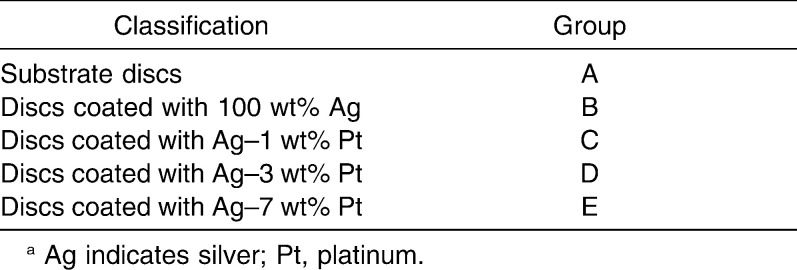
Stainless steel, which is widely used in orthodontics, was cut into 15 mm–diameter, 2 mm–thick samples. All specimen surfaces were polished by barrel polishing and cleaned sonically with alcohol and/or acetone for 15 minutes. Later, one group of cleaned specimens was coated with Ag and the other group with Ag-Pt (Table 1).
Table 1.
Groups of Samplesa
The coating for each group was manufactured using a hybrid coater system (A-Tech Co, Incheon, Korea). After PVD, the specimens were physically cleaned in argon plasma (20 mTorr, 300 W) at a substrate voltage of 100 V for 20 minutes. During this process the specimens were heated to the start temperature of approximately 300°C.
Surface Morphologies and Atomic Composition
The surface morphology of the specimens was examined by field-emission scanning electron microscopy (FE-SEM, Philips Electronics, Eindhoven, The Netherlands) without surface sputtering. The atomic composition was monitored by energy-dispersive x-ray spectroscopy (EDX, Thermo Fisher Scientific Inc, Waltham, Mass)
Surface Hardness and Potentiodynamic Polarization Test
The surface hardness of the specimens was determined using a microhardness tester (Shimadzu Co, Kyoto, Japan) equipped with a Vickers diamond penetrator. Testing was carried out using a 50 g load and a 10-second contact.
To investigate the level of corrosion on the specimen surfaces, an electrochemical potentiodynamic polarization study was carried out in an electrolyte solution (0.9% NaCl, 0.5% KCl, 0.2% CaCl2) at 35°C using potentiostat (Ametek Inc, Paoli, Pa). The salt concentration in the electrolyte solution corresponded to that of body fluids and was de-aerated using high-purity Argon gas for 30 minutes before starting the experiment. Deaeration was continued at a uniform rate during the experiment. A high-purity Pt counter electrode and AgCl reference electrode were used.
Measurement of Ag Ion Released
The release of Ag ions from the coatings was measured with inductively coupled plasma atomic emission spectrometer (ICP-AES System, Perkin Elmer, Rodgau-Jugesheim, Germany). The specimens were immersed in distilled water at 4°C with gentle shaking. The solution was replaced and analyzed after 1, 2, and 8 days by ICP-AES.
Antibacterial Effect
The antimicrobial properties of the Ag-coated specimens or the Ag released from the coated specimens were examined by exposing them to gram-positive Streptococcus mutans (ATCC 25175, designated as Sm), an aerobic microbe involved in tooth decay, and Aggregatibacter actinomycetemcomitans (ATCC 33384, designated as Aa), an anaerobic microbe related to acute periodontitis.
The Sm was grown in brain heart infusion broth (BHI, Difco Laboratories, Livonia, Mich), and Aa was grown in tryptic soy broth (TSB, Difco Laboratories) containing 10% fetal bovine serum (FBS, Gibco Laboratories, Gaithersburg, Md). One hundred microliters of the Sm and Aa suspensions in logarithmic growth phase were seeded (approximately 1 × 10⁸ CFU/mL) to 5 mL of BHI broth for Sm and TSB for Aa containing sterilized specimens or Ag released from the coated specimens. After 16 hours of incubation, the culture fluids were used to measure the absorbance at A630 nm using a spectrophotometer (Bio-Tek Instruments, Winooski, Vt).
Cell Cytotoxicity
To test the effect of Ag ions released from specimens on human gingival fibroblast (HGF) growth, the conditioned media was prepared, as follows: Dulbecco's modified Eagle medium (Gibco Laboratories) and released Ag ion solution with a 1∶1 mixing ratio. The conditioned media contained 10% FBS (Gibco Laboratories), penicillin (100 units/mL), and streptomycin (100 mg/mL). The cells were incubated at 37°C with 5% CO2 and 95% relative humidity. The effects of Ag ions or Pt ions released from the specimens on HGF growth and survival were assessed by a XTT assay using an EZ-Cytox Cell Viability Assay Kit (Daeil Lab Co, Seoul, Korea) according to the manufacturer's instructions. Briefly, 40 µL of EZ-Cytox reagent was added to the HGF culture dish. By the action of mitochondrial dehydrogenases, XTT is metabolized to form a formazan dye that can be spectrophotometrically determined by measuring the absorbance at 450 nm using a microplate reader (Bio-Tek Instrument). The amount of formazan salt formed corresponds to the number of viable cells contained in each well. After 2, 4, 12, and 18 hours of incubation, 10 µL of the assay reagent was added. Subsequently, the optical densities were measured at 450 nm and corrected by subtracting the average absorbance from the wells containing a cell-free medium.
Statistical Analysis
All experiments were performed at least in triplicate and the mean ± standard deviation was determined. The statistical significance was assessed by SPSS 12.0 for Windows (SPSS Inc, Chicago, Ill). The data were analyzed by one-way analysis of variance (ANOVA) with Dunnett's test.
According the authors' institution, this study did not require institutional approval by their institutional review board.
RESULTS
Surface Morphologies and Atomic Composition
The surface morphology of the specimens was examined by FE-SEM. The Ag or Pt coatings had a uniform thickness ranging from 1.03 to 2.34 µm, as determined from cross-sectional image (Figure 1). The atomic composition of the surfaces determined by EDS was as follows: group A, substrate disc; group B, Ag–100.0 wt%; group C, Ag–99.0 wt%; group D, Ag–97.1 wt%; group E, Ag–93.1 wt%.
Figure 1.
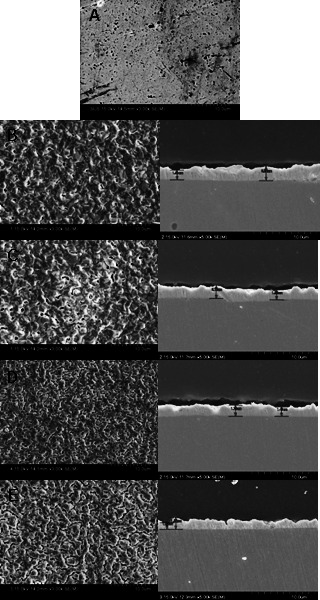
Scanning electron micrographs of the surfaces and cross sections of substrate disc (group A) and discs coated with silver (Ag) or Ag-platinum (Pt) alloys (group B, 100 wt% Ag; group C, Ag–1 wt% Pt; group D, Ag–3 wt% Pt; group E, Ag–7 wt% Pt).
Surface Hardness and Potentiodynamic Polarization Test
A microhardness test was used to investigate the physical properties of the specimens. The hardness of the Ag-Pt–coated specimens (groups C–E) was lower than that of the noncoated specimens (group A) but higher than that of the Ag-coated specimens (group B) (Figure 2A). The level of corrosion on the Ag- or Ag-Pt–coated specimens (groups B–E), as determined by a potentiodynamic test, was lower than that on the noncoated specimens (group A). Moreover, a passive state tendency appeared quickly with increasing Pt composition. This tendency may resist specimen corrosion (Figure 2B).
Figure 2.
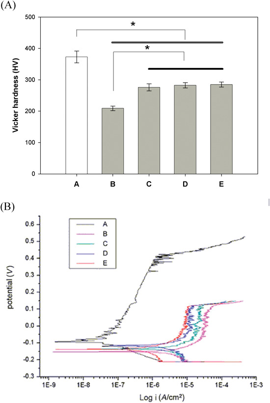
(A) Vickers hardness of the substrate disc surface (group A) and disc surfaces coated with silver (Ag) or Ag-platinum (Pt) alloys (group B, 100 wt% Ag; group C, Ag–1 wt% Pt; group D, Ag–3 wt% Pt; group E, Ag–7 wt% Pt). Bar means no significant difference. * P < .01. (B) Potentiodynamic polarization curve of the substrate disc surface (group A) and disc surfaces coated with Ag or Ag-Pt alloys (group B, 100 wt% Ag; group C, Ag–1 wt% Pt; group D, Ag–3 wt% Pt; group E, Ag–7 wt% Pt).
Measurement of Ag Ion Released
The Ag ions released from the specimens were measured by ICP-AES. After 8 days of incubation, the concentrations of Ag ions were as follows: 1.95 ppm for 100% (group B), 1.94 ppm for 99% (group C), 2.03 ppm for 97% (group D), 2.04 ppm for 93% (group E), and 0 ppm for the 0% Ag-coated specimens (group A) (Figure 3). No iron, nickel, chromium, or Pt ions were detected in any of the specimens.
Figure 3.
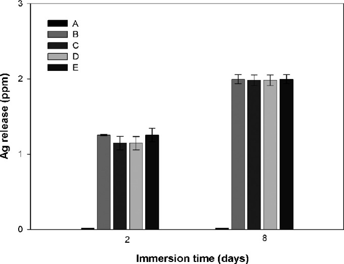
Concentration of released silver (Ag) ions (inductively coupled plasma [ICP] values) of the substrate disc surface (group A) and disc surfaces coated with Ag or Ag-platinum (Pt) alloys (group B, 100 wt% Ag; group C, Ag–1 wt% Pt; group D, Ag–3 wt% Pt; group E, Ag–7 wt% Pt).
Antibacterial Effect
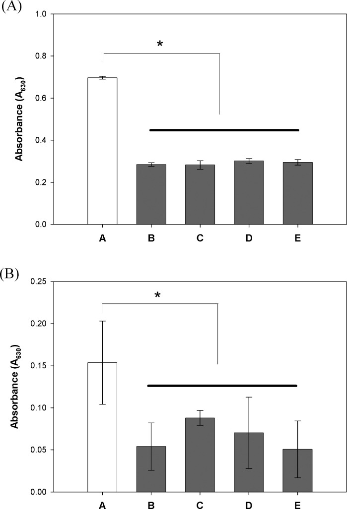
The reaction of Sm and Aa strains with the Ag- or Ag-Pt–coated specimens was compared with that of the noncoated specimens that comprised a control group. The absorbance of the bacterial growth media on the noncoated specimen was set to 100%, and that of test group was calculated accordingly. After an appropriate inoculation period, the level of bacterial growth on the Ag-coated specimens was reduced significantly (approximately 60%). Furthermore, to examine the antibacterial effect of Ag ions released from the specimens, distilled water containing released Ag was added to the media and inoculated with Sm. The growth rate of Sm cultured in the medium containing the released Ag ion was inhibited significantly (Figure 4A,B).
Figure 4.
(A) Survival rate of Streptococcus mutans on substrate disc surface (group A) and disc surfaces coated with silver (Ag) or Ag-platinum (Pt) alloys (group B, 100 wt% Ag; group C, Ag–1 wt% Pt; group D, Ag–3 wt% Pt; group E, Ag–7 wt% Pt) at 8 hours. Bar means no significant difference. * P < .01. (B) Survival rate of Aggregatibacter actinomycetemcomitans on substrate disc surface (group A) and disc surfaces coated with Ag or Ag-Pt alloys (group B, 100 wt% Ag; group C, Ag–1 wt% Pt; group D, Ag–3 wt% Pt; group E, Ag–7 wt% Pt) at 16 hours. The bar means no significant difference. * P < .05.
Cell Cytotoxicity
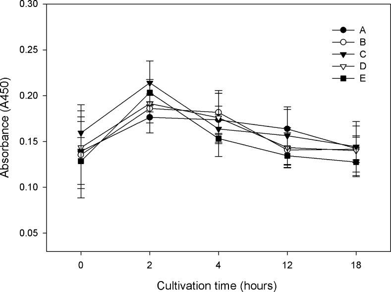
A biocompatibility test was performed using the HGF cell line. HGF cells are an ideal cell source to be used to evaluate their interaction with fabricated material surfaces. The cell viability was assessed using a XTT assay. The absorbance was similar in all groups, indicating that the cells are biocompatible when cultured with the conditioned media (Figure 5). This indicates that the Ag-coated specimens released Ag ions but that the cells in contact with them were apparently unaffected.
Figure 5.
Cell activity of human fibroblast on the substrate disc surface (group A) and disc surfaces coated with silver (Ag) or Ag-platinum (Pt) alloys (group B, 100 wt% Ag; group C, Ag–1 wt% Pt; group D, Ag–3 wt% Pt; group E, Ag–7 wt% Pt).
DISCUSSION
The use of fixed orthodontic appliances can accelerate the rate of plaque accumulation, which can result in enamel decalcification and gingival inflammation without proper maintenance.6,21 Aa or Sm in the plaque are the main bacterial species that cause periodontitis and dental caries, respectively.22,23 The number of oral bacteria can be reduced by brushing or by the additional use of fluoride, chlorhexidine, and antibiotics.24–26 In addition, considerable effort has been made to produce antibacterial dental materials in oral environments.
An antibacterial coating on orthodontic appliances offers a possible strategy for reducing such bacterial damage. In this study, metallic surfaces were coated with Ag-Pt by PVD, and the physical and biological properties of the Ag-Pt coating were evaluated for the purpose of the development of antibacterial orthodontic appliances.
Figure 1 shows SEM images of the Ag- or Ag-Pt–treated samples and pure stainless steel, used as a control (Figure 1A). The thicknesses of the Ag or Ag-Pt coating are highly uniform (Figure 1B–D), as determined from the SEM cross-section image, indicating that the coating method is reliable. Furthermore, the surface topography was analyzed by EDS to evaluate the level of atomic proportion after the Ag-Pt coating was completed. The composition of the coated specimens determined by EDS was the same as the initial proportion of metal sputtered.
The corrosion resistance of the Ag-Pt alloy–coated specimens was higher than that of the Ag-coated one (Figure 2B). This indicates that the hardness and corrosion resistance of Ag coating can be improved by blending with Pt. The antibacterial effect of Ag has been reported in various therapies, including Ag-zeolite in tissue conditioners,27 gargles containing Ag-zeolite,28 and resins containing Ag.29 In this study, the antibacterial effect of Ag was greater in Aa than in Sm (Figure 4). This was related to the different bacterial sensitivity against Ag, and a further study will be needed. Ag ions strongly interact with molecules containing calcium or zinc in living organisms. The antibacterial effect of Ag may cause toxicity in live human cells. Therefore, it is essential to consider the cytotoxicity of Ag ions to human cells. In this study, to investigate the cytotoxicity of Ag, HGF was employed, since HGF is an ideal cell source to be used to evaluate their interaction with fabricated material surfaces. The results showed that the cytotoxicity of Ag in HGF was not significant (Figure 5).
Our data are consistent with current findings30 indicating that the cell compatibility of Ag-coated samples is similar to that of Ag-uncoated samples, although the cell compatibility of Ag is not superior to that of zinc oxide.30 This indicates that the concentration of Ag is suitable for orthodontic materials. Overall, the physical properties of Ag-Pt alloys, such as their strength or corrosion resistance, were superior to those of Ag alone, and the antibacterial effect was maintained. Nevertheless, further studies on the potential discoloration and “drop out” of the coating will be needed.
CONCLUSIONS
Ag-Pt coatings on load-bearing orthodontic bracket surfaces can provide suitable antimicrobial activity and resistance to biofilm formation.
Brackets or archwires containing this Ag-Pt coating can be manufactured for orthodontic patients, especially periodontally compromised or caries-susceptible patients.
Acknowledgments
This study was supported by the Korea Research Foundation Grant funded by the Korean Government to Dr Cho (KRF-313-2008-2-D01310). Dr Bae was supported by a postdoctoral fellowship from the second stage of the Brain Korea 21 program. All bacteria were kindly provided by Dr In-Chol Kang (Department of Oral Microbiology, Chonnam National University, Gwangju, Korea).
REFERENCES
- 1.Fournier A, Payant L, Bouclin R. Adherence of Streptococcus mutans to orthodontic brackets. Am J Orthod Dentofacial Orthop. 1998;114:414–417. doi: 10.1016/s0889-5406(98)70186-6. [DOI] [PubMed] [Google Scholar]
- 2.Brusca M. I, Chara O, Sterin-Borda L, Rosa A. C. Influence of different orthodontic brackets on adherence of microorganisms in vitro. Angle Orthod. 2007;77:331–336. doi: 10.2319/0003-3219(2007)077[0331:IODOBO]2.0.CO;2. [DOI] [PubMed] [Google Scholar]
- 3.De Siate A, Milano V, Laforgia A. A bacteriological study of supragingival bacterial plaque in subjects undergoing orthodontic therapy. Minerva Stomatol. 1991;40:101–105. [PubMed] [Google Scholar]
- 4.Gwinnett A. J, Ceen R. F. Plaque distribution on bonded brackets: a scanning microscope study. Am J Orthod. 1979;75:667–677. doi: 10.1016/0002-9416(79)90098-8. [DOI] [PubMed] [Google Scholar]
- 5.Steinberg D, Eyal S. Initial biofilm formation of Streptococcus sobrinus on various orthodontics appliances. J Oral Rehabil. 2004;31:1041–1045. doi: 10.1111/j.1365-2842.2004.01350.x. [DOI] [PubMed] [Google Scholar]
- 6.Mizrahi E. Enamel demineralization following orthodontic treatment. Am J Orthod. 1982;82:62–67. doi: 10.1016/0002-9416(82)90548-6. [DOI] [PubMed] [Google Scholar]
- 7.Artun J, Brobakken B. O. Prevalence of carious white spots after orthodontic treatment with multibonded appliances. Eur J Orthod. 1986;8:229–234. doi: 10.1093/ejo/8.4.229. [DOI] [PubMed] [Google Scholar]
- 8.van Gastel J, Quirynen M, Teughels W, Pauwels M, Coucke W, Carels C. Microbial adhesion on different bracket types in vitro. Angle Orthod. 2009;79:915–921. doi: 10.2319/092908-507.1. [DOI] [PubMed] [Google Scholar]
- 9.van Gastel J, Quirynen M, Teughels W, Coucke W, Carels C. Influence of bracket design on microbial and periodontal parameters in vivo. J Clin Periodontol. 2007;34:423–431. doi: 10.1111/j.1600-051X.2007.01070.x. [DOI] [PubMed] [Google Scholar]
- 10.Ahn S. J, Lim B. S, Lee S. J. Prevalence of cariogenic streptococci on incisor brackets detected by polymerase chain reaction. Am J Orthod Dentofacial Orthop. 2007;131:736–741. doi: 10.1016/j.ajodo.2005.06.036. [DOI] [PubMed] [Google Scholar]
- 11.Hägg U, Kaveewatcharanont P, Samaranayake Y. H, Samaranayake L. P. The effect of fixed orthodontic appliances on the oral carriage of Candida species and Enterobacteriaceae. Eur J Orthod. 2004;26:623–629. doi: 10.1093/ejo/26.6.623. [DOI] [PubMed] [Google Scholar]
- 12.Tasman A. J, Wallner F, Neumeier R. Antibiotic impregnation of cartilage implants: diffusion kinetics of fluoroquinolones. Laryngorhinootologie. 2000;79:30–33. doi: 10.1055/s-2000-8778. [DOI] [PubMed] [Google Scholar]
- 13.Blaker J. J, Nazhat S. N, Boccaccini A. R. Development and characterisation of silver-doped bioactive glass-coated sutures for tissue engineering and wound healing application. Biomaterials. 2004;25:1319–1329. doi: 10.1016/j.biomaterials.2003.08.007. [DOI] [PubMed] [Google Scholar]
- 14.Berger T. J, Spadaro J. A, Chapin S. E, Becker R. O. Electrically generated silver ions: quantitative effects on bacterial and mammalian cells. Antimicrob Agents Chemother. 1976;9:357–358. doi: 10.1128/aac.9.2.357. [DOI] [PMC free article] [PubMed] [Google Scholar]
- 15.Spadaro J. A, Chase S. E, Webster D. A. Bacterial inhibition by electrical activation of percutaneous silver implants. J Biomed Mater Res. 1986;20:565–577. doi: 10.1002/jbm.820200504. [DOI] [PubMed] [Google Scholar]
- 16.Silver S. Bacterial silver resistance: molecular biology and uses and misuses of silver compounds. FEMS Microbiol Rev. 2003;27:341–353. doi: 10.1016/S0168-6445(03)00047-0. [DOI] [PubMed] [Google Scholar]
- 17.Böswald M, Lugauer S, Regenfus A, et al. Reduced rates of catheter-associated infection by use of a new silver-impregnated central venous catheter. Infection. 1999;(suppl 1):S56–S60. doi: 10.1007/BF02561621. [DOI] [PubMed] [Google Scholar]
- 18.Davenport K, Keeley F. X. Evidence for the use of silver-alloy-coated urethral catheters. J Hosp Infect. 2005;60:298–303. doi: 10.1016/j.jhin.2005.01.026. [DOI] [PubMed] [Google Scholar]
- 19.Viennot S, Dalard F, Lissac M, Grosgogeat B. Corrosion resistance of cobalt-chromium and palladium-silver alloys used in fixed prosthetic restorations. Eur J Oral Sci. 2005;113:90–95. doi: 10.1111/j.1600-0722.2005.00190.x. [DOI] [PubMed] [Google Scholar]
- 20.Ewald A, Glückermann S. K, Thull R, Gbureck U. Antimicrobial titanium/silver PVD coatings on titanium. Biomed Eng Online. 2006;5:22. doi: 10.1186/1475-925X-5-22. [DOI] [PMC free article] [PubMed] [Google Scholar]
- 21.Zachrisson B. U. Cause and prevention of injuries to teeth and supporting structures during orthodontic treatment. Am J Orthod. 1976;69:285–300. doi: 10.1016/0002-9416(76)90077-4. [DOI] [PubMed] [Google Scholar]
- 22.Paolantonio M, Festa F, di Placido G, D'Attilio M, Catamo G, Piccolomini R. Site-specific subgingival colonization by Actinobacillus actinomycetemcomitans in orthodontic patients. Am J Orthod Dentofacial Orthop. 1999;115:423–428. doi: 10.1016/s0889-5406(99)70263-5. [DOI] [PubMed] [Google Scholar]
- 23.Selwitz R. H, Ismail A. I, Pitts N. B. Dental caries. Lancet. 2007;369:51–59. doi: 10.1016/S0140-6736(07)60031-2. [DOI] [PubMed] [Google Scholar]
- 24.Burch J. G, Lanese R, Ngan P. A two-month study of the effects of oral irrigation and automatic toothbrush use in an adult orthodontic population with fixed appliances. Am J Orthod Dentofacial Orthop. 1994;106:121–126. doi: 10.1016/S0889-5406(94)70028-1. [DOI] [PubMed] [Google Scholar]
- 25.Sandham H. J, Nadeau L, Phillips H. I. The effect of chlorhexidine varnish treatment on salivary mutans streptococcal levels in child orthodontic patients. J Dent Res. 1992;71:32–35. doi: 10.1177/00220345920710010501. [DOI] [PubMed] [Google Scholar]
- 26.Slots J, Rams T. E. Antibiotics in periodontal therapy: advantages and disadvantages. J Clin Periodontol. 1990;17:479–493. doi: 10.1111/j.1365-2710.1992.tb01220.x. [DOI] [PubMed] [Google Scholar]
- 27.Abe Y, Ueshige M, Takeuchi M, Ishii M, Akagawa Y. Cytotoxicity of antimicrobial tissue conditioners containing silver-zeolite. Int J Prosthodont. 2003;16:141–144. [PubMed] [Google Scholar]
- 28.Morishita M, Miyagi M, Yamasaki Y, Tsuruda K, Kawahara K, Iwamoto Y. Pilot study on the effect of a mouthrinse containing silver zeolite on plaque formation. J Clin Dent. 1998;9:94–96. [PubMed] [Google Scholar]
- 29.Yoshida K, Tanagawa M, Matsumoto S, Yamada T, Atsuta M. Antibacterial activity of resin composites with silver-containing materials. Eur J Oral Sci. 1999;107:290–296. doi: 10.1046/j.0909-8836.1999.eos107409.x. [DOI] [PubMed] [Google Scholar]
- 30.Chang Y. Y, Lai C. H, Hsu J. T, Tang C. H, Liao W. C, Huang H. L. Antibacterial properties and human gingival fibroblast cell compatibility of TiO2/Ag compound coatings and ZnO films on titanium-based material. Clin Oral Invest. 2011 Jan 14 doi: 10.1007/s00784-010-0504-9. [DOI] [PubMed] [Google Scholar]


