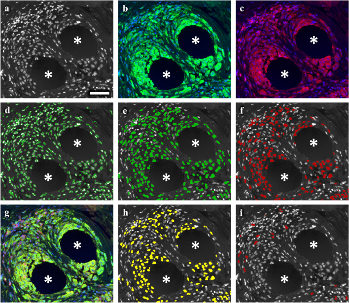
Fig. 2.
Nuclear detection and backward gating of immune cells participating in the foreign body reaction. Immunofluorescence labeling of pan-macrophage marker CD68 (FITC), CD4 (Cy5), and nuclei (DAPI). a Grayscale nuclei shade. b CD68 (green) and nuclei (blue). c CD4 (red) and nuclei (blue). d Backward gating of detected nuclei (green contours). e Backward gating of CD68+ cells (green). f Backward gating of CD4+ cells (red). g Superposition of CD68 (green), CD4 (red) and nuclei (blue). h Backward gating of “hybrid” CD4+CD68+ cells (yellow). i Backward gating of “conventional” CD4+CD68− cells (red). Backward gating is always on the nuclei shade. Locations of mesh fibers are marked with asterisks, scale bar = 50 µm. Images of explant #3 (color figure online)

