

Abstract
Background
Resuscitative endovascular balloon occlusion of the aorta (REBOA) is a helpful adjunct in the control of non-compressible truncal hemorrhage. Concerns regarding ischemia time limits its applicability in transfer. We describe the first reported case of civilian transfer via aeromedical transport to a higher level of care with a zone 3 REBOA catheter deployed.
Case report
We present the case of a patient in hemorrhagic shock with a complex pelvic fracture exceeding the capability of a rural level-two trauma center requiring the use of REBOA catheter to permit aeromedical transport to a level-one trauma center for definitive embolization.
Conclusion
Deployment of REBOA catheter to facilitate aeromedical transport to an appropriate level of care may be considered if travel times can be kept brief and there is a process and training in place to empower flight medics to consider transporting with a REBOA deployed.
Abbreviations: REBOA, Resuscitative Endovascular Balloon Occlusion of the Aorta
Keywords: Aortic occlusion, REBOA, Resuscitative endovascular balloon occlusion of the aorta, Aeromedical transport, Helicopter, Pelvic fracture
Background
Non-compressible truncal hemorrhage (NCTH) after blunt injury can be a source of significant mortality. Controlling NCTH in environments with limited resources can be challenging. Resuscitative endovascular balloon occlusion of the aorta (REBOA) is a temporizing measure that can permit the trauma team to mobilize resources to definitively address the source of hemorrhage. Ground transport to a higher level of care with an inflated REBOA has been described [1], [2]. No descriptions exist of aero-medical transport of a patient with an inflated REBOA catheter outside of swine models [3]. We report the case of a patient transferred via helicopter to a higher level of care with an occlusive REBOA catheter in Zone 3 of the aorta for exsanguinating pelvic hemorrhage.
Case report
A young male helmeted motorcyclist was severely injured following a motorcycle crash. He was brought to a rural level 2 trauma center in shock with declining mental status (see Table 1 for time series). The patient was promptly intubated and central venous access was obtained. FAST exam was negative; however he sustained a number of extremity injuries and a massive perineal laceration contaminated with soil and extending from the perineal body to the lower back (Fig. 1) with significant hemorrhage controlled by packing. Radiographs of the pelvis showed severe pelvic fractures (Fig. 2, Tile C1/Young-Burgess LC III) including comminuted superior and inferior left pubic rami and left sacro-iliac joint disruption with a likely acetabular disruption with concern that this was the etiology of his shock. A pelvic binder was placed.
Table 1.
Time series of events leading to hemorrhage control.
| Time | Events |
|---|---|
| 2156 | Arrival to Level 2 Trauma Center: HR 160, BP 90/42 |
| 2200 | Pelvic wound packed for hemostasis |
| 2203 | Intubated |
| 2206 | Binder placed |
| 2209 | Left Internal Jugular Central Venous sheath placed HR 152, BP 88/53 |
| 2215 | Transport to CT scan |
| 2245 | Completion of CTA – Activation of interventional radiology |
| 2250 | Return to ED HR 138, BP 111/68 |
| 2300 | Placement of right femoral arterial sheath for possible REBOA |
| 2315 | Decision to transfer patient |
| 2335 | Placement and inflation of REBOA catheter |
| 2345 | Slow deflation for 2 min to permit distal flow |
| 2355 | Slow deflation for 2 min to permit distal flow |
| 0012 | Departure from transferring facility |
| 0131 | Arrival in trauma bay at receiving Level 1 Trauma Center HR 143, BP 105/62 |
| 0212 | Removal of REBOA and start of angiography |
Fig. 1.
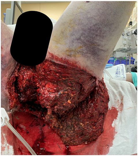
Earliest image of perineal wound in this patient after partial debridement of devitalized tissue demonstrating significant contamination of soft tissue and depth of injury.
Fig. 2.
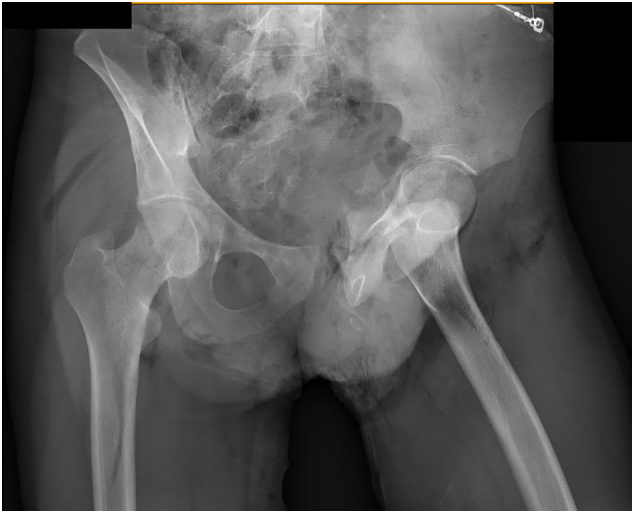
Initial pelvic radiograph demonstrating Tile C1/Young-Burgess LC III fracture pattern.
Massive transfusion was initiated with transient response, during this time, we performed contrasted CT to identify if embolization was indicated. A retroperitoneal hematoma was seen, with arterial extravasation from the left internal iliac artery and the left L4 lumbar artery. Interventional radiology was activated for emergent embolization. In the course of awaiting the angiography team he again became hypotensive and continued to require massive transfusion. He had received 15 units of packed red blood cells (PRBC), 10 units of fresh-frozen plasma, and 3 units of platelets which had essentially depleted the blood bank of its available product. Preperitoneal packing was considered but deferred due to a lack of additional blood product that would be needed during and after undertaking packing as well as difficulty reaching the lumbar artery. It became clear that angiography would not be able to be initiated before exhausting our blood supply and thus, we made the decision to transfer the patient via air to the nearest level one trauma center.
We deployed an ER-REBOA (Prytime Medical, Boerne, TX) catheter [4] into zone 3 to control hemorrhage and permit transfer. We inserted the catheter to 26 cm and confirmed with radiograph. Aortic pressures normalized following inflation. Transport was delayed due to absence of a clear protocol for REBOA management in flight and the need for clearance from the flight medical director. Intermittent deflations of the balloon were performed while on the ground to permit partial perfusion.
The patient was flown via helicopter with the balloon inflated. His heart rate ranged from 133 to 155 beats/min. His blood pressures ranged from 87 to 112/45 to 71 mm Hg. His mean arterial pressure ranged from 59 to 88. He received three units of packed red blood cells enroute. Total flight time was 45 min from take-off to landing. The maximum altitude obtained was 1500 ft. The flight nurses made no adjustments to the REBOA catheter or balloon during flight.
On arrival to the level 1 facility, the patient was transferred emergently to the angiography suite. The REBOA catheter was removed 157 min after insertion. There was a profound drop in mean arterial pressure to 25 mm Hg, this resolved over 3–5 min with bicarbonate and blood transfusion. Bilateral internal iliac embolization was performed with Gelfoam and the bleeding L4 lumbar vessel was coil embolized. The femoral sheath was heparinized and left in place given concern for coagulopathy. Several hours later, neurovascular checks revealed the leg to be cold and pulseless. The patient underwent emergent embolectomy of right external iliac down to the superficial femoral artery and fasciotomy of the calf with return of flow.
His postoperative course was extremely complicated. Notably, an invasive fungal infection related to the soil-contaminated perineal laceration required numerous wide debridements of the trunk, perineum, and legs followed by eventual grafting.
Discussion
Management options of hemorrhage from high-energy pelvic injuries include pelvic binders, pre-peritoneal packing, arterial embolization, and zone 3 REBOA placement. Injuries that exceed a facility's capabilities require rapid transfer to a higher level of care. The authors encountered a situation where the rate of bleeding exceeded the blood bank's capacity during activation of interventional radiology. REBOA provided temporary hemorrhage control and an opportunity to get a patient in hemorrhagic shock to a facility that had adequate blood supply.
Placement of REBOA in zone 3 for pelvic bleeding can be an effective approach to obtain temporary control if the operator is comfortable with the modality [5], [6], [7]. Porcine studies suggest that the ischemic burden and reperfusion related instability is lessened by occlusion of Zone 3 of the aorta when compared to Zone 1 [8]. Ideal duration of occlusion has been proposed to be 60 min for zone 3, however minimal data exists on outcomes if times exceed these intervals. However, Zone 3 aortic occlusion appears to have minimal effect on visceral organ blood flow suggesting that longer periods of occlusion may be tolerated [9].
While prehospital REBOA has been described, we were unable to identify any report of REBOA use in aeromedical transport to permit transfer to a higher level of care [1], [2]. Singer et al. studied application of REBOA in flight in a porcine model. Shock was induced in pigs at ground level, REBOA was deployed and the animals were then placed in a pressure chamber simulating flight at 8000 ft which corresponded to average altitude for evacuation aircraft. Their findings suggested that altitudes experienced in flight did not affect REBOA performance.
The duration of aortic occlusion in our patient was significantly longer than the recommended 60 min – this was multifactorial from delays at the level 2 trauma center related to the time to mobilize angiography staff, readying the patient for transport, and obtaining clearance to fly with an inflated REBOA. A case series exists from the United Kingdom reporting balloon occlusion for time periods exceeding the recommended window, however, they also reported a very high frequency of arterial thrombosis [1]. Curiously, occlusion of the aorta does not appear to lead to complete extinction of perfusion to the lower extremities as shown by Wasicek et al. in CT scans obtained after balloon inflation [10].
We present the first case to be described where a REBOA was deployed allowing a patient to be flown in transfer to a higher level of care. While this pathway should be exceedingly rare, we are hopeful that reporting on this may reduce some of the institutional resistance present when a similar situation presents. Furthermore, as new management practices become standardized, such as intermittent deflation, these practices should be applied in transit to reduce tissue ischemia burden. This challenging case exceeded the capabilities of the transferring facility and once recognized, a REBOA was able to be deployed to good effect permitting flight transfer for definitive care.
References
- 1.Lendrum R., Perkins Z., Chana M., et al. Pre-hospital resuscitative endovascular balloon occlusion of the aorta (REBOA) for exsanguinating pelvic haemorrhage. Resuscitation. 2019;135:6–13. doi: 10.1016/j.resuscitation.2018.12.018. [DOI] [PubMed] [Google Scholar]
- 2.Northern D.M., Manley J.D., Lyon R., et al. Recent advances in austere combat surgery: use of aortic balloon occlusion as well as blood challenges by special operations medical forces in recent combat operations. J. Trauma Acute Care Surg. 2018;85:S98–S103. doi: 10.1097/TA.0000000000001966. [DOI] [PubMed] [Google Scholar]
- 3.Singer K.E., Morris M.C., Blakeman C., et al. Can resuscitative endovascular balloon occlusion of the aorta fly? Assessing aortic balloon performance for aeromedical evacuation. J. Surg. Res. 2020;254:390–397. doi: 10.1016/j.jss.2020.05.021. [DOI] [PubMed] [Google Scholar]
- 4.Bogert J.N., Davis K.M., Kopelman T.R., et al. Resuscitative endovascular balloon occlusion of the aorta with a low profile, wire free device: a game changer? Trauma Case Rep. 2017;7:11–14. doi: 10.1016/j.tcr.2017.01.006. [DOI] [PMC free article] [PubMed] [Google Scholar]
- 5.Harfouche M., Inaba K., Cannon J., et al. Patterns and outcomes of zone 3 REBOA use in the management of severe pelvic fractures: results from the AAST aortic occlusion for resuscitation in trauma and acute care surgery database. J. Trauma Acute Care Surg. 2020;90(4):659–665. doi: 10.1097/TA.0000000000003053. [DOI] [PubMed] [Google Scholar]
- 6.Asmar S., Bible L., Chehab M., et al. Resuscitative endovascular balloon occlusion of the aorta vs pre-peritoneal packing in patients with pelvic fracture. J. Am. Coll. Surg. 2021;232:17–26.e2. doi: 10.1016/j.jamcollsurg.2020.08.763. [DOI] [PubMed] [Google Scholar]
- 7.Bukur M., Gorman E., DiMaggio C., et al. Temporal changes in REBOA utilization practices are associated with increased survival: an analysis of the AORTA registry. Shock Augusta Ga. 2021;55:24–32. doi: 10.1097/SHK.0000000000001586. [DOI] [PubMed] [Google Scholar]
- 8.Tibbits E.M., Hoareau G.L., Simon M.A., et al. Location is everything: the hemodynamic effects of REBOA in zone 1 versus zone 3 of the aorta. J. Trauma Acute Care Surg. 2018;85:101–107. doi: 10.1097/TA.0000000000001858. [DOI] [PubMed] [Google Scholar]
- 9.Halvachizadeh S., Mica L., Kalbas Y., Lipski M., et al. Zone-dependent acute circulatory changes in abdominal organs and extremities after resuscitative balloon occlusion of the aorta (REBOA): an experimental model. Eur. J. Med. Res. 2021;26:10. doi: 10.1186/s40001-021-00485-y. [DOI] [PMC free article] [PubMed] [Google Scholar]
- 10.Wasicek P.J., Shanmuganathan K., Teeter W.A., et al. Assessment of blood flow patterns distal to aortic occlusion using CT in patients with resuscitative endovascular balloon occlusion of the aorta. J. Am. Coll. Surg. 2018;226:294–308. doi: 10.1016/j.jamcollsurg.2017.12.005. [DOI] [PubMed] [Google Scholar]