

Abstract
BACKGROUND:
Laser etching addresses the disadvantages of conventional acid etching technique, such as enamel decalcification and formation of white spot lesions. The aim of the present study was to evaluate and compare the shear bond strength (SBS), adhesive remnant index (ARI), and the surface characteristics of the samples treated with conventional acid etching and Er, Cr: YSGG laser etching with variable output power and time durations.
METHODOLOGY:
The study sample included 78 extracted teeth divided into six groups of 13 teeth each, and 3 samples from each group were utilized for analyzing etch patterns, and the remaining 10 teeth from each group were used for evaluating the shear bond strength. In Group I phosphoric acid etching was done, whereas in Group II– VI Laser etching 1.5 W/10 s, 1.5 W/15 s, 3 W/5 s, 3 W/10 s, 3 W/15 s. Statistical analysis for shear bond strength testing was performed using one-way ANOVA followed by Post HOC tests.
RESULTS:
The mean shear bond strength of Group I was 7.16 Mpa and Group III of 5.43 Mpa. Group II, IV, V, and VI had mean shear bond strength of 4.93 Mpa, 3.88 Mpa, 4.05 Mpa, and 4.88 Mpa, respectively. The ARI scores Group I had a significant number of samples with scores 2 other groups showed increased Score 0. The etch pattern of groups I, II, III showed the combined dissolution of both prism cores, and peripheries were seen. In group IV, the etching pattern was irregular with the pitted type of surface. In groups V and VI, relatively flat and smooth enamel surface was seen.
CONCLUSION:
The bond strength attained by laser etching (1.5 W/10 s and 1.5 W/15 s) was comparable to that obtained by the acid etching technique.
Keywords: Bond strength, Er, Cr:YSGG, laser etching, time
Introduction
Orthodontic treatment is contingent on a force system to be applied to the teeth via brackets bonded to enamel. The basis of bonding materials is bisphenyl-A-glycidyldimethacrylate (Bis-GMA) resin. However, acid pretreatment of enamel using 33% orthophosphoric acid is crucial for improved bond strength.[1] Despite the fact that acid-etching technique is a useful procedure in orthodontics, the bonding procedure needs to be improved to maintain clinically useful bond strengths at the same time minimizing enamel loss. Orthodontics like any other field in dentistry is dynamic, where the hunt for better materials, systems, and innovation is everlasting. Recently, alternative methods for preparing the tooth surfaces, such as laser irradiation, have been developed.[2] Laser etching has been proposed to inhibit caries, which could be of great importance in orthodontics because of the fact that the orthodontic patients are at high risk of developing white spot lesions or caries.[3] Laser etching is accurate, saves time, and avoids procedural errors. Hence, laser irradiation might serve as a suitable technique to etch enamel for orthodontic bonding.[4]
In dentistry, various types of soft tissue and hard tissue lasers have been used. Among these, the family of erbium lasers is the most propitious because their wavelength coincides with the main absorption peak of water and hydroxyapatite. Thus, Er:YAG and Er, Cr:YSGG lasers interact well with all biological tissues, including enamel and dentin surface. Er, Cr:YSGG laser etching produces micro-cracks that are appropriate for resin penetration. Berk N et al.[5] suggested that etching with Er, Cr:YSGG laser is painless and does not produce either heat or vibration. As laser etching eliminates water spraying and air drying, chairside time can also be saved significantly. From a clinical perspective, saving chair time improves adhesion because it significantly reduces the risk of salivary contamination. Also, easy handling of the laser apparatus makes it attractive for clinical use.
Studies show that the use of laser etching to bond orthodontic brackets yields excellent bond strength in a significantly lesser time when compared to the conventional technique.[6] There are studies determining the efficacy of irradiation of enamel with Erbium, chromium: yttrium–scandium–gallium–garnet laser of different output powers (0.5 W, 0.75 W, 1 W, 1.5 W, 1.75 W, and 2 W) with a constant duration time of 15 s for orthodontic bonding. The results show that comparable mean shear bond strength and enamel surface etching can be obtained with Er, Cr:YSGG laser (operated at 1.5 W, 1.75 W, and 2 W for 15 s) as with acid etching.[7]
Laser etching addresses all the disadvantages of conventional acid etching techniques, like enamel decalcification and the formation of white spot lesions. The rationale of this study is to appraise whether laser etching of different output powers and with different time duration can be used as a reliable alternative to conventional acid etching. Hence, the aim of the present study was to evaluate and compare the shear bond strength (SBS), adhesive remnant index (ARI), and the surface characteristics of the samples treated with conventional acid etching and Er, Cr:YSGG laser etching with variable output power and time durations.
Materials and Methods
The study was reviewed and approved by the Institutional Review Board (approval number 200/IHEC/1-19) ethical approval obtained 28/1/20219. The study sample included 78 extracted maxillary first premolars. The samples were collected from the patients who had undergone therapeutic extraction for orthodontic correction of teeth satisfying the following inclusion criteria.
Anatomically and morphologically well-defined maxillary premolar teeth
Intact buccal enamel
Teeth without decalcification, caries, or restoration,
Teeth that were not previously bonded
No cracks caused by extraction forceps.
A total of 78 maxillary premolar teeth that were extracted for orthodontic treatment were stored in 0.1% thymol solution at room temperature. The 78 premolars were divided into six groups of 13 teeth each, and 3 samples from each group were utilized for analyzing etch patterns, and the remaining 10 teeth from each group was used for evaluating the shear bond strength.
Group I - Phosphoric acid etching
Group II - Laser etching (1.5 W/10 s)
Group III - Laser etching (1.5 W/15 s
Group IV - Laser etching (3 W/5 s)
Group V - Laser etching (3 W/10 s)
Group VI - Laser etching (3 W/15 s).
Acrylic resin with a 5 mm × 4 mm hole was placed on the tooth surface to standardize the surface area of the etched enamel. Teeth planned for shear bond strength testing were mounted vertically in self-cure acrylic resin block, so that only the crown was exposed.
In Group I, the buccal enamel surface was etched with 37% ortho phosphoric acid for 15 s and rinsed with water and gentle air spray for 15 s, and dried for another 15 s.
The teeth in the remaining groups were etched with the Er, Cr:YSGG laser, Waterlase MD, BioLase Technology Inc., Irvine, CA, USA. In Group II, enamel was irradiated with Er, Cr:YSGG laser with 1.5 W power output for 10 s. In Group III, enamel was irradiated with Er, Cr:YSGG laser with 1.5 W power output for 15 s. In Group IV, enamel was irradiated with Er, Cr:YSGG laser with 3 W power output for 5 s. In Group V, enamel irradiated with Er, Cr:YSGG laser with 3 W power output for 10 s. In Group VI, enamel was irradiated with Er, Cr:YSGG laser with 3 W power output for 15 s.
After etching, a thin, uniform coat of sealant was applied to the etched surfaces. After the application of the bonding material – Transbond XT, 3 M Unitek, stainless steel premolar brackets – 0.022 inch MBT 3 M Gemini were placed on the tooth surface, adjusted to its final position, and pressed firmly. The excessive adhesive was removed from the borders of the bracket base. Each side of the tooth (mesial, distal, occlusal, and gingival) was light-cured using an LED light-curing unit – Bluephase N - Ivoclar Vivadent curing light for 10 s. Hence, each tooth as a whole was cured for 40 s. Specimens after bonding were stored in deionized water for 24 h before debonding.
The universal testing machine -UNITEK 94100, was used to test the SBS of each tooth. The samples were mounted in the lower arm of a machine in such a way that the applied force was parallel to the tooth surface (occluso-gingivally) with a crosshead speed of 1 mm/min. All the 60 samples were debonded. The force required to debond each bracket was registered in Newtons and converted into megapascals as a ratio of Newton to the surface area of the bracket base (MPa = N/mm2).
After debonding the adhesive remnant, index scoring was done using a magnification glass according to Artun and Bergland.[8]
Three samples from each group which was etched but left unbonded were then sputtered with 10–12 nm thick layer of gold. The specimens were then examined with a scanning electron microscope – JEOL, JSM-5610 LV with INCA EDS, Tokyo, Japan operated at 30 Kv to check the etching pattern. The SEM samples thus obtained were evaluated as per the criteria given by Silverstone et al.[9]
Result
Statistical analysis for shear bond strength testing was performed using one-way ANOVA followed by Post HOC tests. Statistical software IBM SPSS (IBM SPSS Statistics for Windows, Version 20.0. Armonk, NY: IBM Corp) was used for the evaluation of all statistical analysis. The statistical significance level was established at P value ≤ 0.05.
One-way ANOVA showed that the mean shear bond strength of the conventional acid etch group (Group I) was found to be 7.16 Mpa and Group III (1.5 W/15 s) had mean bond strength of 5.43 Mpa. The difference between these two groups (Group I and Group III) was not statistically significant. Group II, IV, V, and VI had mean shear bond strength values of 4.93 Mpa, 3.88 Mpa, 4.05 Mpa, and 4.88 Mpa, respectively [Figure 1]. Turkey's post hoc test showed that in pairwise comparison the bond strength of Group I was statistically higher than that of Group V and VI, and there was no statistical difference between any other groups [Table 1].
Figure 1.
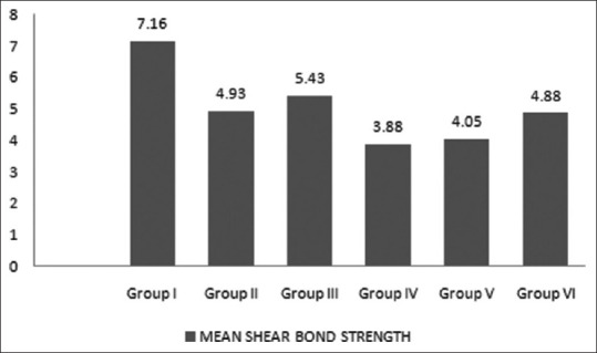
Mean shear bond Strength of the samples
Table 1.
Descriptive statistics and One-way ANOVA for comparison of mean bond strength between the groups
| Groups | Mean | SD | 95% CI for Mean | F | P | |
|---|---|---|---|---|---|---|
|
| ||||||
| Lower | Upper | |||||
| Group I | 7.16*a | 2.77 | 5.17 | 9.14 | 3.2 | 0.012 |
| Group II | 4.93 | 3.31 | 2.56 | 7.31 | ||
| Group III | 5.43 | 1.10 | 4.64 | 6.22 | ||
| Group IV | 3.88 | 1.30 | 2.94 | 4.82 | ||
| Group V | 4.05* | 1.11 | 3.25 | 4.84 | ||
| Group VI | 4.88a | 1.65 | 3.70 | 6.06 | ||
*a=significant difference in pairwise comparison (Tukey’s post hoc test)
The ARI (ADHESIVE REMNANT INDEX) scores were determined for each study sample after debonding [Figure 2]. Chi-square test was done to test the significant difference between the adhesive remnant index score frequency among the groups. Group I has a significant number of samples with a score of 2 when compared to all other groups. Others do not show statistically significant result [Table 2].
Figure 2.

Adhesive remnant score of the samples
Table 2.
Chi-square crosstabs for comparison of Adhesive remnant Index score frequency among the groups
| Groups | Adhesive Remnant Index Score | Chi square statistic | P (Fisher’s exact test) | |||
|---|---|---|---|---|---|---|
|
| ||||||
| No adhesive left on tooth (score 0) | less than 50% adhesive left (score 1) | More than adhesive 50% left (score 2) | Adhesive with impression of the bracket mesh is left (score 3) | |||
| Group I | 3 | 4 | 3* | 0 | 27.28 | 0.004 |
| Group II | 9 | 1 | 0 | 0 | ||
| Group III | 9 | 1 | 0 | 0 | ||
| Group IV | 10 | 0 | 0 | 0 | ||
| Group V | 6 | 4 | 0 | 0 | ||
| Group VI | 6 | 4 | 0 | 0 | ||
Three samples from each group were evaluated for each pattern. The etch pattern varied between different groups. In group I, combined dissolution of both prism cores and peripheries was seen. In group II and III, the same kind of etching patterns was seen. In group IV, the etching pattern was irregular with a pitted type of surface. In group V and VI, relatively flat and smooth enamel surface was seen [Figures 3 and 4].
Figure 3.

SEM examination of samples at 2500 × magnification
Figure 4.

SEM examination of samples at 5000 × magnification
Discussion
This study was undertaken to use laser to overcome the disadvantages of conventional acid etching like enamel decalcification and formation of white spot lesions prone to caries. The laser used in the present study was Er, Cr: YSGG is a hydrokinetic system. It allows precise hard tissue treatment by virtue of laser energy interaction with water above and at the tissue interface. It operates at a wavelength of 2,780 nm and a pulse duration of 140 μs at a rate of 20 Hz. The average output can be varied from 0 W to 6 W.[10] High irradiation outputs from 2.5–6 W could be used for cutting enamel, whereas relatively lower power outputs would be sufficient for etching enamel.[11] In a study conducted by Özer et al.,[12] Er, Cr: YSGG laser irradiation at 0.75 W and 1.5 W was compared with conventional acid etching and self-etching primer for orthodontic bonding. They found out that the varying output power of laser irradiation produces different etching patterns. The 0.75 W laser irradiation group had lower shear bond strength, whereas1.5 W laser irradiation group had equal bond strengths with phosphoric acid and self-etching primer. Hence, the output power chosen for the study was 1.5 W and 3 W.
Another disadvantage of acid etching is increased chair-time. Time duration of 15 s for etching, 15 s for water spraying, and further 15 s for air drying is required for conventional etching. In laser etching, the step of washing and drying the tooth surface after etching is eliminated. A total of 5–10 min could be saved in laser etching which is significant from a clinical point of view.[13] Hence, in order to save clinical time and improve efficacy, different time duration were tested in the study.
The results of the current study show the mean shear bond strength of the conventional acid etch group (Group I) was found to be 7.16 Mpa with the highest bond strength followed by laser etching at 1.5 W for 15 s, Group III with the bond strength of 5.43 Mpa, laser etching at 1.5 W for 10 s group II having mean bond strength of 4.93 Mpa, 3 W for 15 s Group VI with mean bond strength of 4.88 Mpa, 3 W for 10 s Group V with shear bond strength of 4.05 Mpa and 3 W for 5 s (Group IV) having bond strength of 3.88 Mpa. According to the current study, the bond strength of group I was significantly higher than Group V and Group VI. Statistically, no significant difference was seen between any other groups. So the results suggest that the bond strength obtained with laser etching Group II (1.5 W/10 s) and Group III (1.5 W/15 s) were comparable to that of the bond strength obtained with that of the acid etching group. So laser etching with 1.5 W/15 s and 1.5 W/10 s can be used as an alternative to traditional acid etching. Even though the difference between Group I and Group IV was not statistically significant, because of the wide confidence interval and sampling variability, laser etching with 3 W/5 s cannot be equally efficient as acid etching. The findings obtained from the current study were also in agreement with previous studies which reported that the mean shear bond strength after etching with Er, Cr: YSGG laser operated at 1.5 W for 15 s and 1.75 W for 15 s were similar to that of the acid etching group.[14,15]
Scanning electron microscopic analysis of three representative samples from each group which was etched but left unbondeded shows that type 3 pattern which is considered to be ideal for orthodontic bonding[16] was seen in acid etching group and laser etching for 1.5 W for 10 s and 1.5 W for 15 s (Group I, II, III, respectively). This can be correlated to the lack of significant difference in the bond strength between these groups according to the results of the present study. Type 4 pattern of pitted enamel surface wAs seen in laser etching with 3 W for 5 s (Group IV), whereas Type 5 pattern was seen in laser etching with 3 W for 10 s and 3 W for 15 s (Group V and VI, respectively). This can be correlated to the lower mean shear bond strength of Group V and VI.
The adhesive remnant index scoring was carried out as proposed by Artun and Begland. Score 0 is seen more frequently in the acid etching group when compared to the laser etching groups, whereas, score 1 and 2 were seen more frequently in laser etching groups when compared to the acid etching group. Score 3 was not seen in any of the groups.
The result of the current study indicates that in the laser group, bond failure occurs more commonly at the enamel-adhesive interface. In other words, the bond between the enamel and adhesive was much stronger than that between the adhesive and the bracket base. This property has an advantage as well as a disadvantage. As the amount of adhesive left on the tooth is very less, less chair time is needed to remove the adhesive remnant following debonding. But the chance of enamel fracture upon debonding is relatively higher as the bond failure occurs at the enamel-adhesive interface. So studies to evaluate the amount of enamel fracture occurring with debonding after etching with laser is essential to throw more light on this subject.
In orthodontics, stronger is not always better. In certain clinical situations, especially while bonding in mandibular premolars, high bond strength is required to avoid frequent deboning. But contrastingly, in certain scenarios like bonding ceramic brackets require relatively low bond strength to avoid difficulties while debonding at the end of the treatment. In laser etching, depending on the clinical requirement, we can adjust the power output and irradiation duration to manipulate the bond strength. This versatility of lasers where the output power and duration can be changed to suit the clinical situation can also be an advantage.
Conclusion
The bond strength attained by laser etching (1.5 W/10 s & 1.5 W/15 s) was comparable to that obtained by the acid etching technique. So, it can be concluded that laser etching can be a viable alternative to acid etching. Laser etching can even produce superior results because of its numerous advantages like versatility, precision, ease of handling, time-saving, and formation of caries-resistant enamel.
Financial support and sponsorship
Nil.
Conflicts of interest
There are no conflicts of interest.
References
- 1.Li N, Nikaido T, Alireza S, Takagaki T, Chen JH, Tagami J. Phosphoric acid-etching promotes bond strength and formation of acid-base resistant zone on enamel. Oper Dent. 2013;38:82–90. doi: 10.2341/11-422-L. [DOI] [PubMed] [Google Scholar]
- 2.Lasmar MF, Reher VG, Lalloo R, Reher P. Enamel demineralization and bracket bond strength when etching with acid and/or Er: YAG laser. Aust Dent J. 2012;57:190–5. doi: 10.1111/j.1834-7819.2012.01679.x. [DOI] [PubMed] [Google Scholar]
- 3.Ahrari F, Poosti M, Motahari P. Enamel resistance to demineralization following Er: YAG laser etching for bonding orthodontic brackets. Dent Res J. 2012;9:472–7. [PMC free article] [PubMed] [Google Scholar]
- 4.Kang Y, Rabie AB, Wong RW. A review of laser applications in orthodontics. Int J Orthod Milwaukee. 2014;25:47–56. [PubMed] [Google Scholar]
- 5.Berk N, Başaran G, Özer T. Comparison of sandblasting, laser irradiation, and conventional acid etching for orthodontic bonding of molar tubes. Eur J Orthod. 2008;30:183–9. doi: 10.1093/ejo/cjm103. [DOI] [PubMed] [Google Scholar]
- 6.Sallam RA, Arnout EA. Effect of Er:YAG laser etching on shear bond strength of orthodontic bracket. Saudi Med J. 2018;39:922–7. doi: 10.15537/smj.2018.9.22793. [DOI] [PMC free article] [PubMed] [Google Scholar]
- 7.Contreras-Bulnes R, Scougall-Vilchis RJ, Rodríguez-Vilchis LE, Centeno-Pedraza C, Olea-Mejía OF, Alcántara-Galena MD. Evaluation of self-etching adhesive and Er: YAG laser conditioning on the shear bond strength of orthodontic brackets. ScientificWorldJournal 2013. 2013 doi: 10.1155/2013/719182. 719182. [DOI] [PMC free article] [PubMed] [Google Scholar]
- 8.Årtun J, Bergland S. Clinical trials with crystal growth conditioning as an alternative to acid-etch enamel pretreatment. Am J Orthod. 1984;85:333–40. doi: 10.1016/0002-9416(84)90190-8. [DOI] [PubMed] [Google Scholar]
- 9.Silverstone LM, Saxton CA, Dogon IL, Fejerskov O. Variation in the pattern of acid etching of human dental enamel examined by scanning electron microscopy. Caries Res. 1975;9:373–87. doi: 10.1159/000260179. [DOI] [PubMed] [Google Scholar]
- 10.Üşümez S, Orhan M, Üşümez A. Laser etching of enamel for direct bonding with an Er, Cr: YSGG hydrokinetic laser system. Am J Orthod Dentofacial Orthoped. 2002;122:649–56. doi: 10.1067/mod.2002.127294. [DOI] [PubMed] [Google Scholar]
- 11.Anderson AM, Kao E, Gladwin M, Benli O, Ngan P. The effects of argon laser irradiation on enamel decalcification: An in vivo study. Am J Orthod Dentofacial Orthop. 2002;122:251–9. doi: 10.1067/mod.2002.126596. [DOI] [PubMed] [Google Scholar]
- 12.Özer T, Başaran G, Berk N. Laser etching of enamel for orthodontic bonding. Am J Orthod Dentofacial Orthop. 2008;134:193–7. doi: 10.1016/j.ajodo.2006.04.055. [DOI] [PubMed] [Google Scholar]
- 13.Bishara SE, Oonsombat C, Soliman MM, Warren JJ, Laffoon JF, Ajlouni R. Comparison of bonding time and shear bond strength between a conventional and a new integrated bonding system. Angle Orthod. 2005;75:237–42. doi: 10.1043/0003-3219(2005)075<0233:COBTAS>2.0.CO;2. [DOI] [PubMed] [Google Scholar]
- 14.Mirhashemi AH, Chiniforush N, Sharifi N, Hosseini AM. Comparative efficacy of Er, Cr: YSGG and Er: YAG lasers for etching of composite for orthodontic bracket bonding. Lasers Med Sci. 2018;33:835–41. doi: 10.1007/s10103-017-2417-1. [DOI] [PubMed] [Google Scholar]
- 15.Basaran G, Özer T, Berk N, Hamamcı O. Etching enamel for orthodontics with an erbium, chromium: Yttrium-scandium- gallium-garnet laser system. Angle Orthod. 2007;77:117–24. doi: 10.2319/120605-426R.1. [DOI] [PubMed] [Google Scholar]
- 16.Dunn WJ, Davis JT, Bush AC. Shear bond strength and SEM evaluation of composite bonded to Er:YAG laser-prepared dentin and enamel. Dent Mater. 2005;21:616–24. doi: 10.1016/j.dental.2004.11.003. [DOI] [PubMed] [Google Scholar]