ABSTRACT
Background:
Barrier enclosure systems were suggested as the protective equipment for aerosol-generating procedures.
Objective:
The aim of this study was to investigate the efficiency of dental barriers in aerosols and splatters reduction during an ultrasonic scaling.
Materials and Methods:
Two types of dental barriers: (1) metal frame with plastic wrap (MFPW) and (2) plastic shield chamber (PSC) were investigated. Ultrasonic scaling was performed on dental phantom head with and without the use of dental barriers. To detect the splatter contamination, the water system of the scaler was circulated with 0.1% fluorescein dye and filter papers were set at several parts of dental chair, body of an operator, and assistance. For bioaerosol production, water containing 107 colony-forming unit (CFU)/mL of Lactobacillus acidophilus was used as a water coolant system of the scaler.
Results:
The total surface contamination found on the body of the operator was significantly decreased when using both MFPW and PSC barriers (P < 0.05). A significant reduction on the assistant’s body and the dental chair was only observed when PSC was used (P < 0.05). For bacterial aerosols, both barriers significantly reduced the number of bacterial colonies when compared with no barrier used (P < 0.05). The percentages of total colonies reduction for MFPW and PSC were 78.13 (±1.69) and 69.24 (±2.49), respectively. However, no difference in the total number of bacterial colonies was observed between the two types of barriers.
Conclusion:
A dental barrier system was effective in aerosols and splatters reduction during an ultrasonic scaling.
KEYWORDS: Dental aerosols, dental barrier, infection control, ultrasonic scaling
INTRODUCTION
The pandemic spread of coronavirus-19 (COVID-19) has raised a concern on cross-infection and transmission of infectious diseases in the dental office. The viruses and bacteria can be transmitted through direct contact, body fluid, contaminated surfaces, and airborne particles.[1,2] Due to the characteristics of dental procedures that generate a large number of aerosols and splatters, the risk of cross-infection between dental practitioners and patients may be high.[3] As defined by Micik et al.,[4,5] aerosols are particle less than 50 μm in diameter that can remain airborne for an extended period of time, whereas splatters are particles larger than 50 μm that are airborne only briefly prior to hitting a surface or falling to the ground. Dental procedures such as the use of high-speed handpiece for drilling, the use of air-water syringe, and ultrasonic scaling make infected individual’s secretions, saliva, or blood aerosolize to the surroundings. The aerosols could travel as far as 1.5–2.0 m from the oral cavity and remained in the air up to 2 h.[6,7,8,9] The splatters could travel within 30–60 cm range from the treatment site and remained in the air for a shorter period of time.[10] Nevertheless, splatter droplets contain higher number of pathogens due to their large particle sizes increasing the risk of diseases transmission.[11] In order to control the transmission of respiratory tract disease especially during the pandemic of Covid-19, various types of barrier enclosure systems, such as aerosol box, protective shield, plastic drape, and disposable plastic cover, were created and applied during aerosol-generating procedures.[12] It is also suggested that these protective equipment should be applied during the dental procedure as well in order to reduce the spreading of respiratory droplets and airborne particles.[13] However, the effectiveness of barrier enclosure system in reduction of splatter and airborne particles during the dental procedure has never been studied. Therefore, this study aims to investigate the efficacy of dental barriers in aerosols and splatters reduction during an ultrasonic scaling.
MATERIALS AND METHODS
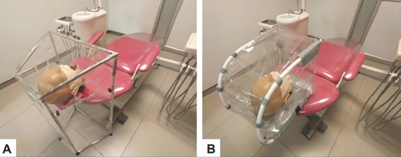
Mock scaling procedures were set on a dental phantom head (KaVo Dental Technologies, Charlotte, NC, USA) fitted on a reclined dental chair 55 cm above the floor in a single-chair operatory room. The magneto strictive ultrasonic scaler was set at 25 kHz, (Superson Merk III, Thai Dental Products Co., Ltd, Bangkok, Thailand), and the scaler handpiece was held by a right-handed operator with a mouth mirror in the other hand, whereas an assistant used a high-volume evacuation (HVE) and intraoral saliva ejector simultaneously. The water coolant for the scaler was set at the highest level. Two types of dental barriers: (1) metal frame with plastic wrap (MFPW) which was designed and constructed by PD10 Company Limited (Bangkok, Thailand) [Figure 1(A), Supplementary Figure 1] and (2) plastic shield chamber (PSC) or Dent Guard which was developed by SCG Co., Ltd. (Bangkok, Thailand) [Figure 1(B), Supplementary Figure 2], were used in this study. The MFPW is a portable floor standing trapezoidal stainless-steel frame covered with disposable plastic wrap around the frame with three 12-cm diameter holes on each side for arm access as illustrated in Figure 1(A) and Supplementary Figure 1. As for the PSC dental barrier, it was constructed from a U-shaped metal and reusable plastic shield with self-closing accesses for operation and patient’s head. The barrier is compact and portable which can be assembled to the head rest of the dental chair and designed to cover the patient’s head [Figure 1(B), Supplementary Figure 2].
Figure 1.

Design and placement of dental barrier during the dental treatment. (A): metal frame with plastic wrap (MFPW), (B): plastic shield chamber (PSC)
The experiment was divided into three groups which were: Group 1 (control group, no barrier was used), Group 2 (the use of MFPW barrier), and Group 3 (the use of PSC barrier). Based on the dissemination of aerosol and splatter that were previously reported from Veena et al.[10] and to achieve the power of 0.80 (effect size =0.20; P < 0.05), five trials in each group were required. The sequence of treatments was randomly allocated.
In order to avoid the airflow that could interfere the spreading characteristic of bioaerosols and splatter droplets, an air flow system in the room was shut down for an hour prior each session until completed.
DETECTION OF SPLATTER DISSEMINATION
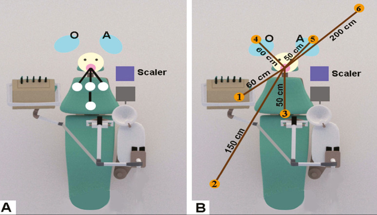
White filter paper discs, which have a diameter of 11 cm and a surface area of 95 cm2 (Double ring, Hangzhou Ocome Technology Co. Ltd, Hangzhou, Zhejiang, China), were placed in various positions: 30 cm at 4, 6, and 8 o’clock and 60 cm in middle of the chair [Figure 2A] to detect the dissemination of the splatter droplets created during an ultrasonic scaling. The filter paper discs were also attached on operator’s and assistant’s body: shoulders, wrists, chest, abdomen, and lap to identify the contamination.
Figure 2.
(A) Placement and position of filter paper on the dental chair. (B) Sampling site for placement of culture plate to detect the bacterial aerosols. O = operator; A = assistant
Fluorescein dye (Sigma-Aldrich, St Louis, MO, USA) was mixed in sterile water into 0.1% (w/v) fluorescein solution and connected to the coolant system of the scaler. Scaling procedures were performed for 5 min. Fluorescent splatter on the papers was detected, and images were captured under ultraviolet light. Then ImageJ (NIH free software, Bethesda, MD, USA) was used to evaluate contaminated areas.
DETECTION OF BACTERIAL AEROSOLS DISSEMINATION
The bioaerosols were generated following the protocol previously reported by Horsophonphong et al.[14] In short, the water coolant system of the scaler containing 107 colony-forming unit (CFU)/mL of Lactobacillus acidophilus was used to produce the bioaerosols while scaling. Then to detect the air contamination, de Man, Rogosa, and Sharpe (MRS) agar plates (Difco, Sparks, MD, USA), which considered selective for Lactobacilli,[15] were placed at six sampling sites in the room. The location and details of each sampling site were described in Table 1 and illustrated in Figure 2(B).
Table 1.
Location and details of each sampling site
| Sampling sites | Site description | |
|---|---|---|
| Horizontal distances from phantom mouth (cm) | Vertical distances from the floor (cm) | |
| 1. Operator’s tray | 60 | 85 |
| 2. Right side of dental chair | 150 | 180 |
| 3. Middle of dental chair | 50 | 60 |
| 4. Operator’s head | 60 | 135 |
| 5. Assistant’s head | 50 | 145 |
| 6. Behind the dental chair | 200 | 220 |
Half an hour before the scaling was performed, the plates were opened and exposed to air in order to establish the baseline room air contamination. After that the new plates were set and ultrasonic scaling was conducted for 10 min. The plates were exposed to the air throughout the scaling procedure and 20 min after the scaling was done. The bacterial cultured plates were incubated at 37± 0.5°C for 48 h. The colonies were counted and reported as colony-forming unit per plate (CFU/plate). At the end of each experiment, the airflow system in the room and two 72-W ultraviolet C germicidal light bulbs with 253.7 nm radiation (G36T8, Philips, Amsterdam, The Netherlands) were operated for half an hour for purification of the air in the room. After that, only air conditioner and exhaust air system continued to work for half an hour more before starting next session.
STATISTICAL ANALYSIS
Statistical analysis was performed using SPSS software version 18 (IBM, Armonk, NY, USA), and all experiments were repeated five times. Differences between groups were analyzed using one-way analysis of variance followed by multiple comparison test or Kruskal–Wallis test, a P-value less than 0.05 (P < 0.05) was considered statistically significant.
RESULTS
DISSEMINATION PATTERN OF THE FLUORESCENT DYE
Surface contamination was presented as the percentage of the stained area on the filter paper discs corresponding to the locations on the dental chair. The contamination was detected at 30 cm distance from the operating site in the 8, 6, and 4 o’clock positions, respectively, and no stain was found at 60 cm distance [Table 2]. Reduction in the percentage of contaminated area was observed when both types of dental barriers were used.
Table 2.
Percentage of contaminated surface area on the filter paper discs corresponding to the location on dental chair
| Position’s direction; distances (from the oral cavity) | 8 o’clock; 30 cm | 6 o’clock; 30 cm | 4 o’clock; 30 cm | 6 o’clock; 60 cm |
|---|---|---|---|---|
| Control | 1.04 (1.01) | 0.70 (0.46) | 0.33 (0.42) | 0 (0) |
| MFPW | 0.35 (0.17) | 0.55 (0.32) | 0.08 (0.07) | 0 (0) |
| PSC | 0.04 (0.04) | 0.05 (0.06) | 0.14 (0.18) | 0 (0) |
Data are presented as mean (SD)
The contaminated surface area on the filter paper corresponding to the part on the operator’s and assistant’s body was reported in Tables 3 and 4, respectively. In the control group, the contaminations were observed in every part of the operator’s body except the right shoulder. The abdomen, lap, and left wrist are the most contaminated parts in order [Table 3]. The use of both dental barriers showed to reduce surface contamination on the operator’s left shoulder, left wrist, chest, abdomen, and lap, as reported in Table 3. However, higher contaminated area on the operator’s right wrist was observed. In contrast to the contaminated area on the operator’s body, there are contaminations on the wrists, abdomen, and lap of the assistant, whereas no contamination was detected on the assistant’s shoulders and chest. The use of both dental barriers revealed contaminated area reduction in every part of the assistant’s body except on the left wrist where the reduction was greater when PSC barrier was used [Table 4]. When considering the percentage of total contaminated surface area, both MFPW and PSC barriers significantly decreased the total contamination detected on the body of the operator comparing with the control group (P < 0.05), as illustrated in Figure 3. A significant reduction in the total contaminated surface area on the assistant and dental chair was only observed when PSC was used (P < 0.05) [Figure 3].
Table 3.
Percentage of contaminated surface area on the filter paper discs corresponding to the part on the operator’s body
| Positions | Right shoulder | Left shoulder | Right wrist | Left wrist | Chest | Abdomen | Lap |
|---|---|---|---|---|---|---|---|
| Control | 0 (0) | 0.11 (0.12) | 0.95 (1.15) | 11.63 (11.80) | 0.37 (0.32) | 18.87 (18.80) | 11.66 (8.96) |
| MFPW | 0 (0) | 0 (0) | 3.48 (1.55) | 0.34 (0.53) | 0 (0) | 0 (0) | 0 (0) |
| PSC | 0 (0) | 0 (0) | 7.034 (2.31) | 0.29 (0.28) | 0 (0) | 0 (0) | 0.02 (0.01) |
Data are presented as mean (SD)
Table 4.
Percentage of contaminated surface area on the paper corresponding to the part on the assistant’s body
| Positions | Right shoulder | Left shoulder | Right wrist | Left wrist | Chest | Abdomen | Lap |
|---|---|---|---|---|---|---|---|
| Control | 0 (0) | 0 (0) | 0.04 (0.04) | 0.16 (0.11) | 0 (0) | 0.01 (0.01) | 8.25 (5.43) |
| MFPW | 0 (0) | 0 (0) | 0 (0.01) | 0.16 (0.11) | 0 (0) | 0 (0) | 0 (0.01) |
| PSC | 0 (0) | 0 (0) | 0.01 (0.01) | 0.01 (0.02) | 0 (0) | 0 (0) | 0.02 (0.02) |
Data are presented as mean (SD)
Figure 3.
Percentage of total contaminated surface area when compared with the control group. (A): operator, (B): assistant, (C): dental chair,*P < 0.05
BACTERIAL AEROSOLS
The baseline room air bacterial contamination detected before each scaling procedure was about 0.67 ± 0.53 CFU/plate. The number of bacterial air contaminations according to each sampling site and type of barrier device used was reported in Table 5. The use of dental barrier was found to significantly reduce bacterial air contamination at every sampling site when compared with the control group (no dental barrier). For each sampling site, no difference in number of bacterial colonies was observed between the two types of barriers [Table 5].
Table 5.
Number of colony-forming units per plate detected corresponding to the location in the room
| Positions | Description | Control | MFPW | PSC |
|---|---|---|---|---|
| 1 | On the operator’s tray | 23.5 (5.447) | 4.6* (1.342) | 5† (0.707) |
| 2 | Next to the dental chair, on the right side | 12.75 (3.593) | 3.4* (0.894) | 3.2† (2.049) |
| 3 | In the middle of the chair | 21.2 (8.983) | 6.2* (1.789) | 13.4† (3.130) |
| 4 | On the head of the operator | 28.25 (11.758) | 6.2* (1.923) | 7.8† (3.033) |
| 5 | On the head of the assistant | 23.75 (14.244) | 4.6* (2.702) | 5.6† (2.408) |
| 6 | Near the wall behind the dental chair | 26.5 (9.256) | 4* (1.225) | 5.8† (2.489) |
Data are presented as mean (SD), *P<0.05 when compared between control and MFPW groups, †P<0.05 when compared between control and PSC groups
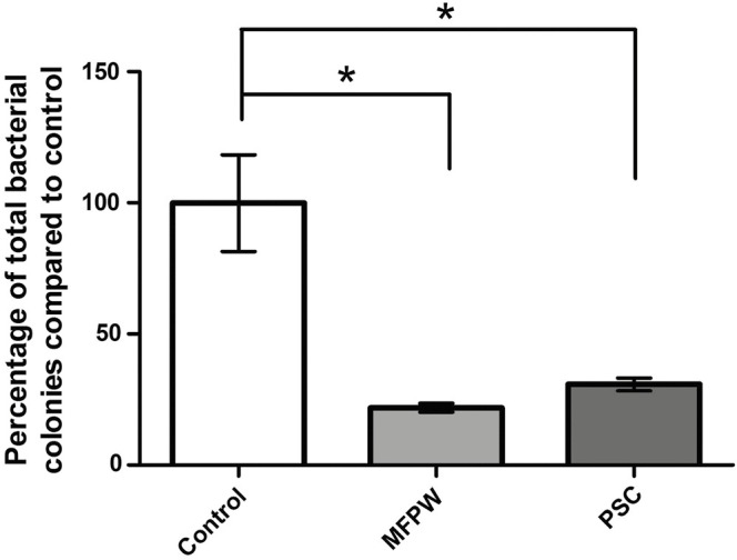
The total number of bacterial colonies detected in the room, represented in percentage of bacterial colonies compared with the control group [Figure 4], was significantly reduced when both MFPW and PSC barriers were used. The percentages of colonies reduction for MFPW and PSC were 78.13 (±1.69) and 69.24 (±2.49), respectively. Overall, no difference in the total number of bacterial colonies was observed between two types of barriers.
Figure 4.

Percentage of total bacterial colonies detected in the room air. MFPW (metal frame with disposable plastic wrap) and PSC (plastic shield chamber), *P < 0.001
DISCUSSION
The spread of the respiratory infectious disease is the main concern in dental clinic considering dental procedures usually generate aerosols and splatters to the surrounding, increasing the risk of cross-infection between dental practitioners and between patients. When carrying out dental procedures such as the use of high-speed handpiece, ultrasonic scaler, and 3-way syringe, special methods are required to mitigate the risk of disease transmission[16] because standard protective measures may not be effective enough to prevent the spread of COVID-19 when masks do not fit snugly.[3] Centers for Disease Control and Prevention recommended the use of HVE and dental dam to minimize aerosol production as much as possible.[17] Moreover, the use of both HVE and intraoral suction was reported to significantly reduce the number of bacterial aerosols.[18] This study used both HVE and intraoral suction device in combination to minimize the number of microbial aerosols generated during the scaling procedure.
In the medical field, barrier enclosure systems were introduced as one of the equipment that could reduce the spreading and dissemination of airborne particles[12] and were suggested to be useful during dental procedures. Nevertheless, the effectiveness of barrier enclosure system for dental procedure has never been studied. This study introduced two types of dental barriers, MFPW and PSC, as the representative of barrier enclosure system that could be used and applied for dental treatments. The MFPW [Figure 1(A) and Supplementary Figure 1] has many advantages including adjustable height, moveable, light weight, easy access, and free arm movement inside. The plastic wrap is disposable and single-use, making it convenient to clean after each patient. In contrast, the disadvantages are instability, more time-consuming to prepare the plastic wrap, and, most importantly, the plastic wrap covered only three sides of MFPW, leaving one side uncovered allowing some bioaerosol to escape. While PSC is able to connect to dental chair’s head rest, steady and all sides are covered including arm accesses with self-closing flaps. On the contrary, PSC being smaller than MFPW may cause discomfort to the patients. Line accesses can limit arm movement and the reusable plastic may raise an issue when cleaning.
Fluorescein dye was used as a tracer for detecting splatter pattern and liquid contamination generated during ultrasonic scaling. The results showed that the splatter droplets generated during scaling could be detected within 30 cm range from the oral cavity. Both types of barriers were able to reduce the surface contamination in most of the areas on dental chair, operator’s and assistant’s body, confirmed with percentage of contaminated surface area compared with control. According to the results, no significant difference in surface contamination of splatter reduction was found between the MFPW and the PSC. A significant reduction in total contaminated surface area on the operator’s body was detected when both types of barriers were used. Whereas a significant reduction of the total contaminated area on the dental chair and assistant was only observed in PSC barrier. This may be because MFPW was designed as one open-end, which causes the dental chair to expose to a greater amount of liquid contamination compared with PSC. The higher surface contamination on operator’s right wrist was observed when using MFPW and PSC as hand action and movement are restricted by the plastic shield on top of the barriers.
The bacterial colonies represented the aerosol contamination detected on each sampling site, which was found to be significantly reduced when both barriers were used. Moreover, both barriers showed to significantly reduce the colony numbers without any difference between the barriers. The results confirmed the efficacy of MFPW and PSC in reducing aerosols during the dental procedure. The advantage of these barriers is that they prevent close proximity to the patient’s mouth where heavy contamination was found within 100 cm distance and contamination levels reduced with increasing distance, according to Innes et al.[19] Both dental barriers are easy to produce, made-to-order producible, low cost, and movable. Nevertheless, some of the designs may need improvement for better access during dental procedures in the future.
The limitation of this study is that only bacterial contamination was determined. Viruses that are smaller and probably spread in greater distances were not investigated.
During the COVID-19 crisis, preventive measures are required to minimize the risks of disease transmission during dental treatments. Besides the standard personal protective protocol, MFPW and PSC dental barriers could be additional options for the dental team.
CONCLUSION
Both MFPW and PSC dental barriers are useful and effective in reducing the spatial distribution of dental splatters and aerosols generated during the ultrasonic scaling.
ACKNOWLEDGEMENTS
Not applicable.
FINANCIAL SUPPORT AND SPONSORSHIP
The study did not receive any funding.
CONFLICTS OF INTEREST
There are no conflicts of interest.
AUTHORS CONTRIBUTIONS
Chestsuttayangkul Y contributed to study design, investigation, data acquisition, analysis, writing-review, and editing of the manuscript. Lertsooksawat W contributed to investigation, data acquisition, and analysis. Horsophonphong S contributed to conceptualization, investigation, supervision, writing-reviewing, and editing of the manuscript.
ETHICAL POLICY AND INSTITUTIONAL REVIEW BOARD STATEMENT
Not applicable.
PATIENT DECLARATION OF CONSENT
Not applicable.
DATA AVAILABILITY STATEMENT
Not applicable.
Supplementary Material
Metal frame with plastic wrap (MFPW). Trapezoidal stainless steel frame with one open-end, whereas other sides are covered with single-used plastic wrap. Floor standing and adjustable-height knobs. Three 12-cm diameter arm accesses on each side
Plastic shield chamber (PSC). U-shaped metal form with reusable plastic covered all sides. Three arm accesses with self-closing flap lids. Assembled to the head rest of dental unit with plastic buckle lock underneath
REFERENCES
- 1.Harrel SK, Molinari J. Aerosols and splatter in dentistry: A brief review of the literature and infection control implications. J Am Dent Assoc. 2004;135:429–37. doi: 10.14219/jada.archive.2004.0207. [DOI] [PMC free article] [PubMed] [Google Scholar]
- 2.Ge ZY, Yang LM, Xia JJ, Fu XH, Zhang YZ. Possible aerosol transmission of COVID-19 and special precautions in dentistry. J Zhejiang Univ Sci B. 2020;21:361–8. doi: 10.1631/jzus.B2010010. [DOI] [PMC free article] [PubMed] [Google Scholar]
- 3.Meng L, Hua F, Bian Z. Coronavirus disease 2019 (COVID-19): Emerging and future challenges for dental and oral medicine. J Dent Res. 2020;99:481–7. doi: 10.1177/0022034520914246. [DOI] [PMC free article] [PubMed] [Google Scholar]
- 4.Micik RE, Miller RL, Mazzarella MA, Ryge G. Studies on dental aerobiology. I. Bacterial aerosols generated during dental procedures. J Dent Res. 1969;48:49–56. doi: 10.1177/00220345690480012401. [DOI] [PubMed] [Google Scholar]
- 5.Miller RL, Micik RE, Abel C, Ryge G. Studies on dental aerobiology. II. Microbial splatter discharged from the oral cavity of dental patients. J Dent Res. 1971;50:621–5. doi: 10.1177/00220345710500031701. [DOI] [PubMed] [Google Scholar]
- 6.Singh A, Shiva Manjunath RG, Singla D, Bhattacharya HS, Sarkar A, Chandra N. Aerosol, a health hazard during ultrasonic scaling: A clinico-microbiological study. Indian J Dent Res. 2016;27:160–2. doi: 10.4103/0970-9290.183131. [DOI] [PubMed] [Google Scholar]
- 7.Grenier D. Quantitative analysis of bacterial aerosols in two different dental clinic environments. Appl Environ Microbiol. 1995;61:3165–8. doi: 10.1128/aem.61.8.3165-3168.1995. [DOI] [PMC free article] [PubMed] [Google Scholar]
- 8.Rautemaa R, Nordberg A, Wuolijoki-Saaristo K, Meurman JH. Bacterial aerosols in dental practice—A potential hospital infection problem? J Hosp Infect. 2006;64:76–81. doi: 10.1016/j.jhin.2006.04.011. [DOI] [PMC free article] [PubMed] [Google Scholar]
- 9.Chuang CY, Cheng HC, Yang S, Fang W, Hung PC, Chuang SY. Investigation of the spreading characteristics of bacterial aerosol contamination during dental scaling treatment. J Dent Sci. 2014;9:294–6. [Google Scholar]
- 10.Veena HR, Mahantesha S, Joseph PA, Patil SR, Patil SH. Dissemination of aerosol and splatter during ultrasonic scaling: A pilot study. J Infect Public Health. 2015;8:260–5. doi: 10.1016/j.jiph.2014.11.004. [DOI] [PubMed] [Google Scholar]
- 11.Wang Y, Xu G, Huang YW. Modeling the load of SARS-CoV-2 virus in human expelled particles during coughing and speaking. PLoS One. 2020;15:e0241539. doi: 10.1371/journal.pone.0241539. [DOI] [PMC free article] [PubMed] [Google Scholar]
- 12.Sorbello M, Rosenblatt W, Hofmeyr R, Greif R, Urdaneta F. Aerosol boxes and barrier enclosures for airway management in COVID-19 patients: A scoping review and narrative synthesis. Br J Anaesth. 2020;125:880–94. doi: 10.1016/j.bja.2020.08.038. [DOI] [PMC free article] [PubMed] [Google Scholar]
- 13.Babu B, Gupta S, Sahni V. Aerosol box for dentistry. Br Dent J. 2020;228:660. doi: 10.1038/s41415-020-1598-3. [DOI] [PubMed] [Google Scholar]
- 14.Horsophonphong S, Chestsuttayangkul Y, Surarit R, Lertsooksawat W. Efficacy of extraoral suction devices in aerosol and splatter reduction during ultrasonic scaling: A laboratory investigation. J Dent Res Dent Clin Dent Prospects. 2022;15:197–202. doi: 10.34172/joddd.2021.033. [DOI] [PMC free article] [PubMed] [Google Scholar]
- 15.De Man JC, Rogosa M, Sharpe ME. A medium for the cultivation of Lactobacilli. J Appl Bacteriol. 1960;23:130–5. [Google Scholar]
- 16.Han P, Li H, Walsh LJ, Ivanovski S. Splatters and aerosols contamination in dental aerosol generating procedures. Appl Sci. 2022;11:1914. [Google Scholar]
- 17.Centers for Disease Control and Prevention. Summary of CDC COVID-19 Guidance for Dental Services. 2020. [Last accessed on 2021 Jun 25]. Available from: https://www.cdc.gov/oralhealth/infectioncontrol/statement-COVID.html .
- 18.Suprono MS, Won J, Savignano R, Zhong Z, Ahmed A, Roque-Torres G, et al. A clinical investigation of dental evacuation systems in reducing aerosols. J Am Dent Assoc. 2022;152:455–62. doi: 10.1016/j.adaj.2021.02.013. [DOI] [PMC free article] [PubMed] [Google Scholar]
- 19.Innes N, Johnson IG, Al-Yaseen W, Harris R, Jones R, Kc S, et al. A systematic review of droplet and aerosol generation in dentistry. J Dent. 2022;105:103556. doi: 10.1016/j.jdent.2020.103556. [DOI] [PMC free article] [PubMed] [Google Scholar]
Associated Data
This section collects any data citations, data availability statements, or supplementary materials included in this article.
Supplementary Materials
Metal frame with plastic wrap (MFPW). Trapezoidal stainless steel frame with one open-end, whereas other sides are covered with single-used plastic wrap. Floor standing and adjustable-height knobs. Three 12-cm diameter arm accesses on each side
Plastic shield chamber (PSC). U-shaped metal form with reusable plastic covered all sides. Three arm accesses with self-closing flap lids. Assembled to the head rest of dental unit with plastic buckle lock underneath
Data Availability Statement
Not applicable.


