

Abstract
Blood flow and shear stresses were quantified using 4-dimensional flow cardiac magnetic resonance and 3-dimensional particle velocimetry before and after transcatheter aortic valve replacement (TAVR). TAVR reduced turbulent kinetic energy by 47% and shear stresses by 33%, illustrating that the benefit of TAVR extends beyond a simple reduction in transvalvular gradients. (Level of Difficulty: Advanced.)
Key Words: 3-dimensional imaging, aortic valve, bicuspid aortic valve, hemodynamics, valve replacement
Abbreviations and Acronyms: CMR, cardiac magnetic resonance; TAVR, transcatheter aortic valve replacement
Central Illustration
Patients with severe aortic stenosis present not only with elevated pressure gradients and high blood flow velocities, but also with increased turbulence and shear stresses. These suboptimal blood flow patterns result in energy loss and decreased availability of the von Willebrand factor, and they may contribute to progressive dilatation of the ascending aorta.1
We present a 69-year-old man with severe, symptomatic bicuspid aortic stenosis and late-stage multiple myeloma. The heart team opted for transcatheter aortic valve replacement (TAVR). An Evolut Pro 26-mm transcatheter heart valve (Medtronic Inc) was implanted transfemorally, resulting in a reduction of the mean transvalvular gradient from 37 to 8 mmHg and an increase of the aortic valve area from 0.9 to 2.1 cm2. The in-hospital course was uneventful, and the patient was discharged home 2 days after the procedure.
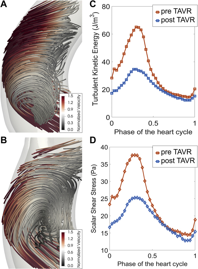
With little evidence available for visualization and quantification of blood flow before and after TAVR in (bicuspid) valve stenosis, both in vivo and in vitro measurements were performed. For in vivo measurements, an electrocardiogram- and respiratory navigator–gated gadolinium-free 4-dimensional flow cardiac magnetic resonance (CMR) tomography was performed before (Figure 1A) and after TAVR (Figure 1B), as approved by the local ethics committee. For in vitro measurements, the computed tomography scan, which is routinely performed for the planning of TAVR, was used to 3-dimensionally print an anatomically correct, optically transparent, and flexible silicone model of the patient’s diseased aortic valve, aortic root, ascending aorta, and aortic arch (ACEO). Physiologic pulsatile flow conditions were mimicked using a waveform generator (Berlin Heart) and an 80-mL ventricular assist device (MEDOS), closely matching the patient’s calculated stroke volume of 73 mL. For postprocedural measurements, the valve was manually placed in the model at the same implantation depth and orientation that was achieved during TAVR. Measurements were performed using 3-dimensional particle-tracking velocimetry (Hi-D Imaging).2 TAVR reduced the average turbulent kinetic energy by 47%, from 65.1 J/m3 to 34.6 J/m3 (Figure 1C), and shear stress by 33%, from 37.7 Pa to 25.3 Pa (Figure 1D).
Figure 1.

Flow, Turbulent Kinetic Energy, and Shear Stresses Before and After Transcatheter Aortic Valve Replacement
Visualization of flow and turbulence by 4-dimensional flow cardiac magnetic resonance (A) before and (B) after transcatheter aortic valve replacement (TAVR). (C) Turbulent kinetic energy and (D) shear stresses were quantified using 3-dimensional particle-tracking velocimetry in an anatomically adequate silicone model.
CMR offers the possibility to quantify phase-averaged, mean blood flow–related hemodynamics parameters, such as mean velocity, mean kinetic energy, and shear stress due to the mean flow.3 However, CMR cannot accurately assess the turbulence-related quantities. Particle-tracking velocimetry has high temporal and spatial resolution and minimizes artifacts from the transcatheter aortic valve, allowing the study of the hemodynamics along the entire heart cycle and assessment of turbulence-related quantities, that is, turbulent kinetic energy and scalar shear stress (composed of stresses from both mean and fluctuating flow fields).
This report illustrates that the benefit of TAVR may well extend beyond an improvement in transvalvular gradient, blood flow velocities, and aortic valve area. Indeed, TAVR improved blood flow patterns, leading to a reduction in turbulent kinetic energy loss and shear stresses, thus improving the efficiency of the left ventricle. These favorable changes may contribute to the symptomatic and prognostic benefits of transcatheter and surgical aortic valve replacement.
Funding Support and Author Disclosures
Dr Toggweiler is a consultant and proctor for Medtronic, Boston Scientific, and Biosensors/New Valve Technology; is a proctor for Abbott Vascular; is a consultant for Shockwave, Teleflex, Medira, AtHeart Medical, and VeoSource; has received institutional research grants from Boston Scientific and Fumedica; and holds equity in Hi-D Imaging. Dr Karakas holds equity in Hi-D Imaging. Dr Gülan holds equity in Hi-D Imaging. Dr De Boeck has reported that he has no relationships relevant to the contents of this paper to disclose.
Footnotes
The authors attest they are in compliance with human studies committees and animal welfare regulations of the authors’ institutions and Food and Drug Administration guidelines, including patient consent where appropriate. For more information, visit the Author Center.
References
- 1.Garcia J., Barker A.J., Markl M. The role of imaging of flow patterns by 4D flow MRI in aortic stenosis. J Am Coll Cardiol Img. 2019;12:252–266. doi: 10.1016/j.jcmg.2018.10.034. [DOI] [PubMed] [Google Scholar]
- 2.Gulan U., Appa H., Corso P., et al. Performance analysis of the transcatheter aortic valve implantation on blood flow hemodynamics: an optical imaging-based in vitro study. Artif Organs. 2019;43:E282–E293. doi: 10.1111/aor.13504. [DOI] [PubMed] [Google Scholar]
- 3.Komoriyama H., Kamiya K., Nagai T., et al. Blood flow dynamics with four-dimensional flow cardiovascular magnetic resonance in patients with aortic stenosis before and after transcatheter aortic valve replacement. J Cardiovasc Magn Reason. 2021;23:81. doi: 10.1186/s12968-021-00771-y. [DOI] [PMC free article] [PubMed] [Google Scholar]