

Abstract
Objective: To analyze the surgical treatment of patients with cervical brucellosis with osteoporosis over a 4-year period in Northwest China. Methods: From 2013 to 2018, 22 patients (12 males and 10 females) with lower cervical spine brucellosis (C3-C7) underwent anterior lesion debridement, decompression, bone grafting and internal fixation combined with posterior bone graft fusion and internal fixation (ADDF+PIF). The follow-up period averaged 37.4 months (ranging from 24 to 57 months). Results: Involvement of 1 vertebra was observed in 3 patients, involvement of 3 vertebrae was observed in 9 patients, and involvement of 3 vertebrae was observed in 10 patients. Before surgery, 1 patient had Frankel grade B, 2 had grade C, 9 had grade D, and 10 had grade E. In the final follow-up, 12 patients had neurological deficits, 10 patients improved by one grade, 6 patients improved by two grades, and the neurological status of 6 patients remained unchanged. In all cases, it was observed that bone fusion required 6.8 months on average. The kyphosis Cobb angle was enhanced from an average of 11.5° preoperatively (range 0°-24°) to 0.13° postoperatively (range 1°-5°), and there was no vital loss of correction in the follow-up. Conclusions: ADDF+PIF is an effective and safe treatment for patients with lower cervical brucellosis with osteoporosis.
Keywords: Brucellosis, osteoporosis, lower cervical, surgical treatment
Introduction
Brucellosis is a systemic disease that frequently occurs in Northwest China. This endemic zoonotic disease can affect various tissues and organs [1]. Brucellosis is mostly caused by cattle and sheep feeders or by the consumption of unpasteurized milk or dairy products by people [2]. Bone and joint involvement is a common issue, while spondylitis symptoms are common in elderly individuals [3]. Brucellosis of the spine affects the vertebra, intervertebral space, and/or paravertebral area. We cannot only rely on basic evaluations to diagnose this disease. If a patient with early stage deformity and neurological dysfunction accepts chemotherapy and improved nutrition, good outcomes can occur. However, conservative treatment methods commonly lead to kyphosis, spinal instability and neurological dysfunction [4,5]. Vertebral body lesions require surgical treatment [6].
Surgical treatment of cervical brucellosis has rarely been reported due to the rarity of the disease. Khan reported on a 27-year-old male with cervical brucellosis treated with anterior lesion debridement decompression and instrumented fusion in 2020 [6]. Ekici reported two patients: a 61-year-old male who had anterior cervical lesion debridement without internal fixation and a 63-year-old male who had an anterior cervical lesion debridement procedure with cage implantation in the intervertebral space in 2012 [7]. Previously articles have reported that surgical treatment based on clinical experience is for anterior lesion removal with cervical brucellosis. However, we searched the relevant literature and found no surgical treatment for osteoporotic lower cervical brucellosis. Further exploration is still needed to develop treatment measures for patients with lower cervical spine issues combined with abscess formation and cervical spinal instability [8]. As China’s population is increasing in age, osteoporosis has become a serious social problem. Osteoporosis brings serious sequelae to the patient, reduces the patient’s survival satisfaction, and even leads to paralysis, which brings a heavy burden to the patient’s psychology and finances. There are no guidelines for treating lower cervical spondylosis combined with osteoporosis, and each hospital treats the disease according to its own clinical experience. Our study tries to explore how to treat this type of patient and fills a vacancy in the literature about the treatment of these patients.
The aim of this study was to analyze the efficiency of ADDF+PIF in patients with lower cervical brucellosis with osteoporosis.
Materials and methods
Patient population
Research based at the largest orthopedic center in Northwest China, namely, the Spinal Surgery Center of Xi’an Honghui Hospital, the study included patients treated with ADDF+PIF at this institution from June 2013 to June 2018. All patients with brucellosis of the lower cervical spine (C3-C7) were treated with ADDF+PIF. Twenty-two patients (12 males and 10 females) ranged in age from 40 to 70 years (average 58 years). The hospital medical records and X-rays were reviewed. The diagnosis was based on all available medical materials, including medical history, radiographic images, and drug reactions. A definite diagnosis was confirmed by histological examination and laboratory examination. Patients were excluded if they had upper cervical spine brucellosis, jumping brucellosis, or cervical brucellosis according to surgical records.
Our study was approved by the ethics committee of the Honghui Hospital, Xi’an Jiaotong University (No. 201004002).
Symptoms and signs
The symptoms and signs were neck pain and stiffness. Seven patients developed obvious physical symptoms, such as fever, sweating and muscle and joint pain. Cervical radiculopathy was observed in 9 patients (Table 1). Neck pain was analyzed via the VAS score. The neurological status was graded in accordance with the Frankel scoring system [9]. Two cases were grade C, 9 were grade D, and 10 were grade E before the operation (Table 2).
Table 1.
Clinical features at presentation
| Features | Number of patients (%) |
|---|---|
| Neck pain | 22 (100.00) |
| Neck stiffness | 15 (68.18) |
| Spastic quadriparesis | 3 (13.64) |
| Cervical radiculopathy | 9 (40.91) |
| Constitutional symptom | 7 (31.82) |
| Sphincteric disturbance | 2 (9.09) |
Table 2.
Summary of clinical data of 22 patients with lower cervical brucellosis with osteoporosis
| Patient no. | Sex | Age (y) | T-score | Neurological status | Affected segments | ADDF+PIF level | Kyphosis (°) | Surgical time (min) | Blood loss (ml) | Duration of hospital stay (day) | Postop. complication | Follow-up (mo) | |||
|---|---|---|---|---|---|---|---|---|---|---|---|---|---|---|---|
|
|
|
||||||||||||||
| Preop. | FFU | Preop. | Postop. | FFU | |||||||||||
| 1 | M | 40 | -2.6 | D | E | C4-6 | C4-7 | 10 | 0 | 0 | 65 | 90 | 7 | 30 | |
| 2 | M | 50 | -3.5 | E | E | C5-6 | C5-7 | 20 | 0 | 0 | 50 | 120 | 9 | 35 | |
| 3 | F | 62 | -2.7 | D | E | C4-6 | C4-7 | 15 | -11 | -8 | 80 | 250 | 5 | 29 | |
| 4 | M | 63 | -3.2 | D | E | C4-6 | C4-7 | 0 | 5 | 4 | 96 | 305 | 9 | 25 | |
| 5 | M | 69 | -2.9 | D | E | C5-7 | C4-7 | 3 | 1 | 1 | 60 | 450 | 11 | 30 | |
| 6 | F | 60 | -3.0 | E | E | C3-4 | C3-5 | 10 | 3 | 2 | 104 | 350 | 5 | 26 | |
| 7 | F | 52 | -3.2 | D | E | C6-7 | C5-7 | 13 | -3 | -3 | 99 | 450 | 7 | Harvest site pain | 33 |
| 8 | M | 60 | -2.6 | E | E | C5 | C5-6 | 18 | 2 | 2 | 93 | 400 | 9 | 32 | |
| 9 | M | 61 | -2.8 | C | D | C3-5 | C3-6 | 19 | 0 | 1 | 90 | 300 | 7 | 31 | |
| 10 | F | 52 | -2.6 | E | E | C3-5 | C3-6 | 16 | 3 | 2 | 84 | 210 | 9 | 43 | |
| 11 | M | 64 | -2.6 | E | E | C4-5 | C4-6 | 13 | 4 | 3 | 100 | 180 | 10 | 30 | |
| 12 | M | 69 | -2.9 | C | E | C6 | C5-6 | 24 | -11 | -10 | 112 | 500 | 12 | Delayed wound healing | 32 |
| 13 | M | 57 | -2.7 | E | E | C4-7 | C4-7 | 20 | 5 | 2 | 102 | 280 | 11 | 36 | |
| 14 | F | 46 | -2.8 | D | D | C4 | C4-6 | 15 | 4 | 2 | 98 | 175 | 11 | 34 | |
| 15 | M | 56 | -2.6 | E | E | C5-6 | C5-6 | 17 | 2 | 0 | 88 | 130 | 12 | 32 | |
| 16 | F | 67 | -2.9 | E | E | C5-6 | C4-6 | 12 | 1 | 1 | 83 | 80 | 4 | Harvest site pain | 33 |
| 17 | M | 46 | -2.6 | D | E | C5-7 | C5-7 | 9 | 3 | 3 | 104 | 150 | 6 | 31 | |
| 18 | M | 56 | -2.9 | B | D | C6-7 | C5-7 | 3 | 1 | 1 | 89 | 250 | 18 | 30 | |
| 19 | F | 64 | -2.9 | E | E | C5-6 | C5-6 | 2 | 0 | 0 | 107 | 390 | 15 | Delayed wound healing | 32 |
| 20 | F | 70 | -3.9 | D | E | C6-7 | C6-7 | 14 | 0 | 0 | 100 | 350 | 16 | 35 | |
| 21 | F | 65 | -3.8 | D | E | C5-7 | C5-7 | 0 | -2 | -1 | 92 | 450 | 15 | 38 | |
| 22 | F | 47 | -2.7 | E | E | C4-6 | C4-6 | 0 | -3 | -3 | 84 | 300 | 12 | 40 | |
ADDF+PIF, anterior lesion debridement, decompression, bone grafting and internal fixation combined with posterior bone graft fusion and internal fixation; FFU, final follow-up; postop., postoperative; preop., preoperative. Neurological grade was evaluated according to Frankel et al. [8].
Preoperative management
All the patients underwent cervical X-ray film, CT and MRI examinations before the operation. The kyphosis angle was determined by evaluating the angle between the first normal vertebral body above the lesion and below the lesion on the lateral X-ray film [5].
One vertebral body was involved in 3 patients, 2 were involved in 9 patients, and 3 were involved in 10 patients. The preoperative kyphosis angle was 0°-24°, with an average of 11.5°. MRI showed 18 cases of epidural abscesses with obvious spinal cord compression. In selected cases, CT of the cervical spine was used to confirm the extent of bone destruction.
Routine laboratory tests, including chest CT and standard serum test tube agglutination (SAT), were performed to detect bacterial titer, tiger red plate agglutination test (RBP), complete blood count, ESR, CRP, blood sugar, urinary examination, and liver and kidney function tests. Eight patients were malnourished (100 g/L hemoglobin and 30 g/L albumin), and nutritional supplementation was provided before the operation. In this study, none of the patients were HIV- or tuberculosis-positive.
Active medication (doxycycline 100 mg bid + gentamicin 5 mg/kg qd + rifampicin 10 mg/kg up to 900 mg once a day or doxycycline 100 mg bid, rifampicin 10 mg/kg up to 900 mg once a day ceftriaxone 2 g intravenously every 12 hours) was administered for 2 weeks [4,10].
Operative indications
Indications included severe neck and/or radicular pain that could not be treated conservatively, neurological dysfunction, obvious kyphosis, or progressive deformity [11].
Surgical skills
Surgery was performed under general anesthesia. Patients were placed in the supine position, and we used the anterolateral approach. We used curette and pituitary forceps to remove the necrotic material from the intervertebral disc and affected vertebrae. The paravertebral abscess was subsequently identified and drained. We adequately debrided all the infected tissue. The vertebral expander was used between adjacent normal vertebral bodies to perform progressive distraction to correct the previous kyphotic deformity and the auxiliary deformity. After confirming that the anterior dura was protruding and the pulsation was good, the height of the anterior gap was measured, and autogenous iliac bone grafting was performed. Autogenous iliac bone grafts were used to repair the defect of the anterior column. A locking plate screw system of an appropriate length (American pivot model system) was used to complete the anterior cervical fixation. Patients were turned over and placed the prone position. The posterior median approach was performed and the vertebral bodies were fixed with pedicle screws.
Postoperative treatment
Oral administration of the following regimes was resumed: (doxycycline 100 mg bid + gentamicin 5 mg/kg qd + rifampicin 10 mg/kg up to 900 mg, once a day for at least 3 months), (doxycycline 100 mg bid + gentamicin 5 mg/kg, max. 900 mg), or (Cyclin 100 mg bid 2 months rifampicin 10 mg/kg, max. 900 mg, once a day for at least 3 months, ceftriaxone 2 g intravenous bolus every 12 hours, at least 1 month). Each patient received medication compliance education. Clinical results, normal erythrocyte sedimentation rate and signs of radiological healing determined when the treatment could be stopped [12].
The patient was allowed to stay in bed on the first postoperative day and sit with a brace on the bed the next day. We encouraged the patient to walk on the third day. Patients with neurological deficits immediately began active rehabilitation.
After treatment, the follow-up was carried out regularly. The follow-up times were 3 months, 6 months, 9 months, 1 year, 2 years, 3 years and then once a year thereafter. Patients received follow-up care consisting of erythrocyte sedimentation rate and CRP value measurements, physical examination and radiological examination, and neurological status assessment. A specially assigned doctor conducted the follow up. The successful fusion of clinical and radiological evidence included the absence of local pain and tenderness at the fusion site, the lack of abnormal motion and the lack of internal fixation failure, and bony fusion was shown on CT scan [13,14].
Statistical analysis
Quantitative data are expressed as the mean ± standard deviation (x̅±s). Preoperative and postoperative data of the previous endpoints were compared by using the single factor analysis of variance, then by using LSD-t test. Significance level was set α=0.05. All the statistical analyses were performed by using Statistical Product and Service Solution Version 18.0 (SPSS, Inc., Chicago, IL, USA).
Results
These operations were carried out by the same group of surgeons in our hospital, and all patients tolerated the operations well. The average surgical time was 90 min (ranging from 50 to 120 min). The average intraoperative blood loss was 280 ml (ranging from 80 to 500 ml). The average hospital stay was 10 days (ranging from 4 to 18 days). Two patients had a superficial infection after the operation, which was cured without sequelae after treatment with systemic antibiotics and debridement. Two people felt pain at the surgical site, but the symptoms disappeared one week after the operation. No nerve or vascular structure damage was seen during the operation.
The histopathological study of surgical specimens revealed brucellosis. The average preoperative erythrocyte sedimentation rate was 55 mm/h (35-80 mm/h), and the average C-reactive protein level was 23 mg/dL (17-38 mg/dL); these values gradually decreased after 7 months and returned to normal. At the last follow-up, the average erythrocyte sedimentation rate was 14.6 mm/h (5-17 mm/h), and the average CRP was 0.93 mg/dL (0.10-2.86 mg/dL) (P<0.05).
The follow-up lasted 24 to 36 months, with an average of 29 months. The kyphosis Cobb angle improved from an average of 11.5° (0°-24°) before the operation to an average of 0.13° (-11°-5°) after the operation (P<0.05). At the final follow-up, the mean kyphotic Cobb angle was -0.05° (-10°-4°) (P<0.05). Pain relief was observed after operation. The preoperative VAS score averaged 8 points (6-10 points), and the average was 1.5 points (0-3 points) at the last follow-up (P<0.05). The neurological function of patients with neurological deficits recovered within 4 days after the operation. At the last follow-up, two patients improved from grade C to grade D, 8 patients improved to grade E, and 10 patients remained at grade E (Table 3). For patients with postoperative neurological deficits, deficits with respect to writing and grasping were the most obvious. The X-rays showed that all patients had bony fusion. The fusion period is 3-10 months, with an average of 6.8 months. No patients had internal fixation failure or imaging manifestations of false joints. There was no recurrence of brucellosis infection.
Table 3.
Neurological recovery according to Frankel grade
| Preoperative Frankel grade | No. of patients | Frankel grade at last follow-up | ||||
|---|---|---|---|---|---|---|
|
| ||||||
| A | B | C | D | E | ||
| A | 0 | 0 | 0 | 0 | 0 | 0 |
| B | 0 | 0 | 0 | 0 | 0 | 0 |
| C | 2 | 0 | 0 | 0 | 2 | 0 |
| D | 9 | 0 | 0 | 0 | 1 | 8 |
| E | 10 | 0 | 0 | 0 | 0 | 10 |
A typical case
The patient was a man aged 40 years with neck pain and stiffness and numbness and weakness in his limbs. The neurological examination was graded as D level based on the Frankel scoring system. Preoperative digital radiography showed that the damaged vertebral body was C5-6 (Figure 1). Preoperative computed tomography examination showed that the C4-6 vertebral body was destroyed (Figure 2). Preoperative magnetic resonance imaging examination showed that the C4-6 vertebral body was destroyed and that the spinal cord was compressed (Figure 3). After 4 weeks of conservative treatment, his neck pain and limb numbness and weakness worsened. The patient underwent C4 partial resection, C5-C6 subtotal resection of the vertebral body, autogenous iliac bone graft, and anterior plate internal fixation combined with posterior bone graft fusion and internal fixation (Figure 4). After the surgery, pain relief was quickly observed, and the patient fully recovered his athletic ability after 3 months. CT showed that the patient achieved bony fusion one year after surgery (Figure 5). Magnetic resonance imaging examination showed that the spinal canal was patent one year after surgery (Figure 6).
Figure 1.

In a 40-year-old male patient, preoperative digital radiography examination showed that the C5-6 vertebral body was destroyed.
Figure 2.
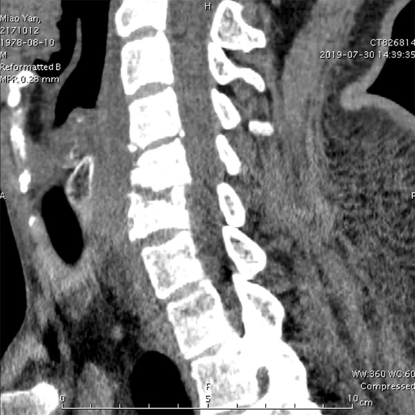
In a 40-year-old male patient, preoperative computed tomography imaging showed destruction of the C4-C6 vertebrae.
Figure 3.

In a 40-year-old male patient, preoperative magnetic resonance imaging examination showed that the C4-6 vertebral body was destroyed and that the spinal cord was compressed.
Figure 4.

In a 40-year-old male patient, the patient underwent C4 partial resection, C5-C6 subtotal resection of the vertebral body, autogenous iliac bone graft, and anterior plate internal fixation + posterior bone graft fusion and internal fixation.
Figure 5.

In a 40-year-old male patient, CT showed that the patient achieved bony fusion one year after surgery.
Figure 6.

In a 40-year-old male patient, postoperative magnetic resonance imaging examination showed that the spinal canal was patent.
Discussion
Brucellosis is a threat to health globally. There are more than 500,000 new cases every year [1,5]. The incidence rate is higher in Northwest China. Xi’an Red Cross Hospital is the orthopedic center in Northwest China. Brucellosis is directly or indirectly transmitted to humans. Direct transmission requires contact with animals that carry the pathogenic bacteria Brucella or with infectious substances in the livestock industry and meat processing. Indirect transmission is achieved through the consumption of nonpasteurized dairy products. Cervical brucellosis is less common than thoracic and lumbar spine brucellosis. The emergence of effective drugs makes most brucelloses a treatable medical disease [15]. Our patients with cervical brucellosis who did not meet the indications for surgery received nonsurgical treatment on an outpatient basis. After 3 to 6 months of chemotherapy, good healing can be obtained.
Although drugs are effective in treating brucellosis of the spine [16,17], people with multiple vertebral body lesions, refractory diseases, severe kyphosis, or neurological dysfunction still require surgical treatment [5]. We performed this study to analyze anterior lesion debridement, decompression, bone grafting and internal fixation combined with posterior bone graft fusion and internal fixation for multilevel cervical brucellosis, including the improvement of postoperative nerve function, the progression of intervertebral fusion, the degree of correction and maintenance of kyphosis, and the incidence of problems related to bone grafting and internal fixation. The curative effect was evaluated in 22 patients with the shortest follow-up time of 2 years. At the last follow-up, 15 of the 20 patients with neurological deficits recovered completely. The kyphotic Cobb angle improved from an average of 16.58° before surgery to an average of 4.6° after surgery. There were no reports of implant failure or false joints, and all the patients achieved bony fusion. It is suggested that this method can promote local bone healing and stability of the spine and prevent deformities.
As the population of China ages, the number of patients with osteoporosis is increasing. Additionally, patients with osteoporosis with cervical brucellosis have caused the medical field challenges in finding effective treatment. No reports about the surgical treatment of lower cervical brucellosis have been found in the literature, especially reports about the treatment of elderly patients with osteoporosis. We suggest that anterior combined with posterior surgery is necessary; that is, the use of anterior lesion debridement, decompression, bone grafting and internal fixation combined with posterior bone graft fusion and internal fixation can increase the stability of the spine and fully fuse the bone graft. To correct kyphosis, distraction was performed between adjacent normal vertebrae during the operation. Here, autologous iliac bone grafting was the first choice since we believe that vascularization and ossification will occur faster. We are concerned about the sinking of the titanium cage after surgery. In our study, 2 patients reported pain at the surgical site, but the symptoms disappeared within 1 week after surgery.
The main disadvantage of this study is its retrospective nature. Our next step is to perform a prospective, multicenter, randomized controlled trial with a long-term follow-up study in the later period. This study will compare the treatment of osteoporosis with brucellosis with the anterior approach alone and with a combination of the anterior and posterior approaches. In this study, all the patients had disease that involved one or more vertebral bodies. Usually, anterior combined with posterior internal fixation is required to effectively correct kyphosis and provide adequate fixation [18,19].
Conclusion
ADDF+PIF is a safe and effective treatment for patients with lower cervical brucellosis with osteoporosis. The clinical and imaging results are good. The advantages of this type of surgery include one-stage surgery, anterior combined with posterior surgery, thorough debridement, adequate spinal cord decompression, and sufficient spinal stability.
Acknowledgments
We thank the patients included in this study and their families for allowing us to describe these cases in detail. This work was supported by the National Natural Science Foundation of China (No. 81830077) and Natural Science Research Plan of China (No. 81772357).
Disclosure of conflict of interest
None.
References
- 1.Liang C, Wei W, Liang X, De E, Zheng B. Spinal brucellosis in Hulunbuir, China, 2011-2016. Infect Drug Resist. 2019;12:1565–1571. doi: 10.2147/IDR.S202440. [DOI] [PMC free article] [PubMed] [Google Scholar]
- 2.Reşorlu H, Saçar S, Inceer BŞ, Akbal A, Gökmen F, Zateri C, Savaş Y. Cervical spondylitis and epidural abscess caused by brucellosis: a case report and literature review. Folia Med (Plovdiv) 2016;58:289–292. doi: 10.1515/folmed-2016-0035. [DOI] [PubMed] [Google Scholar]
- 3.Alyousef M, Aldoghaither R. First case of cervical epidural abscess caused by brucellosis in Saudi Arabia: a case report and literature review. IDCases. 2018;12:107–111. doi: 10.1016/j.idcr.2018.04.003. [DOI] [PMC free article] [PubMed] [Google Scholar]
- 4.Unuvar GK, Kilic AU, Doganay M. Current therapeutic strategy in osteoarticular brucellosis. North Clin Istanb. 2019;6:415–420. doi: 10.14744/nci.2019.05658. [DOI] [PMC free article] [PubMed] [Google Scholar]
- 5.Zhang Q, Yang X, Yu H, Jia B, Meng B. Spinal epidural abscess caused by brucella species: a review of 17 cases. J Neurosurg Sci. 2017;61:271–276. doi: 10.23736/S0390-5616.16.03185-X. [DOI] [PubMed] [Google Scholar]
- 6.Khan MM, Babu RA, Iqbal J, Batas SN, Raza A. Cervical epidural abscess due to Brucella treated with decompression and instrumentation: a case report and review of literature. Asian J Neurosurg. 2020;15:440–444. doi: 10.4103/ajns.AJNS_358_19. [DOI] [PMC free article] [PubMed] [Google Scholar]
- 7.Ekici MA, Ozbek Z, Gökoğlu A, Menkü A. Surgical management of cervical spinal epidural abscess caused by brucella melitensis : report of two cases and review of the literature. J Korean Neurosurg Soc. 2012;51:383–387. doi: 10.3340/jkns.2012.51.6.383. [DOI] [PMC free article] [PubMed] [Google Scholar]
- 8.Song KJ, Yoon SJ, Lee KB. Cervical spinal brucellosis with epidural abscess causing neurologic deficit with negative serologic tests. World Neurosurg. 2012;78:375, e15–9. doi: 10.1016/j.wneu.2011.12.081. [DOI] [PubMed] [Google Scholar]
- 9.Vanek P, Bradac O, Trebicky F, Saur K, de Lacy P, Benes V. Influence of the preoperative neurological status on survival after the surgical treatment of symptomatic spinal metastases with spinal cord compression. Spine. 2015;40:1824–1830. doi: 10.1097/BRS.0000000000001141. [DOI] [PubMed] [Google Scholar]
- 10.Dreshaj S, Shala N, Dreshaj G, Ramadani N, Ponosheci A. Clinical manifestations in 82 neurobrucellosis patients from Kosovo. Mater Sociomed. 2016;28:408–411. doi: 10.5455/msm.2016.28.408-411. [DOI] [PMC free article] [PubMed] [Google Scholar]
- 11.Roushan MRH, Ebrahimpour S, Afshar ZM, Babazadeh A. Cervical spine spondylitis with an epidural abscess in a patient with brucellosis: a case report. J Crit Care Med (Targu Mures) 2019;5:103–106. doi: 10.2478/jccm-2019-0013. [DOI] [PMC free article] [PubMed] [Google Scholar]
- 12.Zhou Y, Xie S, Zheng R, Dai Q, Xu Z, Zuo W, Ding J, Zhang Y. Brucellar reproductive system injury: a retrospective study of 22 cases and review of the literature. J Int Med Res. 2020;48:300060520924548. doi: 10.1177/0300060520924548. [DOI] [PMC free article] [PubMed] [Google Scholar]
- 13.Baghi MAM, Al-Aani FK, Rahil A, Ayari B. Brucellar cervical epidural abscess-a rare cause of neck pain. IDCases. 2021;24:e01101. doi: 10.1016/j.idcr.2021.e01101. [DOI] [PMC free article] [PubMed] [Google Scholar]
- 14.Campero M, Espinoza R, Silva C. Aseptic meningitis caused by brucellosis. Report of one case. Rev Med Chil. 2020;148:1844–1847. doi: 10.4067/S0034-98872020001201844. [DOI] [PubMed] [Google Scholar]
- 15.Sarrou S, Skoulakis C, Hajiioannou J, Petinaki E, Bizakis I. Brucella melitensis as causative agent for neck abscess in an endemic area. Balkan Med J. 2017;34:78–80. doi: 10.4274/balkanmedj.2015.1143. [DOI] [PMC free article] [PubMed] [Google Scholar]
- 16.Ackelsberg J, Liddicoat A, Burke T, Szymczak WA, Levi MH, Ostrowsky B, Hamula C, Patel G, Kopetz V, Saverimuttu J, Sordillo EM, D’Souza D, Mitchell EA, Lowe W, Khare R, Tang YW, Bianchi AL, Egan C, Perry MJ, Hughes S, Rakeman JL, Adams E, Kharod GA, Tiller R, Saile E, Lee S, Gonzalez E, Hoppe B, Leviton IM, Hacker S, Ni KF, Orsini RL, Jhaveri S, Mazariegos I, Dingle T, Koll B, Stoddard RA, Galloway R, Hoffmaster A, Fine A, Lee E, Dentinger C, Harrison E, Layton M. Brucella exposure risk events in 10 clinical laboratories, New York city, USA, 2015 to 2017. J Clin Microbiol. 2020;58:e01096–01019. doi: 10.1128/JCM.01096-19. [DOI] [PMC free article] [PubMed] [Google Scholar]
- 17.Soares Filho PM, Dias AS, Castro ISP, de Souza PG, de Freitas Galvão M, Xavier FG. Bovine cervical bursitis co-infection caused by brucella abortus and onchocerca sp. J Parasit Dis. 2019;43:730–732. doi: 10.1007/s12639-019-01135-1. [DOI] [PMC free article] [PubMed] [Google Scholar]
- 18.Yahyaoui Azami H, Ducrotoy MJ, Bouslikhane M, Hattendorf J, Thrusfield M, Conde-Álvarez R, Moriyón I, Zúñiga-Ripa A, Muñoz Álvaro PM, Mick V, Bryssinckx W, Welburn SC, Zinsstag J. The prevalence of brucellosis and bovine tuberculosis in ruminants in Sidi Kacem province, Morocco. PLoS One. 2018;13:e0203360. doi: 10.1371/journal.pone.0203360. [DOI] [PMC free article] [PubMed] [Google Scholar]
- 19.Khalafalla AI, Rashid J, Khan RA, Alamin KM, Benkhelil A, De Massis F, Calistri P, Giovannini A, Khan IA, Al Hosani MA, Al Muhairi SS. Preliminary comparative assessment of brucellergene skin test for diagnosis of brucellosis in dromedary camels (camelus dromedarius) Vector Borne Zoonotic Dis. 2020;20:412–417. doi: 10.1089/vbz.2019.2537. [DOI] [PubMed] [Google Scholar]