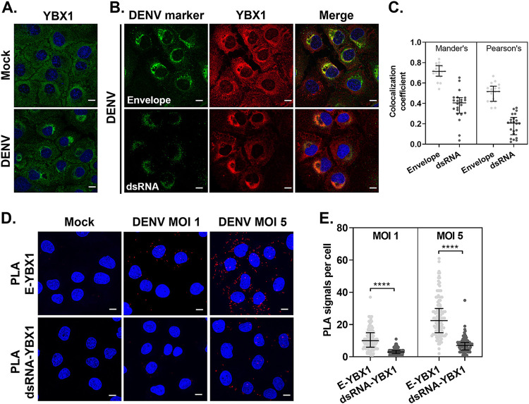
FIG 2.
YBX1 localizes to DENV assembly sites. (A, B) Representative immunofluorescence of Huh7 cells mock-infected or infected with DENV at MOI 5 for 24 hpi and stained for the indicated markers. (C) Pearson's and Manders' correlation coefficients were extracted from the data set generated by the coloc2 plugin for imageJ. (D) At 24 hpi, Huh7 cells were processed for in situ proximity ligation assay (PLA) of E and YBX1 (upper panels) and dsRNA-YBX1 (lower panels). Nuclei were stained with DAPI (blue) and PLA signals are detected as red puncta. (E) The number of PLA signals per cell were counted in at least 30 cells per experiment using an in-house macro for ImageJ. Data are presented as median ± IQR from two independent experiments.

