Abstract
We experienced a case of pregnancy after hysterosalpingogram and residual lipiodol in the extraperitoneal space. Initially, we suspected a metallic remnant; however, analysis by mass spectrometer confirmed that it was a remnant of lipiodol.
Keywords: hysterosalpingogram, lipiodol, mass spectrometry, remnant
We experienced a case of pregnancy after hysterosalpingogram and residual lipiodol in the extraperitoneal space. Initially, we suspected a metallic remnant; however, analysis by mass spectrometer confirmed that it was a remnant of lipiodol.

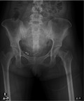
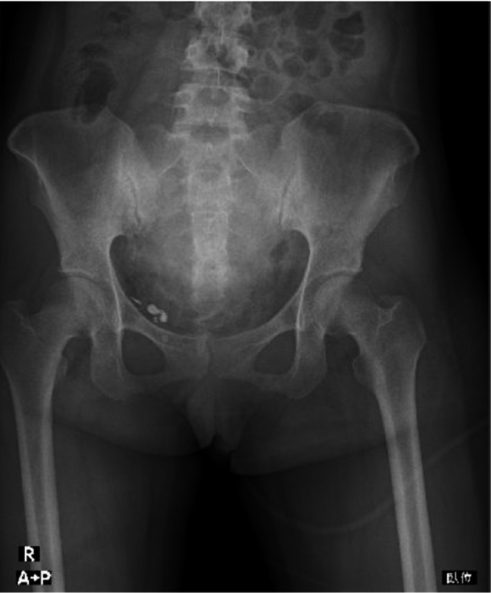
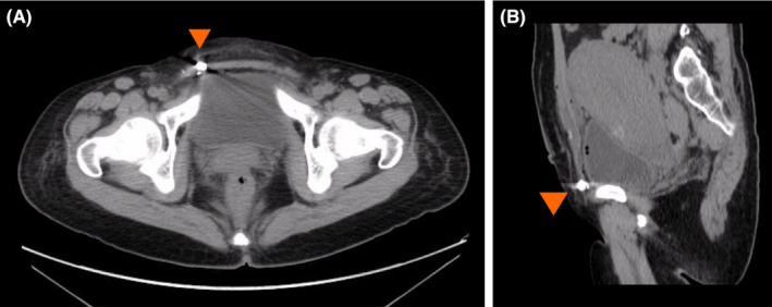
A 36‐year‐old nulliparous woman with 40 weeks of gestation was admitted to our department due to premature rupture of the membranes. She had no prior surgery. The hysterosalpingogram had been performed with 4 ml of lipiodol approximately 10 months prior to her presentation, and the fallopian tubes were normal. An emergency cesarean section was performed as intrauterine infection was suspected. Routinely performed postoperative abdominal X‐ray showed high‐enhancement shadows (Figure 1). It was confirmed that no gauze and instrument were lost after the surgery. CT images showed high‐density shadows in extraperitoneal along the inguinal canal (Figure 2), suggesting the presence of a metal device. Surgically, the cystic mass was located in the abdominal wall, connected to the inguinal canal, and was removed. The removed object was about 2 cm in diameter with a yellow liquid. The only connective tissue was found pathologically. Mass spectrometry showed the substance was highly consistent with the same lot number of lipiodol used at the previous clinic. There are a few cases of intraperitoneal remnants of lipiodol. 1 , 2 In our case, it was found outside the abdominal cavity. Gynecologists should keep in mind the possibility of contrast agent remnants when examining X‐ray on the patient with a history of hysterosalpingogram.
FIGURE 1.

Abdominal X‐ray image showed high‐density shadows
FIGURE 2.

Abdominal CT images showed high‐density object with metal artifact outside the abdominal cavity. (A) Transverse plane and (B) sagittal plane
CONFLICTS OF INTEREST
The authors declare no conflicts of interest associated with this manuscript.
AUTHOR CONTRIBUTIONS
CK: managed the patient, collected the data, and wrote the paper. SG: managed the patient and wrote the paper. YY: collected the data and performed the analysis. FC: collected the data and performed the analysis. HI: collected the data and performed the analysis. EK: managed the patient and wrote the report.
CONSENT
Written informed consent was obtained from the patient to publish this report in accordance with the journal's patient consent policy.
ACKNOWLEDGMENTS
We greatly thank Yuichi Kodaira for English language editing, and our hospital's medical staff for the assistance. We would like to acknowledge the patient for providing written consent to share her case prior to the submission of the manuscript.
Kurimoto C, Goto S, Yamagishi Y, Chiba F, Iwase H, Kato E. Lipiodol remnants misinterpreted as a metal device on postoperative abdominal X‐ray images. Clin Case Rep. 2022;10:e05553. doi: 10.1002/ccr3.5553
REFERENCES
- 1. Wakabayashi Y, Hashimura N, Kubouchi T. Retained Lipiodized Oil Misdiagnosed as residual metallic material. Radiat Med. 2004;22(5):362‐363. [PubMed] [Google Scholar]
- 2. Roest I, Rosielle K, van Welie N, et al. Safety of oil‐based contrast medium for hysterosalpingography: a systematic review. Reprod BioMed Online. 2021;42(6):1119‐1129. [DOI] [PubMed] [Google Scholar]


