Abstract
Rho guanine nucleotide exchange factor 10 (ARHGEF-10) is a RHO GTPase that has a role for neural morphogenesis, however its effect on the eyes remains unknown. Here, we report a 44-year-old man who presented with eyelid swelling along with a history of bilateral hand contractures, high-arched feet and muscle wasting, who was found to have an ARHGEF-10 mutation. Neuroimaging was significant for numerous nerve-based cystic abnormalities in the bilateral orbits and throughout the neuraxis, and an orbital biopsy revealed S-100 and SOX-10 positive lesion consistent with pseudocysts. While the role of ARHGEF-10 remains unclear, further research is warranted to further describe its clinical manifestations.
Keywords: eye, neurology (drugs and medicines)
Background
Rho guanine nucleotide exchange factor 10 (ARHGEF-10) is a Rho GTPase that stimulates the exchange of guanine diphosphate for guanine triphosphate. Laboratory data showed that ARHGEF-10 has a potential role in myelination control and neural morphogenesis.1 2 However, clinical manifestations of ARHGEF-10 mutation remain unknown. Literature has related this gene to Charcot-Marie-Tooth (CMT) disease, although a study of patients with autosomal dominant mutation of this gene presented no features of CMT.3 Here we report a case of a patient presenting with eyelid swelling who was found to have hypertrophic neural lesions associated with ARHGEF-10 mutation.
Case presentation
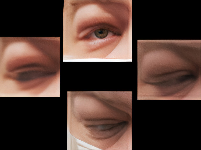
A man in his 40’s was referred to the oculoplastic surgery clinic for right upper eyelid swelling of 1 month. He was initially treated by an outside provider for presumed preseptal cellulitis with oral antibiotics without improvement. On examination, visual acuity was 20/20 in each eye with normal intraocular pressures. External examination revealed fullness in the bilateral upper eyelids, right greater than left eye, with full extraocular motility (figure 1). Anterior and posterior segment examinations were normal.
Figure 1.
External photo demonstrating eyelid fullness with normal extraocular motility.
Further history revealed that the patient had a ‘locking’ sensation in his fingers and joints since age 20s with progressive worsening to the point of contractures. Neurological examination demonstrated bilateral muscle wasting, thin distal ‘stork’ legs and pes cavus.
Investigations
Electromyography/nerve conduction studies demonstrated bilateral absent responses in median, ulnar and radial motor responses, and ulnar-below-elbow nerve velocity to be 29.3 m/s. This prompted genetic testing, which revealed a variant of uncertain significance in the ARHGEF-10 gene.
CT of the face demonstrated multiple elongated fusiform hypodense structures along multiple cranial nerves in the orbits and skull base concerning for multiple nerve sheath tumours (figure 2). T2-weighted MRI demonstrated numerous large minimally enhancing hyperintense lesions along nerves within the face, exiting along nerve roots of the cervical, thoracic and lumbar spine concerning for neural cysts (figure 2).
Figure 2.
CT of the head (left) and MRI of the head (middle) and spine (right) demonstrating numerous large hyperintense lesions along nerves.
Differential diagnosis
Orbital biopsy via anterior orbitotomy and ultimately excision of the right frontal nerve (there was no surgical plane between the ‘cyst’ and nerve structure) demonstrated normal nerve components with intraneural myxoid-like material that was S-100, SOX-10 and neurofilament positive but Epithelial membrane antigen (EMA)-stain negative. There was no evidence of malignancy or infection. Despite what grossly appeared like a large clear fluid-filled cystintraoperatively, histopathology did not show an epithelial-lined cyst but rather separated nerve bundles with mucoid degeneration; thus fluid spaces represented pseudocysts. Subsequent cervical spinal laminectomy, performed urgently due to the degree of spinal cord compression, yielded similar biopsy results.
Treatment
Management is mainly support, but may include surgical therapy such as spinal cord decompression.
Outcome and follow-up
We report a case of a patient with neurological abnormalities similar to CMT since young adulthood with multiple neural cystic lesions of essentially all cranial and spinal nerve roots brought to light due to prolonged eyelid swelling that on clinical examination and imaging mimicked orbital inflammatory syndrome. ARHGEF-10 gene mutation may be responsible for the multiple nerve lesions found in this patient. This is the first article to report this gene’s potential ocular/orbital involvement. The gene’s association to CMT requires further elucidation.
Discussion
The ARHGEF-10 gene has not been well studied. According to Mouse Genome Informatics, the gene has been associated with behavioural, neurological and metabolic disorders in mice with no identified ocular involvement.2 The gene has been strongly linked to slowed nerve conduction.1 2 According to animal studies, ARHGEF-10 may be associated with peripheral neuropathies like Charcot-Marie-Tooth (CMT) disease, although CMT patients do not typically have ARHGEF-10 mutations.3 In a study of Leonberger and Saint Bernard dogs with ARHGEF-10 mutations, the dogs showed phenotypic features of CMT.4 A study of cancer patients with this gene mutation demonstrated their nervous system to be highly susceptible to chemotoxic agents and was associated with chemotherapy-induced peripheral neuropathy.5 6 However, a pivotal study done by Verhoeven et al of a four-generation family with autosomal dominant ARHGEF-10 mutation only demonstrated slowing of nerve conduction velocities and no phenotypic symptoms of CMT.3
Our patient has some notable phenotypic characteristics of CMT such as claw hands and stork legs.7 8 Studies of CMT-related ocular findings have been limited. Ma’luf et al described a known CMT patient with phenotypic features such as claw hands who had orbital lesions similar to the presently reported patient, however, contrary to our patient, the pathology of the orbital lesion was a mixture of S-100 positive and S-100 negative cells consistent with neurofibroma.9
Learning points.
Rho guanine nucleotide exchange factor 10 (ARHGEF-10) is a RHO GTPase that may present as orbital inflammation.
ARHGEF-10 patients may present with Charcot-Marie-Tooth phenotype.
ARHGEF-10 may present with nerve based cystic abnormalities in orbits.
Footnotes
Contributors: EKT contributed in the planning, conducting and reporting of the case report. She also critically reviewed the report and was involved in collecting data and patient care for the study patient. NVL served as a scientific advisor. She critically reviewed the report, and was involved in collecting data and patient care for the study patient. MTJ served as a scientific advisor. He critically reviewed the report, and was involved in collecting data and patient care for the study patient. MAM served as a scientific advisor. He critically reviewed the report, and was involved in collecting data and patient care for the study patient. MKD served as a scientific advisor. She critically reviewed the report, and was involved in collecting data for the study patient. DRL contributed in planning, conducting, reporting of the case report. He served as a scientific advisor, and critically reviewed the report. All authors had no conflicts of interest.
Funding: The authors have not declared a specific grant for this research from any funding agency in the public, commercial or not-for-profit sectors.
Case reports provide a valuable learning resource for the scientific community and can indicate areas of interest for future research. They should not be used in isolation to guide treatment choices or public health policy.
Competing interests: None declared.
Provenance and peer review: Not commissioned; externally peer reviewed.
Ethics statements
Patient consent for publication
Consent obtained directly from the patient(s).
References
- 1.The Gene Card: Human Gene Database . ARHGEF-10 gene, 2020. Available: https://www.genecards.org/cgi-bin/carddisp.pl?gene=ARHGEF10 [Accessed 1 Nov 2020].
- 2.Mouse Genome Informatics . Arhgef10 gene detail. Available: http://www.informatics.jax.org/marker/MGI:2444453
- 3.Verhoeven K, De Jonghe P, Van de Putte T, et al. Slowed conduction and thin myelination of peripheral nerves associated with mutant Rho guanine-nucleotide exchange factor 10. Am J Hum Genet 2003;73:926–32. 10.1086/378159 [DOI] [PMC free article] [PubMed] [Google Scholar]
- 4.Ekenstedt KJ, Becker D, Minor KM, et al. An ARHGEF10 deletion is highly associated with a juvenile-onset inherited polyneuropathy in Leonberger and Saint Bernard dogs. PLoS Genet 2014;10:e1004635. 10.1371/journal.pgen.1004635 [DOI] [PMC free article] [PubMed] [Google Scholar]
- 5.Beutler AS, Kulkarni AA, Kanwar R, et al. Sequencing of Charcot-Marie-Tooth disease genes in a toxic polyneuropathy. Ann Neurol 2014;76:727–37. 10.1002/ana.24265 [DOI] [PMC free article] [PubMed] [Google Scholar]
- 6.Boora GK, Kulkarni AA, Kanwar R, et al. Association of the Charcot-Marie-Tooth disease gene ARHGEF10 with paclitaxel induced peripheral neuropathy in NCCTG N08CA (Alliance). J Neurol Sci 2015;357:35–40. 10.1016/j.jns.2015.06.056 [DOI] [PMC free article] [PubMed] [Google Scholar]
- 7.Charcot Marie Tooth Disease . MayoClinic. Available: https://www.mayoclinic.org/diseases-conditions/charcot-marie-tooth-disease/symptoms-causes/syc-20350517
- 8.Bird TD. Charcot-marie-tooth (Cmt) hereditary neuropathy overview. In: Adam MP, Ardinger HH, Pagon RA, eds. GeneReviews®. Seattle: University of Washington, 1993. [Google Scholar]
- 9.Ma'luf RN, Noureddin Baha' N, Ghazi NG, et al. Bilateral, localized orbital neurofibromas and Charcot-Marie-Tooth disease. Arch Ophthalmol 2005;123:1443–5. 10.1001/archopht.123.10.1443 [DOI] [PubMed] [Google Scholar]


