ABSTRACT
Alphaviruses are enveloped viruses transmitted by arthropod vectors to vertebrate hosts. The surface of the virion contains 80 glycoprotein spikes embedded in the membrane, and these spikes mediate attachment to the host cell and initiate viral fusion. Each spike consists of a trimer of E2-E1 heterodimers. These heterodimers interact at the following two interfaces: (i) the intradimer interactions between E2 and E1 of the same heterodimer and (ii) the interdimer interactions between E2 of one heterodimer and E1 of the adjacent heterodimer (E1’). We hypothesized that the interdimer interactions are essential for trimerization of the E2-E1 heterodimers into a functional spike. In this work, we made a mutant virus (chikungunya piggyback [CPB]) where we replaced six interdimeric residues in the E2 protein of Sindbis virus (wild-type [WT] SINV) with those from the E2 protein from chikungunya virus and studied its effect in both mammalian and mosquito cell lines. CPB produced fewer infectious particles in mammalian cells than in mosquito cells, relative to WT SINV. When CPB virus was purified from mammalian cells, particles showed reduced amounts of glycoproteins relative to the capsid protein and contained defects in particle morphology compared with virus derived from mosquito cells. Using cryo-electron microscopy (cryo-EM), we determined that the spikes of CPB had a different conformation than WT SINV. Last, we identified two revertants, E2-H333N and E1-S247L, that restored particle growth and assembly to different degrees. We conclude the interdimer interface is critical for spike trimerization and is a novel target for potential antiviral drug design.
IMPORTANCE Alphaviruses, which can cause disease when spread to humans by mosquitoes, have been classified as emerging pathogens, with infections occurring worldwide. The spikes on the surface of the alphavirus particle are absolutely required for the virus to enter a new host cell and initiate an infection. Using a structure-guided approach, we made a mutant virus that alters spike assembly in mammalian cells but not mosquito cells. This finding is important because it identifies a region in the spike that could be a target for antiviral drug design.
KEYWORDS: Glycoprotein spike, assembly, host-range, alphaviruses
INTRODUCTION
Alphaviruses are transmitted most commonly by arthropod vectors, usually mosquitoes, to vertebrate hosts, including humans, birds, and horses (1, 2). While a majority of alphaviruses have an arthropod vector, a group of alphaviruses have been identified to transmit only between invertebrates (3), and others use a different vector to infect aquatic animals (4). Therefore, for an alphavirus to complete its infection cycle, it must be able to assemble particles in all of these host environments. Virus infection must rely on different host factors since the same viral proteins are synthesized.
The alphavirus genome consists of four nonstructural proteins and six structural proteins, which are required for viral genome replication and particle assembly, respectively (1, 2, 5, 6). The alphavirus particle consists of an inner nucleocapsid core, a host-derived lipid membrane, and 80 trimeric spikes on the surface of the virion (7). The spikes are trimers of E2-E1 heterodimers with each heterodimer forming the edge of a triangular spike. The E2 and E1 proteins each contain a single transmembrane domain and are embedded within the viral membrane. The endodomain of E2 interacts with the capsid protein in the core in a 1:1 ratio (8–12). Thus, the E2 protein transits the entire particle and helps align the core and the spikes, which is a unique feature of alphaviruses compared with other enveloped viruses (7).
Alphavirus spike assembly is a highly regulated process that depends on specific interactions between the viral proteins and host factors. The structural proteins are translated as a capsid-E3-E2-6K-E1 polyprotein (or capsid-E3-E2-TF when frameshifting occurs). The capsid is cleaved autoproteolytically from the rest of the polyprotein (13). E3 serves as the signal sequence to translocate the rest of the polyprotein into the endoplasmic reticulum (ER) (14, 15), where the cellular enzyme signalase cleaves the polyprotein into E3-E2 (also known as pE2 or P62), 6K, and E1 (16, 17). When programmed ribosomal frameshifting occurs, the protein pE2 and TF are translated (5). In mammalian cells, the ER chaperones Erp57 and calnexin/calreticulin regulate the folding and disulfide bond formation of E2 (18–20), and BiP and protein disulfide isomerase do the same for E1 (18–22). pE2 and E1 form heterodimers within the ER, before transiting to the Golgi where trimerization of the stable spike complex is predicted to occur. Glycosylation of E2 and E1 occur in the ER and Golgi. In the ER and Golgi, there is a slight decrease in pH, and the E3 proteins act as a clamp and prevent low pH-mediated dissociation of the E2-E1 heterodimer (23–25). These spikes in the stable conformation are transported to the plasma membrane through the host secretory system. In the late secretory pathway, the cellular enzyme furin cleaves E3 from E2, which acts as a priming event and converts the spike from the stable to metastable conformation (26, 27). At the plasma membrane, E2 interacts with the nucleocapsid core and initiates budding and virus release. Particles may also bud from glycoprotein-containing vesicles called cytopathic vesicles II, and this pathway is used more by mosquito cells (28).
Voss et al. solved the chikungunya virus (CHIKV) E2-E1 heterodimer in the metastable conformation and in complex with E3 at neutral pH in the stable conformation (29), and Li et al. solved the Sindbis virus (SINV) E2-E1 heterodimer at acidic pH (30). These structures showed the intradimer contacts within a heterodimer. Both groups fit the atomic heterodimers into cryo-electron microscopy (cryo-EM) structures of alphavirus virions to identify interdimer interactions, or contacts between heterodimers within the spike, and between the E2 and nucleocapsid core. E1 consists of three domains, I to III. Domain II contains the fusion peptide and makes extended contacts with E2 in the intradimer heterodimer (29–31). This intradimer interface contains the acid-sensitive region and has been studied in regard to fusion regulation and mutations that expand vector range (32, 33). E2 has three domains, A to C. Domain B is the most distal domain and acts as a cap of the distal end of E1 protecting the fusion peptide (29, 30). In the low pH structure by Li et al., Domain B was disordered, suggesting it is the first portion of the heterodimer to undergo conformational changes in response to low pH (30). Domain A in E2 is the central domain, and Domain C is the closest domain to the lipid bilayer. Domain C is sandwiched between Domain II of E1 from its intradimer heterodimer and the Domain II of E1 from the adjacent heterodimer, or E1’. Both Voss et al. and Li et al. identified residues in E2 Domain C that contact residues in Domain II of E1’ (29, 30). Based on these two structures, we hypothesized that E2 Domain C plays a key role in spike assembly, and disrupting the interdimer contacts between E2 and E1’ could affect trimer formation.
To test the role of Domain C in assembly, we substituted six amino acids in SINV E2 Domain C to the corresponding residues in CHIKV, an alphavirus in a different clade (2, 34). The E2 residues that were mutated are predicted to interact with E1’ residues in the adjacent dimer or to “piggyback” on the dimers within a spike, and hence, our mutant was named chikungunya piggyback (CPB). We determined that CPB had slower growth and smaller plaques when grown in mammalian cells but not mosquito cells. CPB quickly gained second-site revertants, namely, E2-H333N and E1-S247L, which were each able to restore growth in CPB. CPB grown in mammalian cells forms particles with assembly defects, but the revertants restored their assembly to various degrees. A further analysis of the spike conformations on the CPB by cryo-EM showed the spikes in CPB were more flexible than the spikes in the WT virus.
RESULTS
Domain C of E2 is important for spike trimerization.
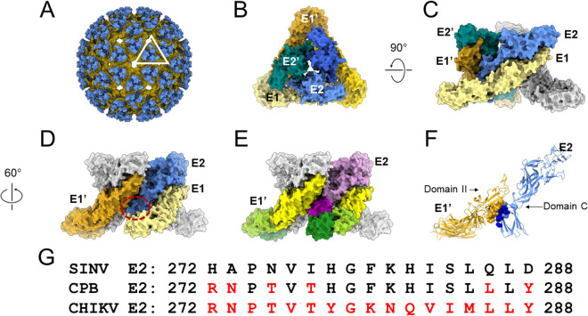
The atomic structure of the CHIKV E2-E1 heterodimer identified the interface between E2 and E1 within a heterodimer and the intradimer contacts that were present. When these heterodimers were placed into the cryo-EM density of intact SINV virions, the potential contacts between heterodimers, or interdimer contacts, were identified (Fig. 1A to C). In the virion, Domain C of the E2 protein was sandwiched between two E1 proteins, namely, one in the same heterodimer, designated E1, and one from the adjacent heterodimer, designated E1’ (Fig. 1D and E) (29). We have colored the different domains of E1 and E2 in Fig. 1E to illustrate these interactions of Domain C of E2. We hypothesized that Domain C would be important for spike trimerization because these interdimer contacts would bridge or connect the individual E2-E1 dimers into trimers.
FIG 1.
Interdimer interface residues in Domain C of E2 may be important for spike assembly. (A) Crystal structure of CHIKV glycoproteins E2 (blue) and E1 (yellow) (PDB: 3N40) (29) fit into the cryo-EM map of SINV (PDB: 1Z8Y) (29). One trimeric spike is outlined. (B) During assembly, E1 (shades of yellow) forms heterodimers with E2 (shades of blue), and these dimers then trimerize. A top view of one of these trimeric spikes, with the individual E2 (in teal, light blue, and blue) and E1 (in gold, light yellow, and saffron) proteins is shown here. A total of 80 spikes are on the surface of the alphavirus particle. (C) The trimer is rotated 90 degrees for a side view. The light-blue E2 and light-yellow E1 are a heterodimer. Intradimer contacts occur between Domains I and II of E1 with Domains A, B, and C of E2. The teal E2’ and gold E1’ form another heterodimer, with similar intradimer contacts. The last dimer in the spike is shown in gray. (D) A 60-degree rotation of C highlights an interdimer interface in the trimer. Interdimer contacts are between the gold E1’ of one heterodimer and the light-blue E2 in the adjacent dimer. Domain C of E2 (red dashed circle) is sandwiched between the adjacent E1’ (gold) and its cognate E1 (light yellow), forming interdimer and intradimer contacts, respectively. (E) The same proteins, namely, E1’, E2, and E1, shown in D are color coded by the domain here. Domain III of E1' and E1 is dark green, Domain I of E1' and E1 is yellow green, and Domain II of E1' and E1 is yellow. Domain C of E2 is in dark purple and Domains A and B are in light purple. The other monomers are colored gray for clarity. (F) Ribbon diagram of the interdimer of E1’-E2. Residues in E1’ that contact E2 are in yellow residues in E2 that contact E1’ are in blue spheres (light and dark). The residues mutated in this study are in dark-blue spheres. (G) Amino acid alignment of CHIKV and SINV E2 in the interdimer region; SINV residues shown in black and CHIKV residues are shown in red. The CHIKV piggyback (CPB) chimera was generated by substituting the nonhomologous E2 CHIKV residues for the corresponding SINV E2 residues (highlighted in red in CPB) in this region.
Voss et al., identified 10 residues in the E2 Domain C that contact 12 residues in E1 Domain II in the adjacent heterodimer (29). In the low pH SINV E2-E1 heterodimer structure, Li et al. identified three residues in the E2 Domain C that contact four residues in E1’ Domain II, and all were also identified in the CHIKV structure (30). Residues E2-272 to E2-288 in Domain C contained a majority of the E2 interdimer contacts (Fig. 1F and G). While there are 13 residues that differ between SINV and CHIKV in the primary amino acid sequence in the E2-272 to E2-288 region, only 6 of these residues are in contact with E1’ in the tertiary structure. Using SINV as our parental virus, we mutated the six residues in this region from SINV to residues found in CHIKV, which belongs to a different clade than SINV (Fig. 1F and G) (2, 34). The resulting mutant was named chikungunya piggyback (CPB), as the SINV E1’ residues “piggyback” on the six E2 residues that were introduced from CHIKV. We opted to focus on the E2 interdimer residues because there are fewer contact residues in E2 than those in E1 and we speculated that mutating a larger number of residues in E1 would have increased the chance of misfolding due to too many disrupted interactions within the E1 protein itself (21).
Growth of CPB is attenuated in mammalian cells compared with growth in mosquito cells.
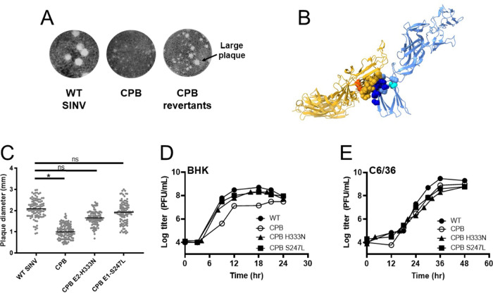
To determine the effect of our CPB mutation, we infected mammalian (BHK-21) and mosquito (C6/36) cells at a multiplicity of infection (MOI) of 1 PFU/cell and quantified infectious virus release over time. In BHK cells, the growth of CPB was attenuated, producing virus at a titer 1 to 1.5 logs lower than that of WT SINV (Fig. 2A) as early as 8 hours postinfection. Additionally, CPB had a mean plaque size of 1 mm compared with the mean plaque size of 2 mm for WT SINV (Fig. 3A and C). In C6/36 cells, however, CPB grew at a similar rate as WT SINV (Fig. 2B). BHK cells are typically grown at 37°C, while C6/36 cells are grown at 28°C. To rule out temperature dependence on growth, we also measured WT SINV and CPB growth in BHK cells grown at 28°C. We found that there was still a 1- to 1.5-log reduction in CPB titer relative to the WT SINV titer (Fig. 2C), suggesting that attenuated CPB growth is mainly an effect of a different host cell environment, rather than solely different temperatures.
FIG 2.
CPB grows slower than WT SINV in mammalian cells than in mosquito cells. Cells were infected at an MOI of 1 PFU/cell. At the indicated time points, the medium was collected and replaced with fresh media. The titers of the collected samples were determined by standard plaque assay on BHK cells. Results are shown for one representative experiment (n = 5). (A) Growth kinetics of infectious virus released from BHK cells at 37°C show that CPB was attenuated by 1 to 1.5 logs relative to WT SINV. (B) Growth kinetics of virus grown in C6/36 cells at 28°C show CPB releases infectious particles at the same rate as WT SINV. (C) Growth kinetics of virus grown in BHK cells at 28°C show that CPB growth is still attenuated by 1 to 1.5 logs relative to WT SINV.
FIG 3.
Two CPB revertants map close to the interdimer interface and grow similarly to WT SINV. (A) CPB collected 20 to 40 hours postelectroporation have smaller plaques than WT SINV plaques (P < 0.0015, see 3C). When CPB is harvested later, or as it is passaged in BHK cells, larger plaques are seen in addition to the small plaques. Five of these larger plaques were isolated, the RNA was sequenced, and two independent second-site revertants were found. (B) The locations of 2 second-site revertant sites, namely, H333N in E2 (cyan) and S247L in E1 (orange), are shown in the E1’-E2 dimer. E1’ and E2 contact residues are colored in yellow and blue, respectively, as they were in Fig. 1F; mutated sites in CPB are in dark blue. (C) CPB E2-H333N and CPB E1-S247L were introduced independently into CPB. The plaque size of CPB E2-H333N and CPB E1-S247L in BHK cells was larger than that of CPB and was not significantly (ns) different from that of the WT. (D and E) Growth kinetics of the two revertants show they have comparable titers as WT SINV in BHK (D) and C6/36 (E) cells. Representative curves are shown (n = 3). Cells were infected at an MOI of 1 PFU/cell, medium was collected at the indicated time points, and the titers of samples were determined on BHK cells.
Two separate second-site revertants identified for CPB.
As we worked with the CPB virus, we noticed that the virus reverted quickly as indicated by the change in plaque size phenotype from small to large. Changes in plaque size were evident often after cells were infected more than 40 hours or passaged more than 2 or 3 times (Fig. 3A). To isolate revertants, we passaged CPB in BHK cells and saw a mixed plaque phenotype. We isolated and plaque-purified larger plaques and then isolated and sequenced the viral RNA. We identified two independent single-site revertants, namely, E2-H333N and E1-S247L (Fig. 3B). E2-H333N was seen four times, and E1-S247L was seen once. Another mutation, E1-P250S, was seen twice but only in combination with E2-H333N. We focused on E2-H333N and E1-S247L since these single sites potentially changed the virus fitness on their own. Both the E2-H333N and E1-S247L mutations were in close proximity to the interdimer interface (Fig. 3B). E2-H333N is spatially near the six-residue cluster we mutated in E2, approximately 13 Å away from the center of the E2 interdimer contacts. E1-S247L is approximately 20 Å away from the center of the 10 residues of E1’ which interact with the 6 residues of E2. SINV E1 is glycosylated at position 245. The E1-S247L mutation abrogates the N-X-S/T motif needed for N-linked glycosylation at residue E1 245 (35), by changing the Ser to Leu (Table 1).
TABLE 1.
Location of glycosylation sites on E2 and E1 proteins in Sindbis and chikungunya virus
| Virus | Protein | Glycosylation site |
|---|---|---|
| SINV | E2 | N196 |
| E2 | N318 | |
| E1 | N139 | |
| E1 | N245 | |
| CHIKV | E2 | N263 |
| E2 | N345 | |
| E1 | N141 |
To determine how these point mutations affect viral assembly, both of these mutations were inserted back into CPB and the WT SINV. The plaque sizes of the two revertants cloned into CPB were larger, which is comparable to that seen with WT SINV. The mean plaque size of CPB E2-H333N was 1.6 mm and of CPB E1-S247L was 1.9 mm; both were larger than CPB which was 1 mm (Fig. 3C). Next, the growth kinetics of CPB E2-H333N and CPB E1-S247L were examined independently by infecting BHK cells (Fig. 3D). Both revertants resulted in a higher virus yield which grew between 1 and 2 logs better than the parental CPB and at a similar rate as WT SINV, indicating that both revertants rescued growth in the CPB background. We also conducted growth kinetics in C6/36 cells (Fig. 3E) and observed no difference between the two revertants in CPB and WT SINV. When the two mutations were independently cloned into the WT SINV backbone and the viral growth examined in both BHK and C6/36 cells, we observed no change in plaque size or growth kinetics. These results indicate that both of the mutations are neither deleterious nor enhancing in the absence of the CPB mutations.
CPB particles show defects in spike incorporation compared with WT SINV.
Alphavirus spikes initially form dimers, and then these dimers trimerize before localizing to the plasma membrane. If the spikes do not trimerize, transport to the plasma membrane is reduced. Spike trimerization is thought to be important for alphavirus budding. In our CPB mutant, we used the alphavirus structures and targeted residues that we thought would disrupt spike trimerization but not dimerization as determined from Voss et al. and Li et al. (29, 30). To determine how particle budding was affected in the CPB mutant, we looked at the composition and morphology of purified virus particles from both BHK and C6/36 cells.
We purified virus particles through a sucrose cushion, ran them on an SDS-PAGE gel, and examined the protein composition. We noticed that BHK-purified CPB particles have reduced amounts of E1 and E2 glycoproteins relative to the capsid protein compared with the amounts of these proteins in WT SINV (Fig. 4A). This finding could suggest that fewer spikes are associated with the virion and/or the associated spikes may easily dissociate during the purification process compared with WT particles. The E1 protein band in CPB E1-S247L migrates faster than E1 in WT SINV. The E1 protein in CPB E1-S247L is at the same position as the E1 band in the SINV E1-N245/246Q virus which is known to have one of the two E1 glycosylation sites disrupted resulting in a faster migrating E1 protein band. As an additional control, the purified virion of SINV E1-N139Q/E2-N318Q, which has one E1 glycosylation site and one E2 glycosylation site disrupted resulting in faster migration in both of those protein bands (Fig. 4A), was also included (35).
FIG 4.
Particle morphology and composition are restored in CPB revertants to various degrees. Cells were infected at an MOI of 0.1 PFU/cell for 1 hour, after which serum-free medium was overlaid. Infected BHK medium was collected after 24 hours, and infected C6/36 medium was collected after 120 hours. The medium was clarified and purified through a sucrose cushion at 140,000 × g for 2.5 h. (A and B) To determine the composition of the virions, purified virus was solubilized in reducing SDS sample buffer, run on an 8% SDS-PAGE gel, and imaged by stain-free 2,2,2-trichloroethanol (TCE). CPB particles have a reduced level of E2 and E1 glycoproteins in particles purified from BHK cells. The E1 deglycosylation control is SINV E1-N245/246Q, which has one of its two E1 glycosylation sites deglycosylated resulting in a faster migrating E1 protein band, and the E1/E2 deglycosylation control is E1-N139Q/E2-N318Q and has a deglycosylation site in both E1 and E2 resulting in faster migration in each of those protein bands. (C and D) Purified particles were applied to a Formvar- and carbon-coated 400-mesh copper grid, stained with 2% uranyl acetate, and imaged at ×20,000 magnification with the JEOL 1010 transmission electron microscope. Scale bar for TEM, 200 nm.
In contrast to BHK cells, C6/36-purified CPB particles showed similar amounts of E1 and E2 glycoproteins to WT SINV (Fig. 4B), suggesting qualitatively that the amounts of spike proteins in released and purified particles were similar to those of the WT SINV. The revertants also had a similar protein composition to WT SINV, with CPB E1-S247L having a faster migrating E1 protein band (Fig. 4B). In both CPB and CPB E2-H333N virions, the E2 protein is a smeared band suggesting heterogenous glycosylation on the E2 protein (36).
CPB particles from mammalian cells show defects in particle morphology, which are partially or fully rescued by revertants.
The reduced amount of glycoproteins in CPB particles suggested that the CPB particle may be morphologically different compared with WT particles. To test this hypothesis, we stained our particles with uranyl acetate and imaged them using transmission electron microscopy (TEM). WT SINV is approximately 70 nm in size and spherical, and this result is observed in WT particles purified from BHK (Fig. 4C) and C6/36 cells (Fig. 4D). CPB, however, made almost no identifiable virus particles when purified from BHK cells (Fig. 4C). We also noticed that any visible particles were not spherical. CPB E2-H333N partially rescued particle morphology, while CPB E1-S247L fully recovered particle morphology (Fig. 4C). CPB E2-H333N made more particles than CPB, and some of them looked to be around 70 nm in size; however, many nonspherical particles were still seen. On the other hand, CPB E1-S247L made primarily spherical particles of a 70-nm diameter, which were similar to those of WT SINV. In both CPB E2-H333N and CPB E1-S247L, there are also small particles ranging in size of approximately 10 to 40 nm, which could be assembly intermediates or disassembled fragments. These smaller particles suggest that CPB E2-H333N and CPB E1-S247L particles may still be fragile compared with WT SINV, despite being just as infectious.
WT SINV, CPB, and revertant virus particles purified from C6/36 were spherical and homogenous in shape, which is consistent with no major assembly or growth defects (Fig. 4D). CPB particles appeared to have dimples or creases. This result could suggest that even in C6/36 cells the mutations made in CPB are detrimental to proper spike formation or have defects in assembly but are still good enough that infectious particle assembly occurs (28).
Glycoprotein transport is not significantly affected in CPB.
The small amounts of glycoproteins in CPB virions compared with WT SINV virions from BHK cells led us to two hypotheses. One hypothesis was that CPB has defects in spike trimerization and transport to the plasma membrane is diminished. A second hypothesis was that CPB virions are misassembled because interdimer contacts have been disrupted. As a result, CPB may disassemble more than WT SINV during the purification process. These options are not mutually exclusive.
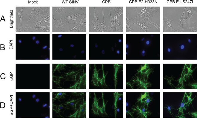
We used immunofluorescence to test if glycoprotein transport was altered in CPB compared that of WT SINV. We infected BHK cells at an MOI of 2 PFU/cell for 10 h, a time when there was a difference in infectious particles released, and probed for glycoproteins at the cell membrane (Fig. 5). Cells were fixed with paraformaldehyde, a nonpermeabilizing fixative, and probed for SINV glycoproteins. Under these infection conditions, cells were not displaying CPE (Fig. 5A and C). There was no drastic difference in glycoprotein levels at the plasma membrane between WT SINV, CPB, and revertant infected cells (Fig. 5C and D). Virus-infected cells were equally healthy and had comparable amounts of detectable glycoproteins.
FIG 5.
CPB glycoprotein spikes are transported to the plasma membrane of BHK cells in approximately equal amounts compared with WT. BHK cells were infected at an MOI of 2 PFU/cell. Ten hours postinfection, the cells were fixed with 4% EM-grade paraformaldehyde and probed for SINV E1/E2. Images were taken at 40× magnification on a Nikon Ni-E microscope. Representative images are shown (n = 4). (A and B) Brightfield images (A) and DAPI staining (B) show cells in field of view and their nuclei in blue. (C) Cell surface E2 and E1 expression is shown in green, and Alexa Fluor 488 was the secondary antibody. (D) Anti-E2 and E1+DAPI merge shows green fluorescence from the surface glycoprotein expression relative to the cells present.
Spikes in CPB particles are distorted compared with WT SINV.
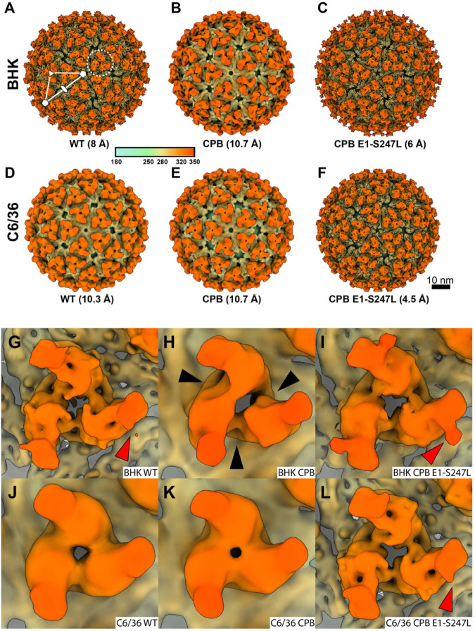
The difference in particle morphology between CPB and WT SINV in mammalian cells was striking when imaged by TEM (Fig. 4C and D). To further examine the particle structure and spike morphology, we purified virions and used cryo-EM to solve their 3D reconstructions. We focused on WT SINV, CPB, and the revertant CPB E1-S247L since they had the most extreme phenotypes and their structures would be most clear by cryo-EM (Fig. 6). We imposed icosahedral averaging (Table 2) during the reconstruction process. The overall structural organizations of all six particles (WT SINV, CPB, and CPB E1-S247L from BHK and C6/36) were preserved (Fig. 6A to F). There are 80 petal-like spikes arranged into a triangulation number of T = 4 surface lattice; the white triangle delineates 1 asymmetric unit (Fig. 6A) (37). No clear E3 density was observed in any of the particles, which is consistent with what has been previously observed with SINV (8). The glycan modification at E2-196, located at the distal end of the E2 protein, was seen clearly when the estimated resolution was better than 10 Å (red arrows). As seen in other alphavirus structures (7, 38–42), the E1 protein contributes to the continuous shell underneath the spikes (Fig. 6A to F, yellow color) with holes at every 2-fold and 5-fold above the membrane. Note the resolution of the particles range from 4.5 Å to 10.7 Å, which reflects both the number of particles used in the reconstruction (Table 2) and the heterogeneity of the particles themselves.
FIG 6.
Cryo-EM 3D reconstructions show that CPB spikes from mammalian cells have an altered interdimer organization. (A to F) Radially colored isosurface rendering of WT SINV, CPB, and SINV E1-S247L from BHK cells (A to C, top row) and C6/36 cells (D to F, bottom row). All views are at the 5-fold axis. All six maps have a similar outer appearance with 80 spikes decorating a fenestrated surface. In A, the white triangle shows one asymmetric unit. The filled oval, triangle, and pentagon indicate locations of 2-fold, 3-fold, and 5-fold axes, respectively. The dotted circle shows one of the spikes on the quasi-three axis. (G to L) Enlarged views of SINV spikes at the quasi-3-fold location, and all images are shown at a 10-Å resolution for comparison purposes. The spike of CPB from BHK (H) shows a distinct organization from other SINV spikes. Black arrowheads indicate the difference in the electron density between each interdimer interface. Red arrowheads show glycan modification at E2-196.
TABLE 2.
Parameters for data collection and 3D image reconstructions
| Reconstruction data | Data by cell type and virus |
|||||
|---|---|---|---|---|---|---|
| BHK |
C6/36 |
|||||
| WT SINV | CPB | CPB E1-S247L | WT SINV | CPB | CPB E1-S247L | |
| Data collection information | ||||||
| Electron microscope | TFS Titan Krios | TFS Titan Krios | TFS Titan Krios | JEOL 3200FS | TFS Titan Krios | TFS Titan Krios |
| Operation voltage (kV) | 300 | 300 | 300 | 300 | 300 | 300 |
| Electron detector | Gatan K3 | Gatan K3 | Gatan K3 | DE-12 | Gatan K3 | Gatan K3 |
| Energy filter (slit width in eV) | 20 | 20 | 20 | 20 | 20 | 20 |
| Data collection mode | Counting | Superresolution counting | Counting | Integrated | Counting | Counting |
| Nominal magnification (×) | 105,000 | 64,000 | 64,000 | 80,000 | 64,000 | 64,000 |
| Pixel size in 3D map (Å [pixel size in the censor]) | 1.68 (0.84) | 2.72 (0.68) | 1.36 (1.36) | 1.9 (1.9) | 2.72 (1.36) | 1.36 (1.36) |
| Total accumulated dose (e−/Å2) | 30 | 30 | 30 | 30 | 30 | 30 |
| Data process statistics | ||||||
| Box size (pixel) | 500 | 300 | 600 | 440 | 300 | 600 |
| No. of particles | 9037 | 15956 | 16133 | 7102 | 63509 | 65053 |
| CTF estimation | CTFFIND4 | CTFFIND4 | CTFFIND4 | CTFFIND4 | CTFFIND4 | CTFFIND4 |
| Data processing software | RELION 3.1 | RELION 3.1 | RELION 3.1 | RELION 3.1 | RELION 3.1 | RELION 3.1 |
| Symmetry imposition | I2 | I2 | I2 | I2 | I2 | I2 |
| B-factor applied (Å2) | −545.7 | N/A | −311.4 | −297.3 | N/A | −237.3 |
| Final resolution (Å) | 8 | 10.7 | 5.9 | 10.3 | 10.7 | 4.5 |
However, when looking more carefully at the enlarged view of each spike at the quasi-3-fold axis, (dotted circle, Fig. 6A), there were discernible differences between the samples (Fig. 6G to L). We chose the quasi-3-fold spike because spikes at 5-, 3-, and 2-fold would, by default of icosahedral symmetry, be identical. The spikes at the quasisymmetry axes would not have any symmetry imposed on them, and differences in morphology would be more evident. To best compare the quasi-3-fold spikes, we show them all at 10.7 Å, which is the resolution of CPB from BHK cells. The irregular density at the interdimer location of the spike was clearest in CPB purified from BHK cells (Fig. 6H, black arrows). Note that the densities of two lobes at the spike petal were fused together, while the other heterodimer within the same spike remained separate (Fig. 6H, BHK CPB). For the other five particles, the E1/E2 dimers within a spike were organized in a trimeric manner with true trimeric symmetry.
DISCUSSION
To investigate the role of the interdimeric contacts in assembly, we used a structure-guided approach and mutated six residues in E2 that, in the stable and metastable structures of the spike, are at the interface with the adjacent E1’ protein (Fig. 1) (29, 30). We called this mutant CPB. We found that CPB growth is attenuated in mammalian cells compared with growth in mosquito cells (Fig. 2). In mammalian cells, CPB glycoprotein expression on the cell surface is similar to that of WT SINV (Fig. 5), but the released particles show that CPB particle composition and morphology are defective in mammalian cells, while they are affected minimally in mosquito cells (Fig. 4). Our cryo-EM structures of CPB show that CPB particle spikes do not have three symmetrical lobes. Fig. 6 shows a visible gap between two of the lobes and the third, which could suggest that two heterodimers are interacting but the third is not. The mutations in E2 disrupt interdimer contacts, and we hypothesized that CPB growth falls behind WT SINV because of its subsequent assembly defects. However, it is difficult to separate assembly defects from downstream entry impacts.
The difference in phenotype in mammalian and mosquito cells suggests that there is a host factor that is involved in the trimerization of E2-E1 heterodimers since the same viral proteins are assembling in each host cell. If this assembly was autonomous, we would not observe a host-specific difference in both infectious particle production and particle morphology. These differences could be due to different posttranslational modifications by the host, different host-protein chaperones affecting virus assembly, or a combination of both. BHK and C6/36 cells are defective in some innate immune responses, so the attenuation in growth is not entirely a host response to infection. The CPB mutant and its revertants will be useful to identify host chaperones important in spike assembly.
Stability, composition, and conformation of CPB and revertant spikes.
Our immunofluorescence results show that glycoprotein transport to the plasma membrane is comparable between CPB and WT SINV. However, nothing about the conformation of the spikes can be concluded. Furthermore, while E2-E1 are most stable as heterodimers, small amounts of the monomeric proteins will localize to the plasma membrane (15). Our results cannot differentiate conformation or oligomeric state of the glycoproteins at the cell surface.
It is perplexing that we can obtain a structure of CPB purified from BHK cells when this same sample shows few particles by negative-stain TEM, and there were low levels of glycoproteins when we look at particle composition on an SDS-PAGE gel (Fig. 4). There are several possibilities for these discrepancies, and likely more than one is a contributing factor.
Due to the trimerization defect, fewer spikes may be incorporated into virus particles, and those that are on the particle may be more heterogenous than those of WT SINV. The CPB trimer has a different conformation than the WT SINV trimer; it could also be that the cytoplasmic domain of E2 is no longer able to interact with the capsid in a 1:1 ratio, which affects titer and particle stability. The reduced number of spikes on the particles explains the lower titers (Fig. 2) and why purified particles show smaller amounts of spike proteins in CPB than those in WT SINV (Fig. 4). Our cryo-EM results also support this hypothesis. We used roughly 16,000 CPB and CPB E1-S247L particles each from BHK-infected cells for our reconstructions. Yet, the resolutions of the final structures were ∼11Å and 6 Å, respectively, suggesting more heterogenous particles for CPB than those for the revertant. WT SINV used only 9,000 particles and a resolution of 8 Å was obtained. From our cryo-EM reconstructions, we can see heterogeneity in the individual spikes of CPB compared with WT SINV and CPB E1-S247L particles. Interestingly, our TEM images show that CPB S247L produces particles that are much more WT like than CPB H333N, despite both revertants restoring CPB growth.
Together, the lower number of spikes and their misassembly and heterogeneity could account for particle instability. Fewer spikes on the surface means there is no E1 lattice that covers the viral envelope. The particles are more fragile and sensitive to chemicals. Particles are dehydrated and treated with an acidic stain when preparing for negative stain imaging, and as a result, particles may disassemble or aggregate. In contrast, flash freezing the samples for cryopreservation has a mild effect on the particle’s integrity, so misassembled particles will be frozen and can be analyzed.
Possible implications of revertant residues.
CPB quickly reverted resulting in two independently fit mutations, namely, E2-H333N and E1-S247L, and one mutation, namely, P250S, which was seen only in combination with E2 H333N. E2-H333N, E1-S247L, and P250S are all in close proximity to the cluster of mutations made at the interdimer interface, although none were identified by Voss or Li as one of the inter- or intradimer contact residues (29, 30). Residue 333 for most viruses is either a histidine or an asparagine. Additionally, the corresponding residue for SINV H333 is N330 in CHIKV. Because the histidine side chain has a pKa of 6.00, it is within the range of different pH environments in the host cell. This histidine residue may affect CPB either during entry, specifically during disassembly in the endosome (43, 44), or during assembly as the E2-E1 heterodimer travels through the ER and Golgi (45). The role in disassembly could be unfavorable in CPB since it may have a less stable trimer and less stable heterodimers even without a low pH environment. During assembly of the CPB E2-E1 heterodimers, there may be improper folding possibly making trimer assembly more sensitive to pH which cannot occur with H333.
S247L interferes with an N-linked glycosylation motif that allows E1-N245 to be glycosylated. In CHIKV, there is only one glycosylation site on E1 at N141 versus two in SINV, one at N139, and one at N245 (Table 1) (35, 38). Although residue 247 is not conserved among alphaviruses, it is clear that some viruses, including SINV, have the N-X-S/T glycosylation motif at N245, while others, including CHIKV, do not. It is interesting that CPB S247L appears more WT like in particle morphology than CPB H333N. It is possible that deglycosylation at this site allows for better folding or removes a steric hinderance that allows for trimerization.
From our revertant sequencing, we also saw that in some instances when E2-H333N was mutated, E1-P250S was also mutated. Residue 250 is a proline residue in SINV and a serine residue in CHIKV. It was seen with H333N but does not seem necessary for restoring growth since H333N was seen alone two times and CPB H333N restored growth independently. However, the proline residue in CPB might disrupt folding since it is unable to form hydrogen bonds. This finding could explain why CPB H333N has less spherical looking particles, despite having growth comparable to that of WT SINV. In all three cases, it appears that CPB is reverting to be more like CHIKV.
Other revertants in spike proteins that show host specificity.
Our work is not the first time a mutation has shown host specificity in alphaviruses. In CHIKV, Ashbrook et al., showed that a mutation at E2 G82R enhanced infectivity in mammalian cells but reduced infectivity in mosquito cells and reduced virulence in a mouse model (46). This residue is present on the exterior of the E2 protein and was determined to be important to GAG binding, entry, and virulence.
Jupille et al. identified Ross River E2 Y18H as having a fitness advantage in mosquito cells and a disadvantage in mammalian cells (47). This residue lies in the intradimer interface of the E2-E1 heterodimer. Interestingly, in the Ross River clade, the viruses have either a tyrosine or a histidine at position 18, and the authors suggest that this residue serves as a regulator of fitness between the mosquito vector and mammalian host (47). In our work, the revertant E2 H333N was isolated. Most alphaviruses are either a histidine or asparagine at this residue emphasizing the structural and functional requirement of this residue.
Our work presented here is the first example of a mutation that has host specificity and mapping to defects in spike assembly. Further work needs to be done to dissect the exact mechanism of where in the spike assembly pathway the CPB mutant fails in mammalian cells and how the revertants overcome these defects. The presented results now allow us to further identify host-specific chaperones and factors necessary for alphavirus glycoprotein folding and oligomerization and possibly extend to other arthropod-borne viruses (arboviruses) that assemble in multiple host environments.
MATERIALS AND METHODS
Viruses and cells.
The virus strains used in this work were the TE12 strain of SINV and 181/21 strain of CHIKV (a gift from Terrence Dermody). BHK-21 cells (BHK) (American Type Culture Collection, Manassas, VA) were grown in minimal essential medium (Mediatech, Manassas, VA) supplemented with 10% fetal bovine serum (FBS; Atlanta Biologicals, Lawrenceville, GA), nonessential amino acids, l-glutamine, and antibiotic-antimycotic solution (Corning, Corning, NY). BHK-21 cells were grown at 37°C in the presence of 5% CO2 or at 28°C in the presence of 5% CO2. C6/36 cells (American Type Culture Collection) were grown in identical medium at 28°C in the presence of 5% CO2.
Generation of wild-type and mutant viruses.
The CPB mutant virus was generated using a two-part QuikChange site-directed mutagenesis (Agilent, Santa Clara, CA) of the TE12 SINV cDNA clone. First, the residues between nucleotides 9444 and 9494 were deleted, and then the chimera sequence was inserted. No additional changes were introduced to the chimera virus during cloning. The mutations were confirmed by sequencing the E2 region. The CPB E2-H333N and CPB E1-S247L were generated by QuikChange site-directed mutagenesis.
Wild-type and mutant cDNA clones were linearized with SacI and in vitro transcribed with SP6 polymerase at 39°C for 2 h. For electroporation of BHK cells, approximately 107 BHK cells were trypsinized, washed two times with phosphate-buffered saline (PBS), and resuspended with PBS to a final volume of 500 μL. The cells were mixed with in vitro-transcribed RNA in a 2-mm-gap cuvette and pulsed once at 1.5 kV, 25 μF, and 200 Ω using a gene pulser Xcell electroporation system (Bio-Rad Laboratories, Hercules, CA). Following a 5-min recovery at room temperature, the cells were diluted 1:10 in cell medium and incubated at 37°C in the presence of 5% CO2. At the indicated time points (around 24 hours postinfection for WT, CPB E2-H333N, and CPB E1-S247L and 40 hours postinfection for CPB), virus was harvested, and the titer was determined using a standard plaque assay procedure (23). Plaques were detected at 48 hours postinfection by formaldehyde fixation and crystal violet staining.
One-step growth analysis.
Confluent 12-well plates of BHK cells were infected with virus at a MOI of 1 PFU/cell at room temperature for 1 h. Following this adsorption period, the cells were washed with PBS to remove any unbound particles, and 400 μL of medium was added. At the indicated time points, 400 μL medium was removed and replaced with fresh media. The titers of these samples were determined by standard plaque assay and plotted against time. P values were calculated using Welch’s unpaired t test.
Identification of second-site revertants.
Large plaques were detected from media of CPB-infected BHK cells more than 40 hours postelectroporation. As previously described (23), large plaques were isolated and used to infect BHK cells. The infected cells were lysed at 14 hours postinfection with TRIzol reagent (Invitrogen, Carlsbad, CA), and cytoplasmic RNA was isolated using chloroform extraction. The coding sequence of the structural polyprotein was amplified from the viral RNA using reverse transcriptase PCR (RT-PCR) and two primers, namely, one specific for the E1 region and the other specific for the E2 region. The region corresponding to the structural polyprotein was sequenced to identify the location of the potential second-site mutation. To verify that the large plaque phenotypes were due to the mutation identified in sequencing, the revertant sites were introduced back into the chimeric viruses, and growth kinetics and plaque sizes were analyzed.
Virus purification.
To purify virus particles, 150-mm dishes were infected with 3 mL virus at an MOI of 0.1 PFU/cell at room temperature for 1 h. After the adsorption period, the cells were overlaid with 15 mL serum-free media (Thermo Fisher Scientific Life Technologies, Waltham, MA) supplemented with nonessential amino acids, l-glutamine, and antibiotic-antimycotic solution (Corning). The medium was collected at approximately 24 hours postinfection for BHK cells. For C6/36 cells, 5 mL fresh serum-free medium was added after 72 hours, and medium was collected after approximately 120 hours postinfection. The medium was then spun down at 1,157 × g for 5 min at 15°C to remove cells and cell debris. Virus particles from the clarified medium were pelleted through a sucrose cushion. The clarified medium was overlaid onto 3 mL 27% sucrose in 20 mM HEPES-NaCl (HN) buffer (20 mM HEPES [pH 7.5] and 150 mM NaCl) and spun at 140,000 × g for 2.5 h at 15°C (36, 48).
SDS-PAGE gel of purified virus particles.
Purified virus particles were loaded onto an 8% SDS gel containing 0.5% 2,2,2-trichloroethylene (vol/vol) in the resolving gel and run for approximately 45 minutes at 200 V. The gel was imaged with the Bio-Rad ChemiDoc MP imaging system using the stain-free image setting. The PageRuler prestained protein ladder (Thermo Fisher Scientific-Invitrogen, Waltham, MA) was used in all studies.
Immunofluorescence analysis of cell surface spike protein expression.
SINV was purified from BHK and C6/36 cells. The purified particles were run on an SDS-PAGE gel, and the glycoprotein bands were excised and sent to Cocalico Biologics, Inc. (Stevens, PA) to generate polyclonal antibodies. The primary antibodies were precleared to reduce background signal from nonspecific cell binding. A confluent well of BHK cells was washed twice with PBS, chilled with PBS for 2 hours, and then incubated at 4°C with 200 μL of a 1:10 dilution of antibody in cold PBS for 2 hours while rocking. The antibody mixture was removed and centrifuged, and the supernatant was saved and used in immunofluorescence studies.
BHK cells were grown on coverslips, and at approximately 75% confluence, they were infected with virus at an MOI of 2 PFU/cell at room temperature for 90 minutes. After this adsorption period, fresh medium was added, and cells were incubated at 37°C in the presence of 5% CO2. Ten hours postinfection, the cells were washed with PBS and then fixed with 4% paraformaldehyde (Thermo Fisher Scientific Life Technologies, Waltham, MA) at room temperature for 10 minutes. EM grade paraformaldehyde was used because it is methanol free. Stocks were diluted in PBS. The cells were then washed, blocked in 2.5% bovine serum albumin (BSA) in PBS for 30 minutes, and incubated with precleared polyclonal anti-SINV glycoprotein (1:50 of precleared) in 2.5% BSA for 45 minutes. Cells were washed and incubated with Alexa 488 goat anti-rabbit secondary antibody (1:5,000) in 2.5% BSA in PBS for 45 minutes in the dark. Cells were then washed and stained with 4′,6-diamidino-2-phenylindole (DAPI). The coverslips were carefully removed, dipped in distilled water and blotted, and inverted onto 5 μL of Aqua-Poly/Mount medium (Polysciences, Warrington, PA) on a slide. Slides were imaged using a IX71 fluorescence microscope (Olympus, Center Valley, PA).
Transmission electron microscopy.
Four microliters of purified virus was applied to a Formvar- and carbon-coated 400-mesh copper grid (Electron Microscopy Sciences, Hatfield, PA) for 25 seconds, washed with 4 μL water for 25 seconds, and stained with 2% uranyl acetate for 25 seconds. The stained grids were analyzed using a JEOL 1010 transmission electron microscope (Tokyo, Japan) operating at 80 kV. Images were recorded using a Gatan UltraScan 4000 charge-coupled-device camera (Pleasanton, CA).
Cryo-EM imaging and 3D reconstruction.
To prepare a frozen-hydrated cryo-EM specimen, approximately 4 μL of a purified virus sample was applied to a glow-discharged 300-mesh copper grid coated with a continuous carbon film (Electron Microscopy Sciences, Hatfield, PA). The grid was plunged into a liquid ethane container that is further cooled by a liquid nitrogen bath. This process was performed using Vitrobot Mark IV under 4 degree and 100% humidity (Thermo Fisher Scientific-Invitrogen, Waltham, MA). Frozen-hydrated cryo-EM grids for all samples except SINV WT (C6/36) were clipped into cartridges and then transferred into a cassette according to the manufacturer’s protocol. The cassette was loading inside a 300-kV Titan Krios G3i instrument equipped with Gatan BioContinuum K3 direct electron detection camera. Data acquisition was set up using Thermo Fisher Scientific EPU (TFS-EPU) under counted superresolution mode. The nominal magnification is 64,000× (equal to 0.7 Å per pixel), and the illumination has a dose rate of 1 e-/Å2 per frame under the exposure preset with a total dose of 30 e-/Å2. The zero-loss peak was aligned every hour with an energy slit opened at 20 eV. Data collection for SINV WT (C6/36) was done by using a 300-kV JEOL JEM-3200FS TEM equipped with a DE-12 complementary metal-oxide semiconductor (CMOS) camera (Direct Electron). The frozen hydrated grid was transferred to a Gatan 626 cryo-holder and inserted into the TEM. The nominal magnification was set to 80,000× (equal to 1.9 e-/Å2). The zero-loss peak was aligned at the beginning of the data collection with a slit opened at 20 eV. The total accumulated dose is ∼30 e-/Å2.
Image analysis was performed using RELION (v3.1) (49). Initial particle picking was done by the Laplacian of Gaussian-filtered autopicking method implemented in RELION (50). Subsequently, 2D classification was used to eliminate the noise density that got picked earlier, and the 3D initial model was built de novo. This initial model was then used as a template for automatic particle picking for all samples. Similarly, multiple runs of 2D classification and 3D refinement were performed to obtain the final 3D models (summarized in Table 2). Each volume was rendered using UCSF ChimeraX (51).
ACKNOWLEDGMENTS
We thank the Indiana University (IU) Virology group for constructive discussions and Jim Powers, the Walczak and Shaw labs, and the IU Light Microscopy Facility.
Funding for this work was provided by the IU Wells Scholars Program, an LS McClung Fellowship to S.C.R., and startup funds from The Pennsylvania State University College of Medicine to J.C.-Y.W. and IU IAS to S.M. We also gratefully acknowledge The Pennsylvania State University College of Medicine for access to the cryo-EM (RRID:SCR_021178) and the high-performance computing (HPC) core facilities.
Contributor Information
Suchetana Mukhopadhyay, Email: sumukhop@indiana.edu.
Rebecca Ellis Dutch, University of Kentucky College of Medicine.
REFERENCES
- 1.Griffin DE. 2013. Chapter 23: alphaviruses, p 652–770. In Knipe DM, Howley PM (ed), Fields’ virology. Lippincott Williams & Wilkins, Philadelphia, PA. [Google Scholar]
- 2.Kuhn RJ. 2013. Chapter 22: Togaviradae, p 629–650. In Knipe DM, Howley PM (ed), Fields’ virology. Lippincott Williams & Wilkins, Philadelphia, PA. [Google Scholar]
- 3.Hermanns K, Marklewitz M, Zirkel F, Overheul GJ, Page RA, Loaiza JR, Drosten C, van Rij RP, Junglen S. 2020. Agua Salud alphavirus defines a novel lineage of insect-specific alphaviruses discovered in the New World. J Gen Virol 101:96–104. 10.1099/jgv.0.001344. [DOI] [PMC free article] [PubMed] [Google Scholar]
- 4.Forrester NL, Palacios G, Tesh RB, Savji N, Guzman H, Sherman M, Weaver SC, Lipkin WI. 2012. Genome-scale phylogeny of the alphavirus genus suggests a marine origin. J Virol 86:2729–2738. 10.1128/JVI.05591-11. [DOI] [PMC free article] [PubMed] [Google Scholar]
- 5.Firth AE, Chung BY, Fleeton MN, Atkins JF. 2008. Discovery of frameshifting in Alphavirus 6K resolves a 20-year enigma. Virol J 5:108. 10.1186/1743-422X-5-108. [DOI] [PMC free article] [PubMed] [Google Scholar]
- 6.Chen R, Mukhopadhyay S, Merits A, Bolling B, Nasar F, Coffey LL, Powers A, Weaver SC, ICTV Report Consortium. 2018. ICTV virus taxonomy profile: Togaviridae. J Gen Virol 99:761–762. 10.1099/jgv.0.001072. [DOI] [PMC free article] [PubMed] [Google Scholar]
- 7.Cheng RH, Kuhn RJ, Olson NH, Rossmann MG, Choi HK, Smith TJ, Baker TS. 1995. Nucleocapsid and glycoprotein organization in an enveloped virus. Cell 80:621–630. 10.1016/0092-8674(95)90516-2. [DOI] [PMC free article] [PubMed] [Google Scholar]
- 8.Tang J, Jose J, Chipman P, Zhang W, Kuhn RJ, Baker TS. 2011. Molecular links between the E2 envelope glycoprotein and nucleocapsid core in Sindbis virus. J Mol Biol 414:442–459. 10.1016/j.jmb.2011.09.045. [DOI] [PMC free article] [PubMed] [Google Scholar]
- 9.Jose J, Przybyla L, Edwards TJ, Perera R, Burgner JW, II, Kuhn RJ. 2012. Interactions of the cytoplasmic domain of Sindbis virus E2 with nucleocapsid cores promote alphavirus budding. J Virol 86:2585–2599. 10.1128/JVI.05860-11. [DOI] [PMC free article] [PubMed] [Google Scholar]
- 10.Lee S, Owen KE, Choi HK, Lee H, Lu G, Wengler G, Brown DT, Rossmann MG, Kuhn RJ. 1996. Identification of a protein binding site on the surface of the alphavirus nucleocapsid and its implication in virus assembly. Structure 4:531–541. 10.1016/s0969-2126(96)00059-7. [DOI] [PubMed] [Google Scholar]
- 11.Skoging U, Vihinen M, Nilsson L, Liljestrom P. 1996. Aromatic interactions define the binding of the alphavirus spike to its nucleocapsid. Structure 4:519–529. 10.1016/s0969-2126(96)00058-5. [DOI] [PubMed] [Google Scholar]
- 12.Zhao H, Lindqvist B, Garoff H, von Bonsdorff CH, Liljestrom P. 1994. A tyrosine-based motif in the cytoplasmic domain of the alphavirus envelope protein is essential for budding. EMBO J 13:4204–4211. 10.1002/j.1460-2075.1994.tb06740.x. [DOI] [PMC free article] [PubMed] [Google Scholar]
- 13.Melancon P, Garoff H. 1987. Processing of the Semliki Forest virus structural polyprotein: role of the capsid protease. J Virol 61:1301–1309. 10.1128/JVI.61.5.1301-1309.1987. [DOI] [PMC free article] [PubMed] [Google Scholar]
- 14.Mayne JT, Rice CM, Strauss EG, Hunkapiller MW, Strauss JH. 1984. Biochemical studies of the maturation of the small Sindbis virus glycoprotein E3. Virology 134:338–357. 10.1016/0042-6822(84)90302-7. [DOI] [PubMed] [Google Scholar]
- 15.Lobigs M, Zhao HX, Garoff H. 1990. Function of Semliki Forest virus E3 peptide in virus assembly: replacement of E3 with an artificial signal peptide abolishes spike heterodimerization and surface expression of E1. J Virol 64:4346–4355. 10.1128/JVI.64.9.4346-4355.1990. [DOI] [PMC free article] [PubMed] [Google Scholar]
- 16.Liu N, Brown DT. 1993. Transient translocation of the cytoplasmic (endo) domain of a type I membrane glycoprotein into cellular membranes. J Cell Biol 120:877–883. 10.1083/jcb.120.4.877. [DOI] [PMC free article] [PubMed] [Google Scholar]
- 17.Harrington HR, Zimmer MH, Chamness LM, Nash V, Penn WD, Miller TF, 3rd, Mukhopadhyay S, Schlebach JP. 2020. Cotranslational folding stimulates programmed ribosomal frameshifting in the alphavirus structural polyprotein. J Biol Chem 295:6798–6808. 10.1074/jbc.RA120.012706. [DOI] [PMC free article] [PubMed] [Google Scholar]
- 18.Mulvey M, Brown DT. 1996. Assembly of the Sindbis virus spike protein complex. Virology 219:125–132. 10.1006/viro.1996.0229. [DOI] [PubMed] [Google Scholar]
- 19.Molinari M, Helenius A. 1999. Glycoproteins form mixed disulphides with oxidoreductases during folding in living cells. Nature 402:90–93. 10.1038/47062. [DOI] [PubMed] [Google Scholar]
- 20.Molinari M, Helenius A. 2000. Chaperone selection during glycoprotein translocation into the endoplasmic reticulum. Science 288:331–333. 10.1126/science.288.5464.331. [DOI] [PubMed] [Google Scholar]
- 21.Mulvey M, Brown DT. 1994. Formation and rearrangement of disulfide bonds during maturation of the Sindbis virus E1 glycoprotein. J Virol 68:805–812. 10.1128/JVI.68.2.805-812.1994. [DOI] [PMC free article] [PubMed] [Google Scholar]
- 22.Mulvey M, Brown DT. 1995. Involvement of the molecular chaperone BiP in maturation of Sindbis virus envelope glycoproteins. J Virol 69:1621–1627. 10.1128/JVI.69.3.1621-1627.1995. [DOI] [PMC free article] [PubMed] [Google Scholar]
- 23.Snyder AJ, Mukhopadhyay S. 2012. The alphavirus E3 glycoprotein functions in a clade-specific manner. J Virol 86:13609–13620. 10.1128/JVI.01805-12. [DOI] [PMC free article] [PubMed] [Google Scholar]
- 24.Uchime O, Fields W, Kielian M. 2013. The role of E3 in pH protection during alphavirus assembly and exit. J Virol 87:10255–10262. 10.1128/JVI.01507-13. [DOI] [PMC free article] [PubMed] [Google Scholar]
- 25.Sjoberg M, Lindqvist B, Garoff H. 2011. Activation of the alphavirus spike protein is suppressed by bound E3. J Virol 85:5644–5650. 10.1128/JVI.00130-11. [DOI] [PMC free article] [PubMed] [Google Scholar]
- 26.Lobigs M, Garoff H. 1990. Fusion function of the Semliki Forest virus spike is activated by proteolytic cleavage of the envelope glycoprotein precursor p62. J Virol 64:1233–1240. 10.1128/JVI.64.3.1233-1240.1990. [DOI] [PMC free article] [PubMed] [Google Scholar]
- 27.Zhang X, Fugere M, Day R, Kielian M. 2003. Furin processing and proteolytic activation of Semliki Forest virus. J Virol 77:2981–2989. 10.1128/jvi.77.5.2981-2989.2003. [DOI] [PMC free article] [PubMed] [Google Scholar]
- 28.Jose J, Taylor AB, Kuhn RJ. 2017. Spatial and temporal analysis of alphavirus replication and assembly in mammalian and mosquito cells. mBio 8:e02294-16. 10.1128/mBio.02294-16. [DOI] [PMC free article] [PubMed] [Google Scholar]
- 29.Voss JE, Vaney MC, Duquerroy S, Vonrhein C, Girard-Blanc C, Crublet E, Thompson A, Bricogne G, Rey FA. 2010. Glycoprotein organization of Chikungunya virus particles revealed by X-ray crystallography. Nature 468:709–712. 10.1038/nature09555. [DOI] [PubMed] [Google Scholar]
- 30.Li L, Jose J, Xiang Y, Kuhn RJ, Rossmann MG. 2010. Structural changes of envelope proteins during alphavirus fusion. Nature 468:705–708. 10.1038/nature09546. [DOI] [PMC free article] [PubMed] [Google Scholar]
- 31.Lescar J, Roussel A, Wien MW, Navaza J, Fuller SD, Wengler G, Wengler G, Rey FA. 2001. The fusion glycoprotein shell of Semliki Forest virus: an icosahedral assembly primed for fusogenic activation at endosomal pH. Cell 105:137–148. 10.1016/s0092-8674(01)00303-8. [DOI] [PubMed] [Google Scholar]
- 32.Tsetsarkin KA, Chen R, Leal G, Forrester N, Higgs S, Huang J, Weaver SC. 2011. Chikungunya virus emergence is constrained in Asia by lineage-specific adaptive landscapes. Proc Natl Acad Sci USA 108:7872–7877. 10.1073/pnas.1018344108. [DOI] [PMC free article] [PubMed] [Google Scholar]
- 33.Tsetsarkin KA, Chen R, Yun R, Rossi SL, Plante KS, Guerbois M, Forrester N, Perng GC, Sreekumar E, Leal G, Huang J, Mukhopadhyay S, Weaver SC. 2014. Multi-peaked adaptive landscape for chikungunya virus evolution predicts continued fitness optimization in Aedes albopictus mosquitoes. Nat Commun 5:4084. 10.1038/ncomms5084. [DOI] [PMC free article] [PubMed] [Google Scholar]
- 34.Weaver SC, Winegar R, Manger ID, Forrester NL. 2012. Alphaviruses: population genetics and determinants of emergence. Antiviral Res 94:242–257. 10.1016/j.antiviral.2012.04.002. [DOI] [PMC free article] [PubMed] [Google Scholar]
- 35.Pletnev SV, Zhang W, Mukhopadhyay S, Fisher BR, Hernandez R, Brown DT, Baker TS, Rossmann MG, Kuhn RJ. 2001. Locations of carbohydrate sites on alphavirus glycoproteins show that E1 forms an icosahedral scaffold. Cell 105:127–136. 10.1016/s0092-8674(01)00302-6. [DOI] [PMC free article] [PubMed] [Google Scholar]
- 36.Dunbar CA, Rayaprolu V, Wang JC, Brown CJ, Leishman E, Jones-Burrage S, Trinidad JC, Bradshaw HB, Clemmer DE, Mukhopadhyay S, Jarrold MF. 2019. Dissecting the components of Sindbis virus from arthropod and vertebrate hosts: implications for infectivity differences. ACS Infect Dis 5:892–902. 10.1021/acsinfecdis.8b00356. [DOI] [PMC free article] [PubMed] [Google Scholar]
- 37.Zandi R, Reguera D, Bruinsma RF, Gelbart WM, Rudnick J. 2004. Origin of icosahedral symmetry in viruses. Proc Natl Acad Sci USA 101:15556–15560. 10.1073/pnas.0405844101. [DOI] [PMC free article] [PubMed] [Google Scholar]
- 38.Basore K, Kim AS, Nelson CA, Zhang R, Smith BK, Uranga C, Vang L, Cheng M, Gross ML, Smith J, Diamond MS, Fremont DH. 2019. Cryo-EM structure of Chikungunya virus in complex with the Mxra8 receptor. Cell 177:1725–1737.e16. 10.1016/j.cell.2019.04.006. [DOI] [PMC free article] [PubMed] [Google Scholar]
- 39.Hasan SS, Sun C, Kim AS, Watanabe Y, Chen CL, Klose T, Buda G, Crispin M, Diamond MS, Klimstra WB, Rossmann MG. 2018. Cryo-EM structures of eastern equine encephalitis virus reveal mechanisms of virus disassembly and antibody neutralization. Cell Rep 25:3136–3147.e5. 10.1016/j.celrep.2018.11.067. [DOI] [PMC free article] [PubMed] [Google Scholar]
- 40.Kostyuchenko VA, Jakana J, Liu X, Haddow AD, Aung M, Weaver SC, Chiu W, Lok SM. 2011. The structure of Barmah Forest virus as revealed by cryo-electron microscopy at a 6-angstrom resolution has detailed transmembrane protein architecture and interactions. J Virol 85:9327–9333. 10.1128/JVI.05015-11. [DOI] [PMC free article] [PubMed] [Google Scholar]
- 41.Ribeiro-Filho HV, Coimbra LD, Cassago A, Rocha RPF, Guerra J, de Felicio R, Carnieli CM, Leme L, Padilha AC, Paes Leme AF, Trivella DBB, Portugal RV, Lopes-de-Oliveira PS, Marques RE. 2021. Cryo-EM structure of the mature and infective Mayaro virus at 4.4 A resolution reveals features of arthritogenic alphaviruses. Nat Commun 12:3038. 10.1038/s41467-021-23400-9. [DOI] [PMC free article] [PubMed] [Google Scholar]
- 42.Chen L, Wang M, Zhu D, Sun Z, Ma J, Wang J, Kong L, Wang S, Liu Z, Wei L, He Y, Wang J, Zhang X. 2018. Implication for alphavirus host-cell entry and assembly indicated by a 3.5A resolution cryo-EM structure. Nat Commun 9:5326. 10.1038/s41467-018-07704-x. [DOI] [PMC free article] [PubMed] [Google Scholar]
- 43.White J, Kielian M, Helenius A. 1983. Membrane fusion proteins of enveloped animal viruses. Q Rev Biophys 16:151–195. 10.1017/s0033583500005072. [DOI] [PubMed] [Google Scholar]
- 44.White J, Helenius A. 1980. pH-dependent fusion between the Semliki Forest virus membrane and liposomes. Proc Natl Acad Sci USA 77:3273–3277. 10.1073/pnas.77.6.3273. [DOI] [PMC free article] [PubMed] [Google Scholar]
- 45.Paroutis P, Touret N, Grinstein S. 2004. The pH of the secretory pathway: measurement, determinants, and regulation. Physiology (Bethesda) 19:207–215. 10.1152/physiol.00005.2004. [DOI] [PubMed] [Google Scholar]
- 46.Ashbrook AW, Burrack KS, Silva LA, Montgomery SA, Heise MT, Morrison TE, Dermody TS. 2014. Residue 82 of the Chikungunya virus E2 attachment protein modulates viral dissemination and arthritis in mice. J Virol 88:12180–12192. 10.1128/JVI.01672-14. [DOI] [PMC free article] [PubMed] [Google Scholar]
- 47.Jupille HJ, Medina-Rivera M, Hawman DW, Oko L, Morrison TE. 2013. A tyrosine-to-histidine switch at position 18 of the Ross River virus E2 glycoprotein is a determinant of virus fitness in disparate hosts. J Virol 87:5970–5984. 10.1128/JVI.03326-12. [DOI] [PMC free article] [PubMed] [Google Scholar]
- 48.Wang JC, Mukhopadhyay S, Zlotnick A. 2018. Geometric defects and icosahedral viruses. Viruses 10:25. 10.3390/v10010025. [DOI] [PMC free article] [PubMed] [Google Scholar]
- 49.Scheres SH. 2016. Processing of structurally heterogeneous cryo-EM data in RELION. Methods Enzymol 579:125–157. 10.1016/bs.mie.2016.04.012. [DOI] [PubMed] [Google Scholar]
- 50.Zivanov J, Nakane T, Forsberg BO, Kimanius D, Hagen WJ, Lindahl E, Scheres SH. 2018. New tools for automated high-resolution cryo-EM structure determination in RELION-3. Elife 7:e42166. 10.7554/eLife.42166. [DOI] [PMC free article] [PubMed] [Google Scholar]
- 51.Pettersen EF, Goddard TD, Huang CC, Meng EC, Couch GS, Croll TI, Morris JH, Ferrin TE. 2021. UCSF ChimeraX: structure visualization for researchers, educators, and developers. Protein Sci 30:70–82. 10.1002/pro.3943. [DOI] [PMC free article] [PubMed] [Google Scholar]


