PLOS ONE logoLink to PLOS ONE
. 2022 Mar 9;17(3):e0249723. doi: 10.1371/journal.pone.0249723

The IgA in milk induced by SARS-CoV-2 infection is comprised of mainly secretory antibody that is neutralizing and highly durable over time

Alisa Fox 1, Jessica Marino 2,3, Fatima Amanat 4,5, Kasopefoluwa Y Oguntuyo 4,5, Jennifer Hahn-Holbrook 2,3, Benhur Lee 4, Susan Zolla-Pazner 1, Rebecca L Powell 1,*
Editor: Etsuro Ito6
PMCID: PMC8906612  PMID: 35263323

Abstract

Approximately 10% of infants infected with SARS-CoV-2 will experience COVID-19 illness requiring advanced care. A potential mechanism to protect this population is passive immunization via the milk of a previously infected person. We and others have reported on the presence of SARS-CoV-2-specific antibodies in human milk. We now report the prevalence of SARS-CoV-2 IgA in the milk of 74 COVID-19-recovered participants, and find that 89% of samples are positive for Spike-specific IgA. In a subset of these samples, 95% exhibited robust IgA activity as determined by endpoint binding titer, with 50% considered high-titer. These IgA-positive samples were also positive for Spike-specific secretory antibody. Levels of IgA antibodies and secretory antibodies were shown to be strongly positively correlated. The secretory IgA response was dominant among the milk samples tested compared to the IgG response, which was present in 75% of samples and found to be of high-titer in only 13% of cases. Our IgA durability analysis using 28 paired samples, obtained 4–6 weeks and 4–10 months after infection, found that all samples exhibited persistently significant Spike-specific IgA, with 43% of donors exhibiting increasing IgA titers over time. Finally, COVID-19 and pre-pandemic control milk samples were tested for the presence of neutralizing antibodies; 6 of 8 COVID-19 samples exhibited neutralization of Spike-pseudotyped VSV (IC50 range, 2.39–89.4ug/mL) compared to 1 of 8 controls. IgA binding and neutralization capacities were found to be strongly positively correlated. These data are highly relevant to public health, not only in terms of the protective capacity of these antibodies for breastfed infants, but also for the potential use of such antibodies as a COVID-19 therapeutic, given that secretory IgA is highly in all mucosal compartments.

Background

Though COVID-19 pathology among children is typically more mild compared to adults, approximately 10% of infants under the age of one year experience severe COVID-19 illness requiring advanced care, and an ever-growing number of children appear to exhibit signs of “Multisystem Inflammatory Syndrome in Children (MIS-C) associated with COVID-19” weeks or months after exposure [1, 2]. Furthermore, infants and young children can also transmit SARS-CoV-2 to others and the efficacy of vaccines available for adults have not yet been evaluated for young children or infants [3]. Certainly, protecting this population from infection is essential [4].

One potential mechanism of protection is passive immunity provided through breastfeeding by a previously infected mother. Mature human milk contains ~0.6mg/mL of total immunoglobulin [5]. Approximately 90% of human milk antibody (Ab) is IgA, nearly all in secretory (s) form (sIgA, which consists of polymeric Abs complexed to J-chain and secretory component (SC) proteins) [6]. Nearly all sIgA derives from the gut-associated lymphoid tissue (GALT), via the entero-mammary link, though there is also homing of B cells from other mucosa (e.g., from the respiratory system), and possibly drainage from local lymphatics of systemic IgA to the mammary gland [6]. Unlike the Abs found in serum, sIgA in milk is highly stable and resistant to enzymatic degradation not only in milk and the infant mouth and gut, but in all mucosae including the gastrointestinal tract, upper airway, and lungs [7]. Notably, it has been shown that after 2 hours in the infant stomach, the total IgA concentration decreases by <50%, while IgG concentration decreases by >75% [8].

Previously we reported on 15 milk samples obtained early in the pandemic from donors recently-recovered from a confirmed or suspected case of COVID-19 [9]. In that preliminary study, it was found that all samples exhibited significant IgA binding activity against the SARS-CoV-2 Spike. Eighty percent of samples further tested for Ab binding reactivity to the receptor binding domain (RBD) of the Spike exhibited significant IgA binding, and all of these samples were also positive for RBD-specific secretory Ab reactivity with only small subsets of samples exhibiting specific IgG and/or IgM activity, strongly suggesting the RBD-specific IgA was sIgA. In the present study, we report on the prevalence and isotypes of Spike-specific milk Ab from a larger cohort of donors obtained 4–6 weeks post-confirmed SARS-CoV-2 infection, on the durability of these Abs up to 10 months post-infection, and on SARS-CoV-2-directed neutralization by Abs in a subset of these samples.

Methods

Study participants

This study was approved by the Institutional Review Board (IRB) at Mount Sinai Hospital (IRB 19–01243). Individuals were eligible to have their milk samples included in this analysis if they were lactating and had a confirmed SARS-CoV-2 infection (by an FDA-approved COVID-19 PCR test) 4–6 weeks prior to the initial milk sample used for analysis. This post-infection window was selected so as to minimize any contact with participants or their samples when they might have been contagious to the research team, while still capturing the reported peak period for SARS-CoV-2 Ab responses [10]. Participants were excluded if they had any acute or chronic health conditions affecting the immune system. Participants were recruited nationally via social media in April-June of 2020 and subject to an informed consent process. Certain participants contributed milk they had previously frozen for personal reasons, while most pumped samples specifically for this research project. All participants were either asymptomatic or experienced mild-moderate symptoms of COVID-19 that were managed at home. Participants were asked to collect approximately 30mL of milk per sample into a clean container using electronic or manual pumps, and if able and willing, to continue to pump and save monthly milk samples after the initial sample as part of our longitudinal analysis. If any of the participants submitted longitudinal samples at least 4 months after their initial sample, those samples were also included in the present analysis. As little longitudinal mucosal Ab data in COVID-19-recovered individuals past 3 months has been reported to date, the ≥4 month time point was selected, and as many samples that were available were used. To estimate the proportion (p) of all COVID-19-recovered milk donors that would exhibit positive IgA titers against SARS-CoV-2 in their milk after infection, we determined based on the reported IgG seroconversion rate of 90% after mild SARS-CoV-2 infection [11], that the precision (d) of the 95% confidence interval (CI) for p (CI = [p+-d]), as a function of the cohort size N of 74 would allow us to estimate p with 6.79% error. In terms of the cohort size N of 20 for milk IgG and secretory Ab analyses, this would allow us to estimate p with 13.15% error.

Milk was frozen in participants’ home freezers until samples were picked up and stored at -80°C until Ab testing. Pre-pandemic negative control milk samples were obtained in accordance with IRB-approved protocol 17–01089 prior to December 2019 from healthy lactating women in New York City, and had been stored in laboratory freezers at -80°C before processing following the same protocol described for COVID-19 milk samples. All demographic information on participant milk samples is shown in Table 1. Given the diversity of participant ages and stages of lactation, this study sample can be considered representative of a larger population. Notably, 67% of COVID-19-recovered participants reported their race/ethnicity as white or Caucasian, and therefore this sample set is not diverse enough to be considered representative of the USA as a whole. More work needs to be done to obtain sufficient samples from non-white participants. Ten COVID-19-recovered (COV101-COV117) and 10 pre-pandemic control (NEG046-NEG059) participants included in the present study also had their Spike IgA ELISA data reported in the our pilot study publication [9].

Table 1. Participant data.

Sample ID Age Race/ethnicity State of residence Months Postpartum (1st sample)
COV101 32 White or Caucasian New York 4
COV102 30 NR New York <1*
COV103c 25 Hispanic or Latino New York 4
COV108b 26 Black or African American New York 5
COV109b 32 Asian or Pacific Islander New York 3
COV110 32 White or Caucasian New York 2
COV112 33 White or Caucasian New York <1*
COV113 27 Hispanic or Latino New York 7
COV116 34 White or Caucasian New York 7
COV117 32 White or Caucasian New York 3
COV119 31 White or Caucasian New York 1*
COV120 32 White or Caucasian New York <1*
COV121 39 Asian or Pacific Islander New York 23
COV122b 36 White or Caucasian New York <1*
COV123b 33 White or Caucasian New Jersey 9
COV124 44 Multiracial or biracial New York 16
COV125b 21 White or Caucasian New York 6
COV126b 26 Hispanic or Latino New York 6
COV127 34 Asian or Pacific Islander New York 8
COV128b 22 Multiracial or biracial New York 7
COV129c 31 Asian or Pacific Islander New York 2
COV130b 24 NR New York 13
COV131a 21 White or Caucasian New York 3
COV132a 32 White or Caucasian Tennessee 10
COV133a 20 Black or African American New York <1*
COV134a 26 White or Caucasian New York <1*
COV135a 35 White or Caucasian New York 6
COV136a 36 White or Caucasian New York <1*
COV137a 32 White or Caucasian New York
COV142a 35 White or Caucasian New York 14
COV143a 37 White or Caucasian Connecticut 8
COV144b 31 White or Caucasian New Jersey 5
COV146b 30 White or Caucasian New York 2
COV147b 25 White or Caucasian New York 2
COV148a 37 Black or African American New York 5
COV150a 32 White or Caucasian New York 4
COV153a 35 White or Caucasian North Carolina 10
COV154a 36 White or Caucasian New York 2
COV155a 40 White or Caucasian New York <1*
COV159a 39 NR New York 11
COV162c 28 White or Caucasian New York 5
COV163a 32 NR New York 2
COV165a 33 White or Caucasian Ohio 8
COV167a 29 Asian or Pacific Islander New York 24
COV168d 23 White or Caucasian New York 4
COV169a 33 White or Caucasian New York 10
COV171b 34 Hispanic or Latino New York 8
COV172b 22 White or Caucasian New York 1
COV175a 32 White or Caucasian New York 2
COV176a 29 White or Caucasian New Jersey <1*
COV177c 37 White or Caucasian New York 8
COV181a 29 Asian or Pacific Islander New York <1*
COV183a 38 White or Caucasian New York 2
COV184a 37 Asian or Pacific Islander New York 3
COV185d 34 White or Caucasian New York 12
COV186a 32 White or Caucasian New Jersey 19
COV187a 35 White or Caucasian New York 8
COV188a 36 White or Caucasian New York 4
COV189a 36 Asian or Pacific Islander California 3
COV190a 34 White or Caucasian New York 2
COV192a 35 White or Caucasian New York <1*
COV204a 36 White or Caucasian Maryland 8
COV207a 27 White or Caucasian New York 16
COV208a 34 White or Caucasian Washington DC 6
COV220a 31 White or Caucasian New York 1*
COV221a 31 White or Caucasian New York 3
COV222a 33 NR New York <1*
COV223a 40 Multiracial or biracial New York 3
COV224a 32 Asian or Pacific Islander New York 5
COV225a 36 White or Caucasian New York <1*
COV226a 38 White or Caucasian New York 11
COV227a 28 White or Caucasian New York 6
COV228a 34 White or Caucasian New York 6
COV229a 33 White or Caucasian New York 2
NEGS032 38 Asian or Pacific Islander New York 3
NEGS034 36 Hispanic or Latino New York 3
NEGS036 32 White or Caucasian New York 5
NEGS046 38 Hispanic or Latino New York 7
NEGS048 31 White or Caucasian New York 10
NEGS050 38 Asian or Pacific Islander New York 9
NEGS051 25 White or Caucasian New York 9
NEGS052 40 White or Caucasian New York 7
NEGS054 28 White or Caucasian New York 6
NEGS055 27 Asian or Pacific Islander New York 7
NEGS056 39 Hispanic or Latino New York 8
NEGS058 39 Hispanic or Latino New York 5
NEGS059 38 White or Caucasian New York 7
NEGS081 28 White or Caucasian New York 3
NEGS086 34 White or Caucasian New York 14
NEGS088 27 White or Caucasian New York 5
NEGS090 34 White or Caucasian New York 1
NEGS092 23 Black or African American New York 8
NEGS093 39 White or Caucasian New York 4
NEGS094 33 White or Caucasian New York 6

*infected antepartum;

NR: not reported.

ELISA

Levels of SARS-CoV-2 Abs in human milk were measured as previously described [9]. Briefly, before Ab testing, milk samples were thawed, centrifuged at 800g for 15 min at room temperature, fat was removed, and the de-fatted milk transferred to a new tube. Centrifugation was repeated 2x to ensure removal of all cells and fat. Skimmed acellular milk was aliquoted and frozen at -80°C until testing. Both COVID-19 recovered and control milk samples were then tested in separate assays measuring IgA, IgG, and secretory-type Abs, in which the secondary Ab used for the latter measurement was specific for free and bound SC. Half-area 96-well plates (Fisher cat# 14-245-153) were coated with the full trimeric recombinant Spike protein produced as described previously [12]. Plates were incubated at 4°C overnight, washed in 0.1% Tween 20/PBS (PBS-T), and blocked in PBS-T/3% goat serum (Fisher cat# PCN5000)/0.5% milk powder (Fisher cat# 50-751-7665) for 1 h at room temperature. Milk was used undiluted or titrated 4-fold in 1% bovine serum albumin (BSA; Fisher cat# 50-105-8877)/PBS and added to the plate. After 2h incubation at room temperature, plates were washed and incubated for 1h at room temperature with horseradish peroxidase-conjugated goat anti-human-IgA, goat anti-human-IgG (Fisher cat# 40-113-5 and #OB201405), or goat anti-human-secretory component (MuBio cat# GAHu/SC/PO) diluted in 1% BSA/PBS. Plates were developed with 3,3’,5,5’-Tetramethylbenzidine (TMB; Fisher cat#PI34028) reagent followed by 2N sufuric acid (Fisher cat# MSX12446) and read at 450nm on a BioTek Powerwave HT plate reader. Assays were performed in duplicate and repeated 2x.

IgA extraction from milk

Total IgA was extracted from 25—100mL of milk using peptide M agarose beads (Fisher cat# NC0127215) following manufacturer’s protocol, concentrated using Amicon Ultra centrifugal filters (10 kDa cutoff; Fisher cat# UFC901008) and quantified by Nanodrop.

Pseudovirus neutralization assay

Neutralization assays were performed using a standardized SARS-CoV-2 Spike-pseudotyped Vesicular Stomatitis Virus (VSV)-based assay with ACE2- and TMPRSS2-expressing 293T cells (clone F8-2; ATCC CRL-3216-derived) as previously described [13]. This cell line was routinely verified for consistent ACE2 and TMPRSS2 expression by flow cytometry as well as by inclusion of assay-to-assay control virus to monitor consistent infection levels. Pseudovirus was produced by transfection of 293T cells with SARS-CoV-2 Spike plasmid, followed 8 h later by infection with a VSVΔG-rLuc reporter virus. Two days post-infection, supernatants were collected and clarified by centrifugation [13]. Cells and viruses were prepared by and obtained from the Benhur Lee lab. A consistent, pre-titrated amount of pseudovirus was incubated with serial dilutions of extracted IgA for 30 min at room temperature prior to infection of cells seeded the previous day. Twenty hours post-infection, cells were processed and assessed for luciferase activity as described [13].

Analytical methods

Control milk samples obtained prior to December 2019 were used to establish positive cutoff values for each assay. Milk was defined as positive for the SARS-CoV-2 Abs if optical density (OD) values measured using undiluted milk from COVID-19-recovered donors were two standard deviations (SD) above the mean ODs obtained from control samples. Endpoint dilution titers were determined from log-transformed titration curves using 4-parameter non-linear regression and an OD cutoff value of 1.0. Endpoint dilution positive cutoff values were determined as above. Percent neutralization was calculated as (1- (average luciferase Relative Light Units (RLU) of triplicate test wells–average luciferase expression RLU of 6 ‘virus only’ control wells) *100. Mann-Whitney U tests were used to assess significant differences between unpaired grouped data. Paired Student’s t-test was used to assess significant differences between longitudinal time points. The concentration of milk IgA required to achieve 50% neutralization (IC50) was determined as described above for endpoint determination. Correlation analyses were performed using Spearman correlations. All statistical tests were performed in GraphPad Prism, were 2-tailed, and significance level was set at p-values < 0.05.

Results

Ab profile in milk from COVID-19-recovered donors 4–6 weeks after infection

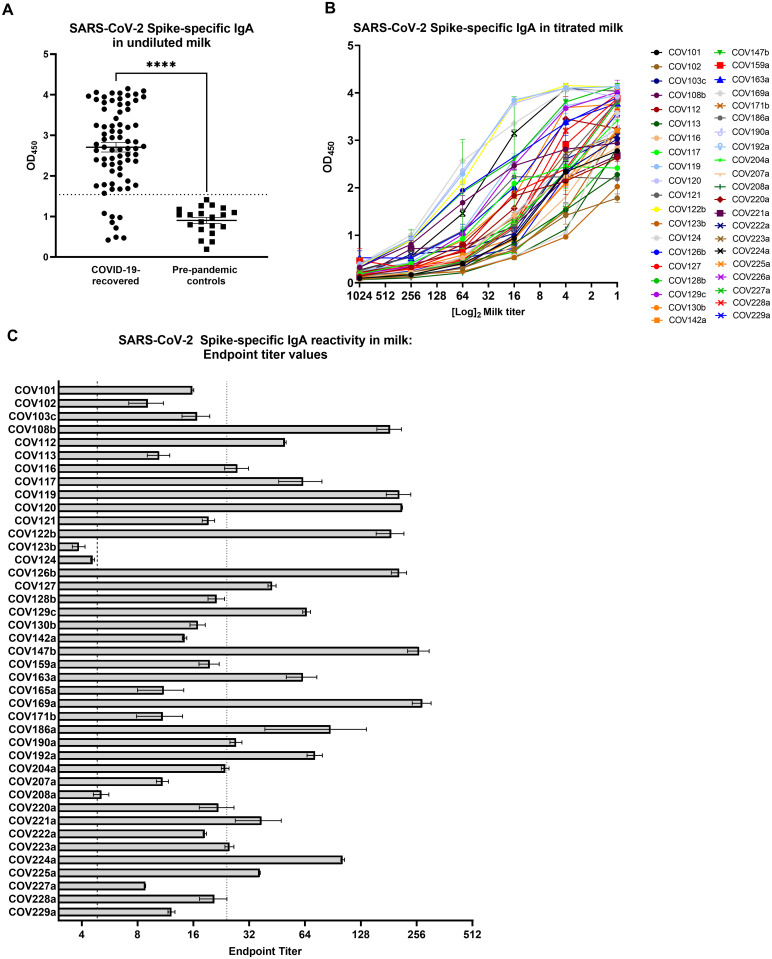
Sixty-six of 74 samples (89%) were positive for Spike-specific IgA, with the COVID-19 samples exhibiting significantly higher Spike-specific IgA binding compared to controls (Fig 1a; p<0.0001). Following this initial screening, 40 of the Spike-positive samples were further titrated to determine binding endpoint titers as an assessment of Ab affinity and/or quantity (Fig 1b). Thirty-eight of 40 (95%) Spike-reactive samples exhibited positive IgA endpoint titers and 19 of these samples (50%) were ≥5 times higher than the endpoint titer of the positive cutoff value, and were therefore designated as ‘high-titer’ (Fig 1c).

Fig 1. A robust, Spike-specific IgA response in milk commonly occurs after SARS-CoV-2 infection.

Fig 1

(A) Screening of undiluted milk samples for specific IgA by ELISA against the full-length Spike trimer. COVID-19 group, N = 74; control group N = 20. Mean values with SEM are shown. Dotted line: positive cutoff value (mean OD of negative control milk samples + 2*SD). ****p<0.0001. Mann-Whitney U test (2-tailed) was used to compare grouped data with significance level set at p < 0.05. (B) Full titration against Spike of 40 milk samples found to be positive by the initial screening. (C) Endpoint dilution titers of the 40 titrated milk samples. Segmented line: positive cutoff value; dotted line: 5x positive endpoint cutoff value, designating samples as ‘high-titer’. Mean values with SEM are shown.

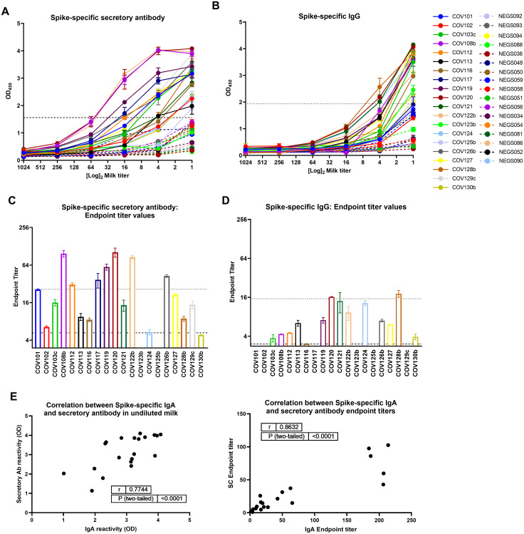
Additionally, 20 samples assayed for Spike-specific IgA were also assessed for Spike-specific secretory Ab (by detecting for SC), and IgG. Nineteen of these undiluted milk specimens (95%) from convalescent COVID-19 donors were positive for Spike-specific secretory Abs compared to pre-pandemic control milk (Fig 2a). One sample (COV125b) was negative for specific IgA but positive for specific secretory Ab, while another sample (COV123b) was positive for specific IgA but negative for specific secretory Ab. Eighteen undiluted milk samples (95%) exhibiting Spike-specific secretory Ab activity also exhibited positive endpoint titers (Fig 2c). Of the samples found to be high-titer for Spike-specific IgA, 7 were also high-titer for specific secretory Ab (70%). Mean OD values for undiluted milk and endpoint titers were used in separate Spearman correlation tests to compare IgA and secretory Ab reactivity (Fig 2e). It was found that IgA and secretory Ab levels were positively correlated (using ODs: r = 0.77, p<0.0001; using endpoint titers: r = 0.86, p<0.0001). Additionally, 15/20 undiluted milk samples from COVID-19-recovered donors were positive for Spike-specific IgG compared to pre-pandemic controls (75%; Fig 2b), with 13/15 of these samples exhibiting a positive endpoint titer (87%; Fig 2d), and 2/15 designated as high titer with values ≥5 times cutoff (13%). No correlation was found between IgG and IgA titers or between IgG and SC titers (S1 Fig).

Fig 2. The dominant Spike-specific IgA response in milk after SARS-CoV-2 infection is strongly correlated with a robust secretory Ab response, while specific IgG activity is relatively modest.

Fig 2

Twenty samples assayed for Spike-specific IgA were also assessed for Spike-specific secretory Ab (by detecting for SC), and IgG. (A, B) Full titration against Spike, detecting (A) secretory Ab, and (B) IgG. NEG (i.e. negative)/segmented lines: pre-pandemic controls. COV/solid lines: milk from COVID-19-recovered donors. Dotted lines: positive cutoff values. (C, D) Endpoint titer values calculated for (A) secretory Ab, and (B) IgG. Segmented lines: positive cutoff values; dotted lines: 5x positive cutoff (high-titer cutoff). (E) IgA and secretory Ab binding OD values or endpoint titers were used in 2-tailed Spearman correlation tests. SC: secretory component.

Durability of the SARS-CoV-2 Spike-specific milk IgA response

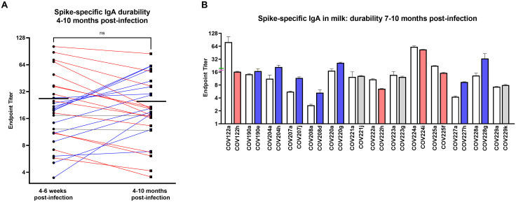
To assess the durability of this sIgA-dominant response, 28 pairs of milk samples obtained from COVID-19-recovered donors 4–6 weeks and 4–10 months after infection were assessed for Spike-specific IgA. All donors exhibited persistently significant Spike-specific IgA titers at the follow-up time point. Mean endpoint titers from the early to the late milk samples grouped were not significantly different (Fig 3a). Fourteen donors (50%) exhibited >10% decrease in IgA titer, 12 donors (43%) exhibited >10% increase in IgA titer, and 2 donors (7%) exhibited no change in titer (Fig 3a). Notably, only 2 donors (7%) exhibited >50% decrease in titer over time. Furthermore, examining a subset of 14 of these samples with the longest follow-up, obtained 7–10 months after infection, mean endpoint titers measured from the early to the late milk samples were also not significantly different (19.8 and 17.8, respectively; Fig 3b). These longest follow-up samples included 4 donors (29%) with >10% decrease in IgA titer, 7 donors (50%) with >10% increase in IgA titer, and 3 donors (21%) with no change in titer (Fig 3b). Only 1 donor (7%) exhibited >50% decrease in titer, and all donors exhibited persistently significant Spike-specific IgA titers.

Fig 3. The Spike-specific IgA response in milk after SARS-CoV-2 infection is highly durable over time.

Fig 3

(A) IgA endpoint titers determined from Spike ELISA for 28 pairs of milk samples obtained from COVID-19-recovered donors 4–6 weeks and 4–10 months after infection are shown. Mean endpoint values for each group are shown. Blue lines: >10% increase, red lines: >10% decrease, grey lines: <10% change. NS: not significant. A paired t-test (2-tailed) was used to assess significance. (B) IgA endpoint titers for a subset of 14 paired samples obtained 4–6 weeks and 7–10 months after infection. Mean with SEM is shown. Mean endpoint values for the 4–6 week and 7–10 month groups are indicated on the y-axis as green and pink ticks, respectively. Blue bars: >10% increase, red bars: >10% decrease, grey bars: <10% change.

SARS-CoV-2 neutralization capacity of total milk IgA from COVID-19-recovered donors

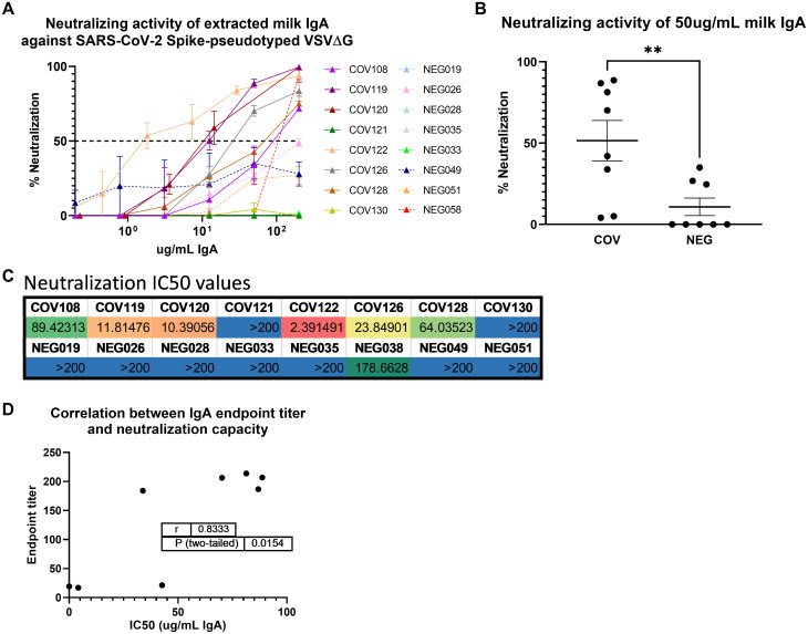
Total IgA was extracted from 8 COVID-19 samples obtained 4–6 weeks after infection and 8 control milk samples previously analyzed for their Spike-specific Ab profile. All 8 COVID-19 samples had been shown to exhibit positive Spike-specific IgA and secretory Ab titers (Figs 1 and 2). Neutralization capacity was tested using a Vesicular Stomatitis Virus (VSV)-based pseudovirus assay, wherein the native VSV surface protein G is replaced by the SARS-CoV-2 Spike, as described previously ((13); Fig 4). At the maximum concentration tested (200ug/mL total purified milk IgA), 6/8 (75%) COVID-19 samples exhibited >50% neutralization (mean, 87% neutralization; range, 70%–100%), while only 1/8 control samples (13%) achieved this benchmark (94% neutralization; Fig 4a). Mean percent neutralization values at 50ug/ml extracted IgA were grouped and compared among COVID-19 and pre-pandemic control samples. COVID-19 samples exhibited significantly greater neutralization compared to controls (p = 0.0064; Fig 4b). As well, when the concentration of IgA required to achieve 50% neutralization (IC50) was determined, 7/8 pre-pandemic controls did not achieve 50% neutralization (IC50>200ug/mL while, for the COVID-19 samples, 2/8 did not achieve 50% neutralization, and the mean IC50 for the 6 COVID19 specimens that displayed neutralizing activity was 33.6ug/mL of total IgA (range, 2.39–89.4ug/mL; Fig 4c). Finally, we compared the neutralization IC50 titers to the IgA endpoint titers measured for these samples (Fig 1). There was a significant positive correlation between IgA binding and neutralization capacities (r = 0.83, p = 0.0154; Fig 4d). Notably, the 2 non-neutralizing COVID-19 IgA samples also exhibited the lowest IgA endpoint titers (COV121, COV130; mean IgA endpoint titers of 19 and 17, respectively), while the 6 neutralizing samples exhibited high Spike-specific IgA binding titers (Figs 1c and 4c).

Fig 4. Extracted milk IgA from COVID-19-recovered donors exhibits SARS-CoV-2 Spike-targeted neutralization potency that is highly correlated with IgA binding activity.

Fig 4

(A) Total IgA was purified from 8 COVID-19 and 8 control milk samples by conventional means using peptide M agarose. IgA was titrated and tested in a VSV-based SARS-CoV-2 pseudovirus neutralization assay. NEG/segmented lines: pre-pandemic controls. COV/solid lines: COVID-19-recovered milk samples. Segmented line: 50% neutralization cutoff value. (B) Percent neutralization achieved using 50ug/mL of total extracted milk IgA. Mean values with SEM are shown. **p = 0.0064. Mann-Whitney U test (2-tailed) was used to compare grouped data with significance level set at p < 0.05. (C) Neutralization IC50 values determined from IgA titration curves. (D) Endpoint titer values determined in Fig 1 and IC50 values were used in a 2-tailed Spearman correlation test.

Discussion

There has been no evidence that SARS-CoV-2 transmits via human milk, with sporadic cases of viral RNA (not infectious particles) detected on breast skin [14]; however, there have been reports of viral RNA in the milk (reviewed in [15]), though collection methods in these reports did not necessarily include masking, cleaning of the breast, or even handwashing to avoid contamination from the donor’s environment. As such, the WHO and CDC recommend that infants not be separated from SARS-CoV-2-infected mothers, and that breastfeeding should be established and not disrupted (depending on the mothers’ desire to do so), in combination with masking and other hygiene efforts [16, 17].

We and others have reported SARS-CoV-2-specific Abs in milk obtained from donors with previously confirmed or suspected infection [9, 14, 18, 19]. Here, we have significantly expanded our earlier work in which we reported on SARS-CoV-2 Ab prevalence among 75 COVID-19-recovered participants whose milk samples were obtained 4–6 weeks after confirmed SARS-CoV-2 infection. Indeed, we have confirmed among this much larger sample set that a SARS-CoV-2 IgA Ab response in milk after infection is very common. Our analysis of a subset of 20 milk samples from COVID-19-recovered participants suggests that this IgA response dominates compared to the measurable but relatively lower titer IgG response. Importantly, a very strong positive correlation was found between Spike-specific milk IgA and secretory Abs, using both ELISA OD values of Ab binding in undiluted milk as well as Ab binding endpoint titers, indicating that a very high proportion of the SARS-CoV-2 Spike-specific IgA measured in milk after SARS-CoV-2 infection is sIgA, confirming our early reports. This is relevant for the effective protection of a breastfeeding infant, given the high durability of secretory Abs in the relatively harsh mucosal environments of the infant mouth and gut [7, 8]. These data are also relevant to the possibility of using extracted milk IgA as a COVID-19 therapy. Extracted milk sIgA used therapeutically would likely survive well upon targeted respiratory administration, with a much lower dose of Ab likely needed for efficacy compared to systemically-administered convalescent plasma or purified plasma immunoglobulin.

All COVID-19 IgA samples analyzed that had been designated as ‘high titer’ for Spike-specific IgA exhibited significant Spike-directed neutralization capacity, wherein IgA binding endpoint titers and neutralization IC50 values were found to be significantly correlated. Of the 3 samples examined for neutralization capacity that exhibited positive but not high titer Spike-specific IgA, 2 were non-neutralizing. It should be noted that these were all samples obtained 4–6 weeks after infection, and future samples may exhibit neutralization as the Ab response matures. These data extend the recent analyses of SARS-CoV-2 neutralization using diluted whole milk [14, 19].

Critically, our IgA durability analysis using 28 paired samples obtained 4–6 weeks and 4–10 months after infection revealed that for all donors, Spike-specific IgA titers persisted for as long as 10 months, a finding that is highly relevant for protection of the breastfeeding infant over the course of lactation, and also pertinent to the size of a potential donor pool for collection of milk from COVID-19-recovered donors for therapeutic use of extracted milk IgA. Notably, even after 7–10 months, only 5 of 14 samples exhibited >10% decrease in specific IgA endpoint titers, while 8 of 14 samples actually exhibited an increase in specific IgA titer. These highly durable or even increased titers may be reflective of long-lived plasma cells in the GALT and/or mammary gland, as well as continued antigen stimulation in these compartments, possibly by other human coronaviruses, or repeated exposures to SARS-CoV-2.

Given the present lack of knowledge concerning the potency, function, durability, and variation of the human milk immune response not only to SARS-CoV-2 infection, but across this understudied field in general, the present data contributes greatly to filling immense knowledge gaps and furthers our work towards in vivo efficacy testing of extracted milk Ab in the COVID-19 pandemic context and beyond.

Limitations of study

One limitation to this study is that all samples were obtained from participants living in the USA, and it should be noted that those in unique geographic areas may exhibit differential immune responses. Notably, 67% of COVID-19-recovered participants reported their race/ethnicity as white or Caucasian, and therefore this sample set is not diverse enough to be considered representative of the USA as a whole. More work needs to be done to obtain sufficient samples from non-white participants. Additionally, the longitudinal and functional components of these data were conducted on small number of samples, and further study will produce a more complete and accurate analysis. Neutralization and other functional analyses for all Ab classes also must be studied in follow-up samples. As well, this study does not demonstrate that the measured milk Ab response is protective for breastfed babies.

Supporting information

S1 Fig. No correlation was found between Spike-specific milk IgG and IgA or IgG and secretory antibody titers.

(A) Secretory Ab versus IgG. (B) IgA versus IgG. Endpoint titers were used in 2-tailed Spearman correlation tests. SC: secretory component.

(PDF)

S1 Data

(XLSX)

Acknowledgments

As always, we are indebted to the milk donors who make this work possible. Spike protein was generously gifted from the Krammer lab.

Data Availability

All relevant data are within the paper and its Supporting information files.

Funding Statement

RP is supported by the NIH/NIAID grant number R01AI158214-01 The funders had no role in study design, data collection and analysis, decision to publish, or preparation of the manuscript.

References

  • 1.Dong Y, Mo X, Hu Y, Qi X, Jiang F, Jiang Z, et al. Epidemiological Characteristics of 2143 Pediatric Patients With 2019 Coronavirus Disease in China. Pediatrics. 2020. [Google Scholar]
  • 2.Jones VG, Mills M, Suarez D, Hogan CA, Yeh D, Bradley Segal J, et al. COVID-19 and Kawasaki Disease: Novel Virus and Novel Case. Hosp Pediatr. 2020. doi: 10.1542/hpeds.2020-0123 [DOI] [PubMed] [Google Scholar]
  • 3.Ludvigsson JF. Children are unlikely to be the main drivers of the COVID-19 pandemic—A systematic review. Acta Paediatr. 2020;109(8):1525–30. doi: 10.1111/apa.15371 [DOI] [PMC free article] [PubMed] [Google Scholar]
  • 4.Li R, Pei S, Chen B, Song Y, Zhang T, Yang W, et al. Substantial undocumented infection facilitates the rapid dissemination of novel coronavirus (SARS-CoV2). Science. 2020. [DOI] [PMC free article] [PubMed] [Google Scholar]
  • 5.Weaver LT, Arthur HM, Bunn JE, Thomas JE. Human milk IgA concentrations during the first year of lactation. Arch Dis Child. 1998;78(3):235–9. doi: 10.1136/adc.78.3.235 [DOI] [PMC free article] [PubMed] [Google Scholar]
  • 6.Brandtzaeg P. The mucosal immune system and its integration with the mammary glands. J Pediatr. 2010;156(2 Suppl):S8–15. doi: 10.1016/j.jpeds.2009.11.014 [DOI] [PubMed] [Google Scholar]
  • 7.Hurley WL, Theil PK. Perspectives on immunoglobulins in colostrum and milk. Nutrients. 2011;3(4):442–74. doi: 10.3390/nu3040442 [DOI] [PMC free article] [PubMed] [Google Scholar]
  • 8.Demers-Mathieu V, Underwood MA, Beverly RL, Nielsen SD, Dallas DC. Comparison of Human Milk Immunoglobulin Survival during Gastric Digestion between Preterm and Term Infants. Nutrients. 2018;10(5). doi: 10.3390/nu10050631 [DOI] [PMC free article] [PubMed] [Google Scholar]
  • 9.Fox A, Marino J, Amanat F, Krammer F, Hahn-Holbrook J, Zolla-Pazner S, et al. Robust and specific secretory IgA against SARS-CoV-2 detected in human milk. iScience. 2020:101735. doi: 10.1016/j.isci.2020.101735 [DOI] [PMC free article] [PubMed] [Google Scholar]
  • 10.Arkhipova-Jenkins I, Helfand M, Armstrong C, Gean E, Anderson J, Paynter RA, et al. Antibody Response After SARS-CoV-2 Infection and Implications for Immunity: A Rapid Living Review. Ann Intern Med. 2021;174(6):811–21. doi: 10.7326/M20-7547 [DOI] [PMC free article] [PubMed] [Google Scholar]
  • 11.Marklund E, Leach S, Axelsson H, Nystrom K, Norder H, Bemark M, et al. Serum-IgG responses to SARS-CoV-2 after mild and severe COVID-19 infection and analysis of IgG non-responders. PLoS One. 2020;15(10):e0241104. doi: 10.1371/journal.pone.0241104 [DOI] [PMC free article] [PubMed] [Google Scholar]
  • 12.Stadlbauer D, Amanat F, Chromikova V, Jiang K, Strohmeier S, Arunkumar GA, et al. SARS-CoV-2 Seroconversion in Humans: A Detailed Protocol for a Serological Assay, Antigen Production, and Test Setup. Curr Protoc Microbiol. 2020;57(1):e100. doi: 10.1002/cpmc.100 [DOI] [PMC free article] [PubMed] [Google Scholar]
  • 13.Oguntuyo KY, Stevens CS, Hung CT, Ikegame S, Acklin JA, Kowdle SS, et al. Quantifying Absolute Neutralization Titers against SARS-CoV-2 by a Standardized Virus Neutralization Assay Allows for Cross-Cohort Comparisons of COVID-19 Sera. mBio. 2021;12(1). doi: 10.1128/mBio.02492-20 [DOI] [PMC free article] [PubMed] [Google Scholar]
  • 14.Pace RM, Williams JE, Jarvinen KM, Belfort MB, Pace CDW, Lackey KA, et al. Characterization of SARS-CoV-2 RNA, Antibodies, and Neutralizing Capacity in Milk Produced by Women with COVID-19. mBio. 2021;12(1). [DOI] [PMC free article] [PubMed] [Google Scholar]
  • 15.Centeno-Tablante E, Medina-Rivera M, Finkelstein JL, Rayco-Solon P, Garcia-Casal MN, Rogers L, et al. Transmission of SARS-CoV-2 through breast milk and breastfeeding: a living systematic review. Ann N Y Acad Sci. 2021;1484(1):32–54. doi: 10.1111/nyas.14477 [DOI] [PMC free article] [PubMed] [Google Scholar]
  • 16.Team W. Breastfeeding and COVID-19. Geneva: WHO; 2020 23 June 2020. Report No.: WHO/2019-nCoV/Sci_Brief/Breastfeeding/2020.1.
  • 17.CDC. Coronavirus Disease (COVID-19) and Breastfeeding. 2020 June 4 2020.
  • 18.Lebrao CW, Cruz MN, Silva MHD, Dutra LV, Cristiani C, Affonso Fonseca FL, et al. Early Identification of IgA Anti-SARSCoV-2 in Milk of Mother With COVID-19 Infection. J Hum Lact. 2020;36(4):609–13. doi: 10.1177/0890334420960433 [DOI] [PMC free article] [PubMed] [Google Scholar]
  • 19.Favara DM, Ceron-Gutierrez ML, Carnell GW, Heeney JL, Corrie P, Doffinger R. Detection of breastmilk antibodies targeting SARS-CoV-2 nucleocapsid, spike and receptor-binding-domain antigens. Emerg Microbes Infect. 2020;9(1):2728–31. doi: 10.1080/22221751.2020.1858699 [DOI] [PMC free article] [PubMed] [Google Scholar]

Decision Letter 0

Xia Jin

15 Jul 2021

PONE-D-21-10237

The Spike-specific IgA in milk commonly elicited after SARS-CoV-2 infection is concurrent with a robust secretory antibody response, exhibits neutralization potency strongly correlated with IgA binding, and is highly durable over time

PLOS ONE

Dear Dr. Powell,

Thank you for submitting your manuscript to PLOS ONE. After careful consideration, we feel that it has merit but does not fully meet PLOS ONE’s publication criteria as it currently stands. Therefore, we invite you to submit a revised version of the manuscript that addresses the points raised during the review process.

It is important that you carefully answer the questions raised by both reviewers reviewers 

Please submit your revised manuscript by Aug 05 2021 11:59PM. If you will need more time than this to complete your revisions, please reply to this message or contact the journal office at plosone@plos.org. When you're ready to submit your revision, log on to https://www.editorialmanager.com/pone/ and select the 'Submissions Needing Revision' folder to locate your manuscript file.

Please include the following items when submitting your revised manuscript:

  • A rebuttal letter that responds to each point raised by the academic editor and reviewer(s). You should upload this letter as a separate file labeled 'Response to Reviewers'.

  • A marked-up copy of your manuscript that highlights changes made to the original version. You should upload this as a separate file labeled 'Revised Manuscript with Track Changes'.

  • An unmarked version of your revised paper without tracked changes. You should upload this as a separate file labeled 'Manuscript'.

If you would like to make changes to your financial disclosure, please include your updated statement in your cover letter. Guidelines for resubmitting your figure files are available below the reviewer comments at the end of this letter.

If applicable, we recommend that you deposit your laboratory protocols in protocols.io to enhance the reproducibility of your results. Protocols.io assigns your protocol its own identifier (DOI) so that it can be cited independently in the future. For instructions see: http://journals.plos.org/plosone/s/submission-guidelines#loc-laboratory-protocols. Additionally, PLOS ONE offers an option for publishing peer-reviewed Lab Protocol articles, which describe protocols hosted on protocols.io. Read more information on sharing protocols at https://plos.org/protocols?utm_medium=editorial-email&utm_source=authorletters&utm_campaign=protocols.

We look forward to receiving your revised manuscript.

Kind regards,

Xia Jin, MD, PhD

Academic Editor

PLOS ONE

Journal Requirements:

1) Please review your reference list to ensure that it is complete and correct. If you have cited papers that have been retracted, please include the rationale for doing so in the manuscript text, or remove these references and replace them with relevant current references. Any changes to the reference list should be mentioned in the rebuttal letter that accompanies your revised manuscript. If you need to cite a retracted article, indicate the article’s retracted status in the References list and also include a citation and full reference for the retraction notice.

2) Please ensure that your manuscript meets PLOS ONE's style requirements, including those for file naming. The PLOS ONE style templates can be found at

https://journals.plos.org/plosone/s/file?id=wjVg/PLOSOne_formatting_sample_main_body.pdf and

https://journals.plos.org/plosone/s/file?id=ba62/PLOSOne_formatting_sample_title_authors_affiliations.pdf

3)  Please provide additional details regarding participant consent. In the ethics statement in the Methods and online submission information, please ensure that you have specified whether consent was informed.

4) PLOS ONE requires experimental methods to be described in enough detail to allow suitably skilled investigators to fully replicate and evaluate your study. See https://journals.plos.org/plosone/s/submission-guidelines#loc-materials-and-methods for more information.

To comply with PLOS ONE submission guidelines, in your Methods section, please provide a more detailed description of your methodology, specifically for assays measuring IgA, IgG, and secretory-type Abs. Please ensure that you describe the sources and catalog numbers (if applicable) of all kits, proteins, antibodies, equipment, etc. in the methods section of your manuscript.

5) In your Methods section, please provide additional information about the participant recruitment method and the demographic details of your participants. Please ensure you have provided sufficient details to replicate the analyses such as:

a) the recruitment date range (month and year),

b) where patients were recruited from,

c) a table of relevant demographic details,

d) a statement as to whether your sample can be considered representative of a larger population, and

e) a description of how participants were recruited.

6)  In your Methods section, please provide additional details regarding the cell lines and reporter viruses used in your study. Please include the source from which you obtained the cells or virus, the catalog numbers if applicable, whether the cell line was verified, and if so, how it was verified. For more information on PLOS ONE's guidelines for research using cell lines, see https://journals.plos.org/plosone/s/submission-guidelines#loc-cell-lines.

7) We note that you have included the phrase “data not shown” in your manuscript. Unfortunately, this does not meet our data sharing requirements. PLOS does not permit references to inaccessible data. We require that authors provide all relevant data within the paper, Supporting Information files, or in an acceptable, public repository. Please add a citation to support this phrase or upload the data that corresponds with these findings to a stable repository (such as Figshare or Dryad) and provide and URLs, DOIs, or accession numbers that may be used to access these data. Or, if the data are not a core part of the research being presented in your study, we ask that you remove the phrase that refers to these data.

[Note: HTML markup is below. Please do not edit.]

Reviewers' comments:

Reviewer's Responses to Questions

Comments to the Author

1. Is the manuscript technically sound, and do the data support the conclusions?

The manuscript must describe a technically sound piece of scientific research with data that supports the conclusions. Experiments must have been conducted rigorously, with appropriate controls, replication, and sample sizes. The conclusions must be drawn appropriately based on the data presented.

Reviewer #1: Yes

Reviewer #2: Yes

**********

2. Has the statistical analysis been performed appropriately and rigorously?

Reviewer #1: Yes

Reviewer #2: Yes

**********

3. Have the authors made all data underlying the findings in their manuscript fully available?

The PLOS Data policy requires authors to make all data underlying the findings described in their manuscript fully available without restriction, with rare exception (please refer to the Data Availability Statement in the manuscript PDF file). The data should be provided as part of the manuscript or its supporting information, or deposited to a public repository. For example, in addition to summary statistics, the data points behind means, medians and variance measures should be available. If there are restrictions on publicly sharing data—e.g. participant privacy or use of data from a third party—those must be specified.

Reviewer #1: No

Reviewer #2: Yes

**********

4. Is the manuscript presented in an intelligible fashion and written in standard English?

PLOS ONE does not copyedit accepted manuscripts, so the language in submitted articles must be clear, correct, and unambiguous. Any typographical or grammatical errors should be corrected at revision, so please note any specific errors here.

Reviewer #1: Yes

Reviewer #2: Yes

**********

5. Review Comments to the Author

Please use the space provided to explain your answers to the questions above. You may also include additional comments for the author, including concerns about dual publication, research ethics, or publication ethics. (Please upload your review as an attachment if it exceeds 20,000 characters)

Reviewer #1: This manuscript reported that secretory IgA response was dominant among the human milk samples and the duration of IgA was 4-10 months. The authors also observed that IgA binding (ELISA) and neutralization capacities (spike-pseudotyped VSV) were positively correlated. These findings have already reported in previous studies. The authors have confirmed the previous studies but the new key findings that increase the knowledge in the field of SARS-CoV-2-specific antibodies in human milk are missed in this study.

-Pace RM, Williams JE, Järvinen KM, Belfort MB, Pace CD, Lackey KA, Gogel AC, Nguyen-Contant P, Kanagaiah P, Fitzgerald T, Ferri R. Characterization of SARS-CoV-2 RNA, antibodies, and neutralizing capacity in milk produced by women with COVID-19. Mbio. 2021 Feb 23;12(1).

-Demers-Mathieu V, DaPra C, Mathijssen GB, Medo E. Previous viral symptoms and individual mothers influenced the leveled duration of human milk antibodies cross-reactive to S1 and S2 subunits from SARS-CoV-2, HCoV-229E, and HCoV-OC43. Journal of Perinatology. 2021 Mar 1:1-9.

Concern about this manuscript:

Abstract

1. The title and abstract are extremely long (PLOS One: 250 characters for title). Authors should summarize the most important findings for the abstracts (PLOS One: 300 word max). In most of peer-reviewed Journals, references are not present in the abstract.

2. “antibodies bearing secretory component” seem a strange and not appropriate term to describe secretory antibodies (line 38).

Background

1. Infants younger than 6 months of postnatal age, cannot be vaccinated due to their immature immune system. It is why influenza and pertussis vaccines are given after 6 months.

2. Mistake “ia” should be “is” (line 74)

Study participants

1. The criteria of inclusion and exclusion for the donors are missing in the methods.

2. How the authors determined the sample size?

3. Do all donors had a clinical /instrumental diagnosis of COVID-19 infection (COVID-19 PCR test)? This information should be presented in the method.

4. Donors and Control groups are not adequately described. All the clinical characteristics of the participants are missing. The demographic description is critical as these maternal factors influence the breast milk antibody titers and neutralizing activity between mothers.

Analytical Methods

1. Control negative with only media and control negative with milk with low/absent SIgA activity (heat-treated human milk) were performed as controls in the experiment? These controls are critical as human milk contains other antimicrobial components that could reduce the viral infectivity.

2. How the sample size calculation was done to obtain good power?

Discussion

1. Authors should explain why they selected 4-6 weeks and 4-10 months post-infection to evaluate antibody titers and neutralizing activity.

2. Limitations of this study is missing and should be included.

Figure.

1. Asterisk to show difference between groups are missing on all Figures.

2. Authors should also add the statistical analysis in the figure legend and the sample size of each group.

Table

Add a table with the demographic description of the participant

Reviewer #2: Summary

In the submitted manuscript, Fox et al report on the milk antibody response specific to the SARS-CoV-2 spike protein following maternal infection. This report greatly expands on the group’s previous report by recruiting a larger cohort of individuals (n=75 participants) and collection of milk samples at 4-6 weeks and 4-10 months post infection. This study provides valuable data on the longer-term durability of the milk antibody response following maternal COVID-19. Further, it also expands on other studies that have examined the ability of human milk to neutralize SARS-CoV-2 by isolating IgA from milk and demonstrating it to be a key factor of milk that effectively neutralizes SARS-CoV-2. This work is of both biological and clinical significance.

Abstract

Per journal guidelines, the abstract should not contain citations. Some of these references are never mentioned in the Introduction (e.g., refs 3-5) and I would suggest including them there.

The concluding sentence is very long and should be restated.

Introduction

Line 63 – should “previously-infected” be hyphenated?

Line 74 – “ia” – typo

Line 71 – “Notably, after two hours in the infant stomach, the total IgA concentration decreases by <50%, while IgG concentration decreases by >75%; importantly, though total SC concentration decreases by ~60%, there ia no decrease in the stomach of infants born pre-term (within the first 3 months of life) – a population highly vulnerable to infection” – This sentence is confusing as it appears to be discussing decreased in IgA, but then ends with stating that there is no decrease in preterm infants (as measured during the first 3 months of life). The reference cited seems to indicate that there is a larger decrease in IgA in preterm infants compared to term infants, but IgG and IgM appear more stable. Please clarify.

Methods

Were any of the participants/data from the group’s previous publication included in this work, if so, can you include that information here. More details should be provided on where (general region is fine) and when sample collections took place, per IRB stipulations.

Line 91 – Can you define what laboratory-confirmed infection means, e.g., PCR test?

Line 92 – Can you expand on why certain participants continued to provide samples 4-10 months after infection? Were these convenience samples?

Line 133 – Please give more information on the antibodies used, e.g., vendor and catalog number.

Line 133/138 – first use of OD and RLU, please define.

Results

Line 119 – Please provide the catalog number for the peptide M agarose beads.

Line 149 – “Skimmed acellular milk was aliquoted and frozen at -80o C until testing. Undiluted milk samples obtained 4-6 weeks post-infection from 75 COVID-19-recovered donors, and 20 pre-pandemic milk samples obtained prior to December, 2019 were screened in our IgA ELISA against recombinant trimeric SARS-CoV-2 Spike.” This is redundant with the methods and can be omitted.

Line 151 – extra comma in “December, 2019”

Lines 171/172 – The samples highlighted are missing the “b” suffix present in the figures. Please clarify.

Line 200 – Should “of the period of follow-up” be “at follow-up”?.

Line 200 – Can the authors comment on the appropriateness of performing statistics comparing mean endpoint titers by pooling samples collected from 4-10 mo post-infection? Were other statistical tests incorporating the interval between collections considered?

Line 201 – Figure 3, panel A would be improved by making it more apparent which donors saw increases, decreases, and no change in IgA over time by coloring them by these factors. Similarly, Panel B would be improved by coloring donors by the groups highlighted in lines 206-208.

Line 204 – Even if not showing the data, please include the number of samples included in the subset. Compared to earlier figures, Fig. 4 could include these data as an additional panel. I would suggest showing these results or highlighting them in panel B (if colored as mentioned above they could be further distinguished with a “#” above the bars or below each joint ID).

Line 209 – “as with the larger durability cohort” – I thought these donors were part of this cohort, but were they not? Please clarify or rephrase.

Lines 220 – Were the milk samples tested the 4-6 week samples or the 4-10 month samples, please clarify.

Discussion

Line 266 – While the milk IgG response is very likely to be less robust than the milk IgA response, this was not demonstrated in the current manuscript, and has yet to be demonstrated with SARS-CoV-2. Consider omitting this statement on the robustness of IgG or provide a citation.

A Limitations section is missing. There is a lack of participant characteristics detailed in this current manuscript. As such it is difficult to determine if these results are generalizable to all lactating women or limited to specific demographics. Another limitation might be that IgG was not isolated and tested for the ability to neutralize SARS-CoV-2.

Figures

The figures would be improved by removing the full sentence descriptions within panels and instead only listing the most pertinent information when necessary, e.g., Fig. 2, panels A and B could be “Spike-specific secretory component” and “Spike-specific IgG”, respectively.

Fig. 1, panel B – the title over the plot indicates that prepandemic controls are included here, but this differs from the legend. Please clarify.

Fig. 1, panel C – Consider ordering the milk samples along the y-axis by endpoint titer (rather than ID). This would greatly aid in easily visualizing the amounts of samples with endpoint titers crossing each threshold.

Error bars should be defined.

Minor

The title could be improved by making it more concise. It is also somewhat circular to state “The Spike-specific IgA in milk elicited after SARS-Cov-2 infection is … strongly correlated with IgA binding”; not much would be lost by omitting some of the more declaratory adjectives.

The v in “Cov” in the title of the manuscript should be capitalized.

**********

6. PLOS authors have the option to publish the peer review history of their article (what does this mean?). If published, this will include your full peer review and any attached files.

If you choose “no”, your identity will remain anonymous but your review may still be made public.

Do you want your identity to be public for this peer review? For information about this choice, including consent withdrawal, please see our Privacy Policy.

Reviewer #1: No

Reviewer #2: No

[NOTE: If reviewer comments were submitted as an attachment file, they will be attached to this email and accessible via the submission site. Please log into your account, locate the manuscript record, and check for the action link "View Attachments". If this link does not appear, there are no attachment files.]

While revising your submission, please upload your figure files to the Preflight Analysis and Conversion Engine (PACE) digital diagnostic tool, https://pacev2.apexcovantage.com/. PACE helps ensure that figures meet PLOS requirements. To use PACE, you must first register as a user. Registration is free. Then, login and navigate to the UPLOAD tab, where you will find detailed instructions on how to use the tool. If you encounter any issues or have any questions when using PACE, please email PLOS at figures@plos.org. Please note that Supporting Information files do not need this step.

PLoS One. 2022 Mar 9;17(3):e0249723. doi: 10.1371/journal.pone.0249723.r002

Author response to Decision Letter 0


7 Oct 2021

Editor comments:

1) Please review your reference list to ensure that it is complete and correct. If you have cited papers that have been retracted, please include the rationale for doing so in the manuscript text, or remove these references and replace them with relevant current references. Any changes to the reference list should be mentioned in the rebuttal letter that accompanies your revised manuscript. If you need to cite a retracted article, indicate the article’s retracted status in the References list and also include a citation and full reference for the retraction notice.

References have been checked.

2) Please ensure that your manuscript meets PLOS ONE's style requirements, including those for file naming.

Manuscript has been formatted.

3) Please provide additional details regarding participant consent. In the ethics statement in the Methods and online submission information, please ensure that you have specified whether consent was informed.

Details have been added in methods to indicate this was informed consent.

4) PLOS ONE requires experimental methods to be described in enough detail to allow suitably skilled investigators to fully replicate and evaluate your study. See https://journals.plos.org/plosone/s/submission-guidelines#loc-materials-and-methods for more information.

To comply with PLOS ONE submission guidelines, in your Methods section, please provide a more detailed description of your methodology, specifically for assays measuring IgA, IgG, and secretory-type Abs. Please ensure that you describe the sources and catalog numbers (if applicable) of all kits, proteins, antibodies, equipment, etc. in the methods section of your manuscript.

Methodology has been more clearly described, with catalogue numbers added.

5) In your Methods section, please provide additional information about the participant recruitment method and the demographic details of your participants. Please ensure you have provided sufficient details to replicate the analyses such as:

a) the recruitment date range (month and year),

b) where patients were recruited from,

c) a table of relevant demographic details,

d) a statement as to whether your sample can be considered representative of a larger population, and

e) a description of how participants were recruited.

Information on all participants has been added as requested in Table 1. Method of recruitment and date range have been added to the methods section. We now state that given the diversity of participant ages and stages of lactation, this study sample can be considered representative of a larger population. Notably, 67% of COVID-19-recovered participants reported their race/ethnicity as white or Caucasian, and therefore this sample set is not diverse enough to be considered representative of the USA as a whole. More work needs to be done to obtain sufficient samples from non-white participants.

6) In your Methods section, please provide additional details regarding the cell lines and reporter viruses used in your study. Please include the source from which you obtained the cells or virus, the catalog numbers if applicable, whether the cell line was verified, and if so, how it was verified. For more information on PLOS ONE's guidelines for research using cell lines, see https://journals.plos.org/plosone/s/submission-guidelines#loc-cell-lines.

Cell line and reporter virus information has been added as requested in the methods section.

7) We note that you have included the phrase “data not shown” in your manuscript. Unfortunately, this does not meet our data sharing requirements. PLOS does not permit references to inaccessible data. We require that authors provide all relevant data within the paper, Supporting Information files, or in an acceptable, public repository. Please add a citation to support this phrase or upload the data that corresponds with these findings to a stable repository (such as Figshare or Dryad) and provide and URLs, DOIs, or accession numbers that may be used to access these data. Or, if the data are not a core part of the research being presented in your study, we ask that you remove the phrase that refers to these data.

References to data not shown have been replaced with supplemental data (S1 Fig) as well as addition of data values to description of Fig 3 and the figure itself.

Reviewer Comments:

Reviewer #1: This manuscript reported that secretory IgA response was dominant among the human milk samples and the duration of IgA was 4-10 months. The authors also observed that IgA binding (ELISA) and neutralization capacities (spike-pseudotyped VSV) were positively correlated. These findings have already reported in previous studies. The authors have confirmed the previous studies but the new key findings that increase the knowledge in the field of SARS-CoV-2-specific antibodies in human milk are missed in this study.

-Pace RM, Williams JE, Järvinen KM, Belfort MB, Pace CD, Lackey KA, Gogel AC, Nguyen-Contant P, Kanagaiah P, Fitzgerald T, Ferri R. Characterization of SARS-CoV-2 RNA, antibodies, and neutralizing capacity in milk produced by women with COVID-19. Mbio. 2021 Feb 23;12(1).

-Demers-Mathieu V, DaPra C, Mathijssen GB, Medo E. Previous viral symptoms and individual mothers influenced the leveled duration of human milk antibodies cross-reactive to S1 and S2 subunits from SARS-CoV-2, HCoV-229E, and HCoV-OC43. Journal of Perinatology. 2021 Mar 1:1-9.

Neither of these studies mentioned by the reviewer measured secretory antibody nor did they measure neutralization of extracted IgA from milk, only the milk itself, therefore not demonstrating the neutralization was IgA-mediated. The Pace et. al. study looked very early after infection with no long-term follow up. The Demers-Mathieu et. al. study seems to not be on any confirmed positive donors, the sample number is very small, and it only has 4 months of follow up.

Concern about this manuscript:

Abstract

1. The title and abstract are extremely long (PLOS One: 250 characters for title). Authors should summarize the most important findings for the abstracts (PLOS One: 300 word max). In most of peer-reviewed Journals, references are not present in the abstract.

Title has been shortened and abstract is under 300 words.

2. “antibodies bearing secretory component” seem a strange and not appropriate term to describe secretory antibodies (line 38).

This has been corrected.

Background

1. Infants younger than 6 months of postnatal age, cannot be vaccinated due to their immature immune system. It is why influenza and pertussis vaccines are given after 6 months.

Infants are immunized against various pathogens within the first 6 months, including hepatitis, diphtheria, pertussis, tetanus, and others. We have not changed the mention of possible infant vaccines. See the CDC vaccine schedule: https://www.cdc.gov/vaccines/schedules/hcp/imz/child-adolescent.html#birth-15

2. Mistake “ia” should be “is” (line 74)

This has been corrected.

Study participants

1. The criteria of inclusion and exclusion for the donors are missing in the methods.

This has been more clearly described in methods

2. How the authors determined the sample size?

This has been added to the methods sections: To estimate the proportion (p) of all COVID-19-recovered milk donors that would exhibit positive IgA titers against SARS-CoV-2 in their milk after infection, we determined based on the reported IgG seroconversion rate of 90% after mild SARS-CoV-2 infection (1), that the precision (d) of the 95% confidence interval (CI) for p (CI=[p+-d]), as a function of the cohort size N of 74 would allow us to estimate p with 6.79% error. In terms of the cohort size N of 20 for milk IgG and secretory Ab analyses, this would allow us to estimate p with 13.15% error.

3. Do all donors had a clinical /instrumental diagnosis of COVID-19 infection (COVID-19 PCR test)? This information should be presented in the method.

This has been added to methods to clarify all participants had a PCR confirmed infection

4. Donors and Control groups are not adequately described. All the clinical characteristics of the participants are missing. The demographic description is critical as these maternal factors influence the breast milk antibody titers and neutralizing activity between mothers.

Now see Table 1, where this information has been added for the COVID-19 and control groups, including race/ethnicity, age, months post-partum at 1st sample, and state of residence. We also now mention in methods that all participants were either asymptomatic or experienced mild-moderate symptoms of COVID-19 that were managed at home.

Please note that in adding this information it was determined that 1 participant (COV166) was originally included in the COVID+ group in error, and this participant never tested positive. This participant’s data was therefore excluded, and the total number of COVID+ participants in this manuscript is now 74.

Analytical Methods

1. Control negative with only media and control negative with milk with low/absent SIgA activity (heat-treated human milk) were performed as controls in the experiment? These controls are critical as human milk contains other antimicrobial components that could reduce the viral infectivity.

Unclear if this is referring to the neutralization data? These experiments were done on extracted milk IgA, not with milk which can exhibit notable ‘background’ neutralization.

2. How the sample size calculation was done to obtain good power?

See above.

Discussion

1. Authors should explain why they selected 4-6 weeks and 4-10 months post-infection to evaluate antibody titers and neutralizing activity.

This post-infection window was selected so as to minimize any contact with participants or their samples when they might have been contagious to the research team, while still capturing the reported peak period for SARS-CoV-2 Ab responses (2). As little longitudinal mucosal Ab data in COVID-19-recovered individuals past 3 months has been reported to date, the ≥4 month time point was selected, and as many samples that were available were used. This info has been added to methods.

2. Limitations of this study is missing and should be included.

This section has been added after the discussion.

Figure.

1. Asterisk to show difference between groups are missing on all Figures.

This has been added in all relevant figures.

2. Authors should also add the statistical analysis in the figure legend and the sample size of each group.

This has been added in all relevant figure legends and sample size has been added in the legends also.

Table

Add a table with the demographic description of the participant

Table has been added (Table 1)

Reviewer #2: Summary

Abstract

Per journal guidelines, the abstract should not contain citations. Some of these references are never mentioned in the Introduction (e.g., refs 3-5) and I would suggest including them there.

Citations have been removed.

The concluding sentence is very long and should be restated.

This sentence has been shortened.

Introduction

Line 63 – should “previously-infected” be hyphenated?

Corrected.

Line 74 – “ia” – typo

Corrected.

Line 71 – “Notably, after two hours in the infant stomach, the total IgA concentration decreases by <50%, while IgG concentration decreases by >75%; importantly, though total SC concentration decreases by ~60%, there ia no decrease in the stomach of infants born pre-term (within the first 3 months of life) – a population highly vulnerable to infection” – This sentence is confusing as it appears to be discussing decreased in IgA, but then ends with stating that there is no decrease in preterm infants (as measured during the first 3 months of life). The reference cited seems to indicate that there is a larger decrease in IgA in preterm infants compared to term infants, but IgG and IgM appear more stable. Please clarify.

This sentence has been clarified with the reference to preterm infants removed as it is not particularly relevant to the present study.

Methods

Were any of the participants/data from the group’s previous publication included in this work, if so, can you include that information here.

10 COVID-19-recovered (COV101-COV117) and 10 pre-pandemic control (NEG046-NEG059) participants included in the present study also had their Spike IgA ELISA data reported in the our pilot study publication (3). This info has been added to the methods section.

More details should be provided on where (general region is fine) and when sample collections took place, per IRB stipulations.

See table 1 which has been added.

Line 91 – Can you define what laboratory-confirmed infection means, e.g., PCR test?

This has been defined now as PCR test confirmation

Line 92 – Can you expand on why certain participants continued to provide samples 4-10 months after infection? Were these convenience samples?

This has been clarified in methods. Participants were asked to if able and willing, to continue to pump and save monthly milk samples after the initial sample as part of our longitudinal analysis. If any of the 75 participants included in this study submitted longitudinal samples at least 4 months after their initial sample, those samples were also included in the present analysis.

Line 133 – Please give more information on the antibodies used, e.g., vendor and catalog number.

This information has been added

Line 133/138 – first use of OD and RLU, please define.

This is now defined in the text.

Results

Line 119 – Please provide the catalog number for the peptide M agarose beads.

This information has been added

Line 149 – “Skimmed acellular milk was aliquoted and frozen at -80o C until testing. Undiluted milk samples obtained 4-6 weeks post-infection from 75 COVID-19-recovered donors, and 20 pre-pandemic milk samples obtained prior to December, 2019 were screened in our IgA ELISA against recombinant trimeric SARS-CoV-2 Spike.” This is redundant with the methods and can be omitted.

This has been removed

Line 151 – extra comma in “December, 2019”

Corrected

Lines 171/172 – The samples highlighted are missing the “b” suffix present in the figures. Please clarify.

Corrected

Line 200 – Should “of the period of follow-up” be “at follow-up”?.

Clarified to say ‘All donors exhibited persistently significant Spike-specific IgA titers at the follow-up time point’

Line 200 – Can the authors comment on the appropriateness of performing statistics comparing mean endpoint titers by pooling samples collected from 4-10 mo post-infection? Were other statistical tests incorporating the interval between collections considered?

Samples were not pooled. These are individual values obtained at 2 discreet time points. The data from these time points were grouped to perform an appropriate paired t-test of significance. If the kinetics of the response are further examined for intervening time points, other statistical methods will be employed.

Line 201 – Figure 3, panel A would be improved by making it more apparent which donors saw increases, decreases, and no change in IgA over time by coloring them by these factors. Similarly, Panel B would be improved by coloring donors by the groups highlighted in lines 206-208.

The lines and bars have been color coded as suggested.

Line 204 – Even if not showing the data, please include the number of samples included in the subset. Compared to earlier figures, Fig. 4 could include these data as an additional panel. I would suggest showing these results or highlighting them in panel B (if colored as mentioned above they could be further distinguished with a “#” above the bars or below each joint ID). The number of samples in 7-10 month subset has been added. Not clear what data the reviewer is referring to to include in Fig 4?

Line 209 – “as with the larger durability cohort” – I thought these donors were part of this cohort, but were they not? Please clarify or rephrase.

This has been clarified in the text.

Lines 220 – Were the milk samples tested the 4-6 week samples or the 4-10 month samples, please clarify.

This has been clarified in the text to indicate the 4-6 week samples were used.

Discussion

Line 266 – While the milk IgG response is very likely to be less robust than the milk IgA response, this was not demonstrated in the current manuscript, and has yet to be demonstrated with SARS-CoV-2. Consider omitting this statement on the robustness of IgG or provide a citation.

This statement has been clarified to indicate it is based on our IgG data described in the paper for a subset of 20 participants.

A Limitations section is missing. There is a lack of participant characteristics detailed in this current manuscript. As such it is difficult to determine if these results are generalizable to all lactating women or limited to specific demographics. Another limitation might be that IgG was not isolated and tested for the ability to neutralize SARS-CoV-2.

Section was added as was participant data.

Figures

The figures would be improved by removing the full sentence descriptions within panels and instead only listing the most pertinent information when necessary, e.g., Fig. 2, panels A and B could be “Spike-specific secretory component” and “Spike-specific IgG”, respectively.

Titles have been shortened for all figures as recommended.

Fig. 1, panel B – the title over the plot indicates that prepandemic controls are included here, but this differs from the legend. Please clarify.

This was an error, it has been removed.

Fig. 1, panel C – Consider ordering the milk samples along the y-axis by endpoint titer (rather than ID). This would greatly aid in easily visualizing the amounts of samples with endpoint titers crossing each threshold.

We have kept the sample ID order as this makes it easier to locate samples and compared to data in other figures

Error bars should be defined.

This has been added where missing in the legends.

Minor

The title could be improved by making it more concise. It is also somewhat circular to state “The Spike-specific IgA in milk elicited after SARS-Cov-2 infection is … strongly correlated with IgA binding”; not much would be lost by omitting some of the more declaratory adjectives.

Title has been made more concise and clarified.

The v in “Cov” in the title of the manuscript should be capitalized.

Fixed.

Attachment

Submitted filename: PloS response to review.docx

Decision Letter 1

Etsuro Ito

2 Feb 2022

The IgA in milk induced by SARS-CoV-2 infection is comprised of mainly secretory antibody that is neutralizing and highly durable over time

PONE-D-21-10237R1

Dear Dr. Powell,

We’re pleased to inform you that your manuscript has been judged scientifically suitable for publication and will be formally accepted for publication once it meets all outstanding technical requirements.

Within one week, you’ll receive an e-mail detailing the required amendments. When these have been addressed, you’ll receive a formal acceptance letter and your manuscript will be scheduled for publication.

An invoice for payment will follow shortly after the formal acceptance. To ensure an efficient process, please log into Editorial Manager at http://www.editorialmanager.com/pone/, click the 'Update My Information' link at the top of the page, and double check that your user information is up-to-date. If you have any billing related questions, please contact our Author Billing department directly at authorbilling@plos.org.

If your institution or institutions have a press office, please notify them about your upcoming paper to help maximize its impact. If they’ll be preparing press materials, please inform our press team as soon as possible -- no later than 48 hours after receiving the formal acceptance. Your manuscript will remain under strict press embargo until 2 pm Eastern Time on the date of publication. For more information, please contact onepress@plos.org.

Kind regards,

Etsuro Ito

Academic Editor

PLOS ONE

Reviewers' comments:

Reviewer's Responses to Questions

Comments to the Author

1. If the authors have adequately addressed your comments raised in a previous round of review and you feel that this manuscript is now acceptable for publication, you may indicate that here to bypass the “Comments to the Author” section, enter your conflict of interest statement in the “Confidential to Editor” section, and submit your "Accept" recommendation.

Reviewer #2: All comments have been addressed

**********

2. Is the manuscript technically sound, and do the data support the conclusions?

The manuscript must describe a technically sound piece of scientific research with data that supports the conclusions. Experiments must have been conducted rigorously, with appropriate controls, replication, and sample sizes. The conclusions must be drawn appropriately based on the data presented.

Reviewer #2: Yes

**********

3. Has the statistical analysis been performed appropriately and rigorously?

Reviewer #2: Yes

**********

4. Have the authors made all data underlying the findings in their manuscript fully available?

The PLOS Data policy requires authors to make all data underlying the findings described in their manuscript fully available without restriction, with rare exception (please refer to the Data Availability Statement in the manuscript PDF file). The data should be provided as part of the manuscript or its supporting information, or deposited to a public repository. For example, in addition to summary statistics, the data points behind means, medians and variance measures should be available. If there are restrictions on publicly sharing data—e.g. participant privacy or use of data from a third party—those must be specified.

Reviewer #2: Yes

**********

5. Is the manuscript presented in an intelligible fashion and written in standard English?

PLOS ONE does not copyedit accepted manuscripts, so the language in submitted articles must be clear, correct, and unambiguous. Any typographical or grammatical errors should be corrected at revision, so please note any specific errors here.

Reviewer #2: Yes

**********

6. Review Comments to the Author

Please use the space provided to explain your answers to the questions above. You may also include additional comments for the author, including concerns about dual publication, research ethics, or publication ethics. (Please upload your review as an attachment if it exceeds 20,000 characters)

Reviewer #2: The authors have addressed all of my comments. The current version of the manuscript reads well and I have no further comments.

**********

7. PLOS authors have the option to publish the peer review history of their article (what does this mean?). If published, this will include your full peer review and any attached files.

If you choose “no”, your identity will remain anonymous but your review may still be made public.

Do you want your identity to be public for this peer review? For information about this choice, including consent withdrawal, please see our Privacy Policy.

Reviewer #2: No

Acceptance letter

Etsuro Ito

14 Feb 2022

PONE-D-21-10237R1

The IgA in milk induced by SARS-CoV-2 infection is comprised of mainly secretory antibody that is neutralizing and highly durable over time

Dear Dr. Powell:

I'm pleased to inform you that your manuscript has been deemed suitable for publication in PLOS ONE. Congratulations! Your manuscript is now with our production department.

If your institution or institutions have a press office, please let them know about your upcoming paper now to help maximize its impact. If they'll be preparing press materials, please inform our press team within the next 48 hours. Your manuscript will remain under strict press embargo until 2 pm Eastern Time on the date of publication. For more information please contact onepress@plos.org.

If we can help with anything else, please email us at plosone@plos.org.

Thank you for submitting your work to PLOS ONE and supporting open access.

Kind regards,

PLOS ONE Editorial Office Staff

on behalf of

Prof. Etsuro Ito

Academic Editor

PLOS ONE

Associated Data

    This section collects any data citations, data availability statements, or supplementary materials included in this article.

    Supplementary Materials

    S1 Fig. No correlation was found between Spike-specific milk IgG and IgA or IgG and secretory antibody titers.

    (A) Secretory Ab versus IgG. (B) IgA versus IgG. Endpoint titers were used in 2-tailed Spearman correlation tests. SC: secretory component.

    (PDF)

    S1 Data

    (XLSX)

    Attachment

    Submitted filename: PloS response to review.docx

    Data Availability Statement

    All relevant data are within the paper and its Supporting information files.


    Articles from PLoS ONE are provided here courtesy of PLOS

    RESOURCES