Figure 5.
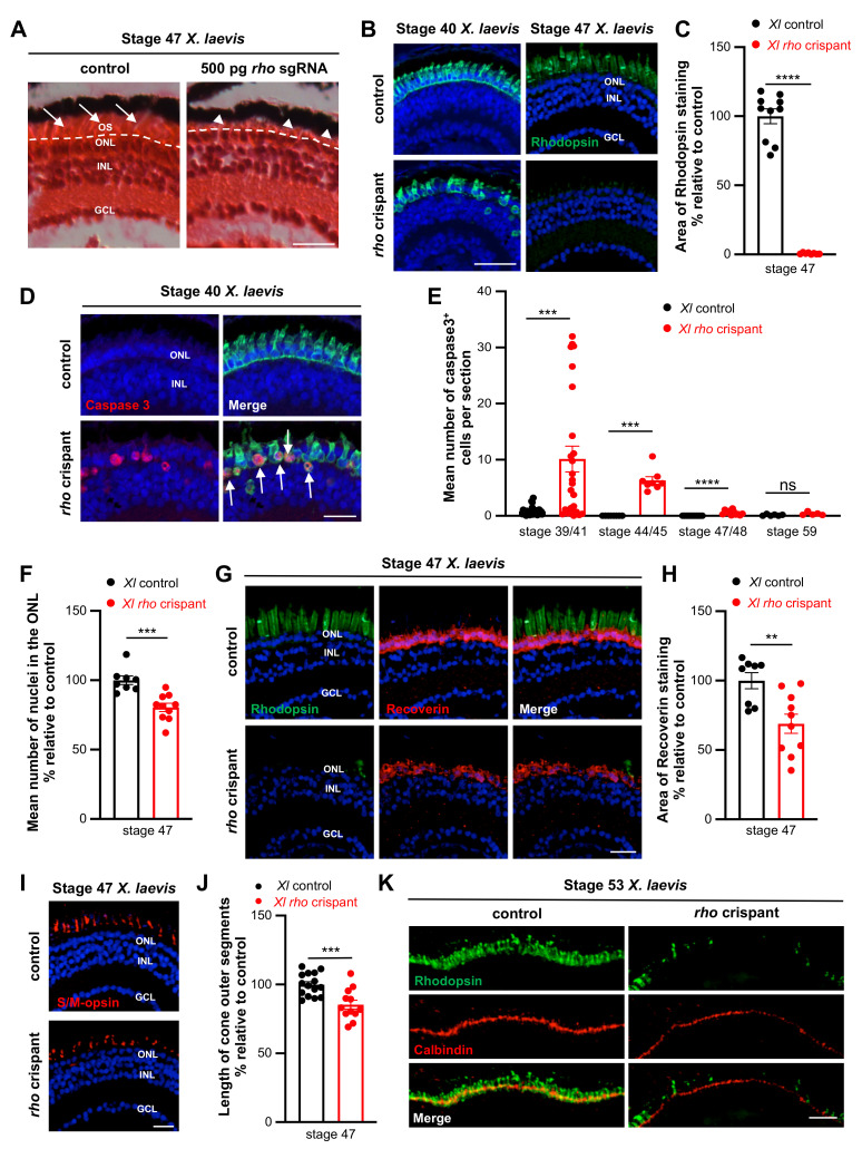
Rod degeneration and cone defects in X. laevis rho crispants. (A) Hematoxylin and Eosin staining on retinal sections from stage 47 control and rho crispant X. laevis tadpoles. The dotted line delineates the border between the outer nuclear layer and photoreceptor outer segments (arrows). Arrowheads point to shortened outer segments. (B,C) Immunofluorescence analysis of Rhodopsin expression on retinal sections from control and crispant X. laevis tadpoles at stage 40 or 47. The area of Rhodopsin staining at stage 47 is quantified in (C). (D) Typical retinal sections from stage 40 control and crispant X. laevis embryos, immunostained for cleaved Caspase 3 and Rhodopsin. Arrows point to double positive cells. (E) Quantification of Caspase 3-positive cells from stage 39/41 to stage 59. (F) Quantification of ONL nuclei on retinal sections from stage 47 control and crispant X. laevis tadpoles. Nuclei were counted in one standard rectangle field per section. (G,H) Immunofluorescence analysis of Recoverin expression on retinal sections from stage 47 control and crispant X. laevis tadpoles. Sections were co-stained for Rhodopsin for comparison. The area of Recoverin staining is quantified in (H). (I,J) Immunofluorescence analysis of S/M-opsin expression on stage 47 control and crispant X. laevis tadpoles. The length of S/M opsin-labelled outer segments is quantified in (J). (K) Typical retinal sections from stage 53 control and crispant X. laevis tadpoles, immunostained for calbindin. Note the continuous but thinner staining in the outer nuclear layer of crispant tadpoles. In (B,D,G,I), cell nuclei are counterstained with Hoechst (blue). In graphs, data are represented as mean ± SEM and each point represents one retina. ** p < 0.01; *** p < 0.001; **** p < 0.0001; ns: non-significant (Mann–Whitney tests). GCL: ganglion cell layer, INL: inner nuclear layer, ONL: outer nuclear layer, OS: outer segment. Scale bar: 25 µm in (A,B,D,H,I) and 50 µm in (K).

