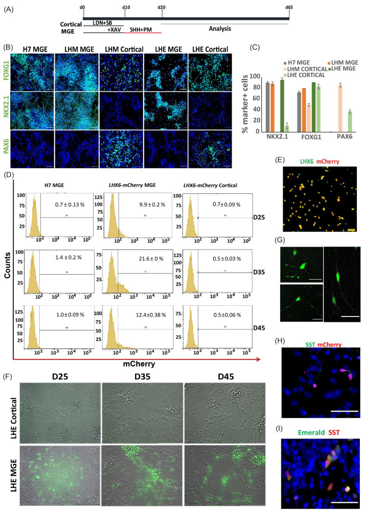
Figure 3.
mCherry and mEmerald expression is restricted to MGE derivatives. (A) Schematic illustration of hPSC interneuron and cortical differentiation protocol. (B) Day 21 LHM. LHE and H7 differentiation cultures were immunostained for neural progenitor markers representing pan-forebrain (FOXG1), MGE (NKX2.1) and developing cortex (PAX6) with DAPI counter stain (blue). (C) Quantification of antibody staining exemplified in B. Bar graphs represents mean ± SEM of three independent differentiation runs. (D) mCherry signal detected by flow cytometry during MGE differentiation of LHM cells; virtually no signal was detected in H7 MGE- or LHM-cortical differentiation. Data represent mean ± SEM of three independent experiments. (E) Double immunostaining for LHX6 and mCherry in LHM MGE-differentiated cultures showing nearly complete colocalization of both proteins. (F) Epifluorescence microscopy of MGE and cortical differentiated LHE cells showing specific mEmerald signal in MGE cultures only. (G) Confocal microscopy of day 50 live cultures of LHE MGE differentiation reveals neuronal process of cells of different morphology. (H) Double immunostaining of SST and mCherry. (I) Double immunostaining of GFP and SST. Scale bars (B): 100µ; (E,G–I): 50 µ.

