Abstract
Therapeutics with activity specifically at the inflamed sites throughout the gastrointestinal tract (GIT) would be a major advance in our therapeutic approach to inflammatory bowel disease (IBD). We aimed to develop the prodrug approach that can allow such site-specific drug delivery. Currently, using cyclosporine as a drug of choice in IBD is limited to the most severe cases due to substantial systemic toxicities and narrow therapeutic index of this drug. Previously, we synthesized a series of a phospholipid-linker-cyclosporine (PLC) prodrugs designed to exploit the overexpression of phospholipase A2 (PLA2) in the inflamed intestinal tissues, as the prodrug-activating enzyme. Nevertheless, the extent and rate of prodrug activation differed significantly. In this study we applied in-vitro and modern in-silico tools based on molecular dynamics (MD) simulation, to gain insight into the dynamics and mechanisms of the PLC prodrug activation. We aimed to elucidate the reason for the significant activation change between different linker lengths in our prodrug design. Our work reveals that the PLC conjugate with the 12-carbon linker length yields the optimal prodrug activation by PLA2 in comparison to shorter linker length (6-carbons). This optimized length efficiently allows cyclosporine to be released from the prodrug to the active pocket of PLA2. This newly developed mechanistic approach, presented in this study, can be applied for future prodrug optimization to accomplish optimal prodrug activation and drug targeting in various conditions that include overexpression of PLA2.
Keywords: inflammatory bowel disease, drug targeting, oral drug delivery, prodrug, cyclosporine, phospholipase A2
1. Introduction
Inflammatory bowel diseases (IBD) are a group of chronic inflammatory diseases of the gastrointestinal tract (GIT) and include Crohn’s disease and ulcerative colitis [1]. In the past decade, these diseases have emerged as a public health challenge worldwide [2,3]. To date, therapeutic strategies in IBD are largely based on anti-inflammatory drugs, steroids, and biological therapy [4,5,6,7]. Due to severe side effects in some patients with IBD, these therapies eventually need to be discontinued [8].
Cyclosporine has been extensively studied and is well-known for its anti-inflammatory effects and immunosuppressive activity [8,9,10,11,12]. It has been used to treat refractory or severely active IBD [13,14]. Cyclosporine’s mechanism of action is binding to cyclophilin and blocking the phosphatase activity of calcineurin, which in turn inhibits T-cell mediated cytokine production [11,15]. The treatment with cyclosporine is restricted to the most severe cases due to substantial systemic toxicities and the narrow therapeutic index [16,17]. Despite numerous side effects of cyclosporine, the treatment with cyclosporine for short-term use in patients that are hospitalized with severely active ulcerative colitis is still maintained, due to its efficiency [8,15,18]. To circumvent the serious side effects, particularly for long-term treatment, there is a strong need for an alternative, safer drug delivery of cyclosporine and improved site targeting to minimize systemic exposure.
Expression and activity of phospholipase A2 (PLA2) enzyme is considerably increased in the inflamed intestinal tissues of patients with IBD [19,20,21,22,23]. This enzyme recognizes the sn-2 acyl bond of a phospholipid (PL) and catalytically hydrolyzes the bond, releasing arachidonic acid and lysophospholipid (LPL). In our previous work, we have developed a new drug targeting approach, a PL-based prodrug approach [24,25]. Most recently, we have developed a library of PL-prodrugs containing PL linked to the cyclosporine through an alkyl linker [26]. These prodrugs differ in the number of the CH2- units (i.e., the length of the linker). The synthesis of the phospholipid-linker-cyclosporine (PLC) prodrugs includes two step condensation of the PL to the cyclosporine through diacyl chloride linkers with diverse lengths [26]. In these PLC prodrugs the fatty acid within the sn-2 position of the PL is replaced by cyclosporine-linker moiety. This approach uses PLA2 as the prodrug-activating enzyme, that allows releasing of the free drug from the PLC complex. In this approach the significantly elevated levels of the enzyme specifically in the inflammation sites, allow release of the free cyclosporin from the PLC prodrug specifically, at the inflamed sites. Thus, this approach effectively targets the regions of intestinal inflammation.
This work elucidates mechanistic background of prodrug activation and dynamics. Our preliminary in-vitro studies demonstrated that the chemical link (carbonic linker) between the cyclosporine and the PL affects the level of recognition and activation by PLA2 [26]. In this work, we aimed to further explore the effect of different levels of the PLA2 enzyme on the activation of different PLC prodrugs. This finding might demonstrate the differences in the rate of PLC prodrugs in healthy vs. diseased tissues. Nevertheless, the exact mechanistic reasoning behind PLC activation by PLA2 remains unknown. In addition, by applying molecular modeling tools, we provide insights into the molecular mechanism and the interpretation of in-vitro results. The main finding from the joined in-silico/in-vitro studies, is that the PLA2-mediated activation of the prodrug highly depends on the prodrug structure and linker length. The best activation efficiency occurs for the 12- carbon linker PLC prodrug that binds effectively to the pocket of the PLA2. On the other hand, with the shorter PL-linker-cyclosporine prodrugs steric hindrance disrupts the prodrugs entry into the enzyme pocket.
The enzyme PLA2 plays an important role in the inflammation. It is responsible for releasing a free arachidonic acid from the PL, and initiating arachidonic acid metabolic pathway, and consequent synthesis of lipid inflammatory mediators, such as prostaglandins, thromboxanes and leukotrienes [27]. Taking this into account, we anticipate that this study can serve as a basis for use of cyclosporine prodrugs (or any other PL-based prodrugs), as well as control of their activation in several other conditions which include inflammation and thus, overexpression of PLA2 [28,29].
2. Results and Discussion
2.1. Design and Activation of PLA2-Trigerred PLC Prodrug Depends on the Length of the Linker
The traditional prodrug approach focuses on altering diverse physicochemical features of the parent drug by binding to the hydrophilic/lipophilic functional groups to enhance the solubility or the passive permeability of the drug [30,31]. Recent modern prodrug strategies are based on promoieties that are attached to the parent drug to target specific membrane transporters or enzymes [32,33]. To provide specific drug targeting, these strategies consider molecular or cellular parameters, such as membrane transporter influx/efflux, enzyme expression and distribution [34,35]. Some approaches utilize lipids, such as PL, as carriers [36]. Such prodrugs have several advantages. First, they can accompany the physiological lipid trafficking pathways [37]. Second, they could target the specific step in lipid processing, particularly if the pathway is changed in the disease. Third, they may facilitate drug release at the specific target site [38].
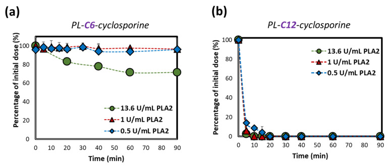
We have previously used this approach in the in-vitro proof-of-concept studies for PL-based prodrugs of diclofenac and indomethacin [24,25]. The structure of PLC prodrugs and preliminary in-vitro studies are described in our previous work [26]. The PLC designed prodrug consists of cyclosporine, bound to the sn-2 position of the PL through a linker that mimics the fatty acid substrate. The synthesis of four PLC prodrug is detailed in our previous work [26]. The NMR data specification of the 2 PLC prodrugs used in this study are seen in Figures S1 and S2. The in-vitro activation profile for three different concentrations of PLA2, was evaluated for both shortest and longest linker lengths (Figure 1). The PLC with the shorter linker length (6-CH2) demonstrated PLA2 concentration-dependent activation. Simultaneously PL-C6-cyclosporine lacks extensive PLA2-mediated hydrolysis (following the incubation in the solution) with the lowest PLA2 hydrolysis at 0.5 U/mL concentration. At concentrations above 1 U/mL of PLA2, PLA2-mediated activation is higher. Longer linker length, PL-C12-cyclosporine, resulted in complete, rapid hydrolysis that was entirely independent on the concentration of the PLA2.
Figure 1.
Activation rate (% of initial dose remaining) of PLC prodrugs that differ by the linker lengths, (a) 6-, and (b) 12-CH2 units, following incubation with 0.5, 1 and 13.6 U/mL bee venom PLA2. Data are presented as average ± SD; n = 3.
In summary, the PLA2 hydrolysis of the PLC prodrugs showed clear evidence that the linker length is crucial for the ability of the enzyme to hydrolyze the ester bond. It was also shown that the extent of hydrolysis is highly dependent upon the concentrations of the PLA2 enzyme. This confirms our hypothesis that enzyme over-expression in the inflamed tissues will selectively activate the prodrug, as opposed to low concentrations of the enzyme in the healthy tissues.
2.2. Insights into the Molecular Mechanisms of the Activation of the PLC Prodrug
To provide insights and interpretation into the prodrug activation, molecular dynamics (MD) simulations were performed for two PLC prodrugs, (1) PL-C6-cyclosporine and (2) PL-C12-cyclosporine, were bound to the pocket of PLA2 enzyme. The specific binding to the PLA2 relies on the sn-2 acyl bond of the PLC conjugates and surrounding water molecules that play a role in the enzymatic hydrolysis. Therefore, the interaction in the pocket occurs between the oxygen atom from the prodrug carbonyl group and His34 residue within PLA2 that is activated by surrounding water molecules.
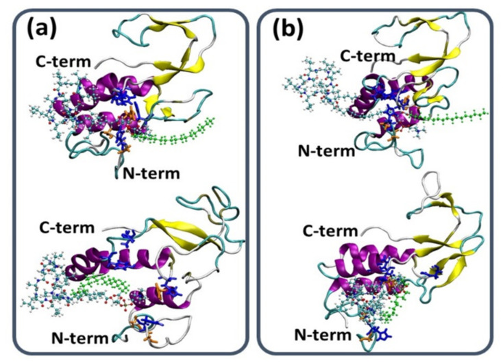
The simulations demonstrate that the shorter, PL-C6-cyclosporine linker prodrug has been embedded into the binding site pocket of the PLA2, thus blocking the activation and drug liberation (Figure 2a). The longer, PL-C12-cyclosporine linker prodrug was exposed to the solution for the entire duration of the simulations. Hence, the longer linker (12-CH2) did not allow the binding site to be blocked with the long fatty acid chain in the sn-1 position; it is evident that the PL-C12-prodrug can easily contact the binding site pocket and consequently the activation occurs (Figure 2b). It is important to note that it is not the fatty acid in the sn-1 position, and the nature of this chain that plays a role in the activation, but rather the length of the linker in the sn-2 position. Previously, it was shown that His34 residue of the PLA2 enzyme is part of the binding site in the PLA2 [39].
Figure 2.
(a) Initial (top) and final simulated (bottom) model of the PL-C6-cyclosporine prodrug-PLA2 complex. (b) Initial (top) and final simulated (bottom) model of the PL-C12-cyclosporine prodrug-PLA2 complex. The hydrophobic chain in the sn-1 position of PL-prodrugs are colored in green. The hydrophobic chain of the PL-C6-cylosporine prodrug blocks the binding pocket of the PLA2. The opposite is true for the hydrophobic chain of the PL-C12-cylosporine, whose longer linker length allows access to the binding pocket. The illustration of the structures was performed by the vmd program [40].
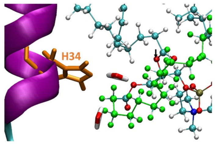
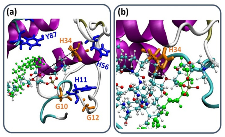
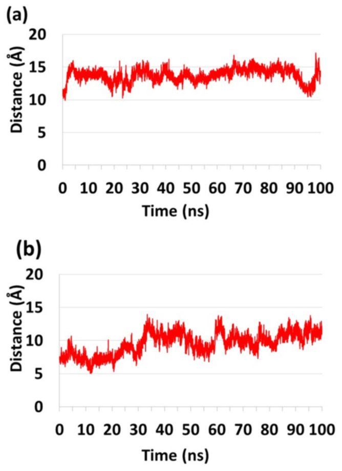
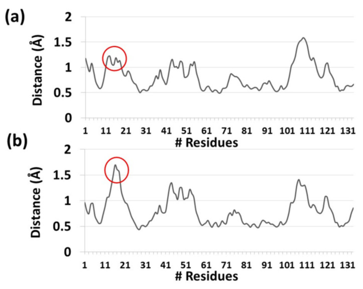
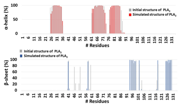
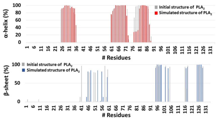
It was proposed that His34 probably operates as a Brønsted base and plays crucial role in the deprotonation of a water molecule [39]. Therefore, the His34 is responsible for the nucleophilic attack on the acyl bond. Indeed, our MD simulations showed that His34 is a crucial residue that plays a role in the activation of the PL-prodrug molecules, as well as water molecules which were observed in the proximity of the binding site pocket (Figure 3). The PL-C12-cyclosporine prodrug is completely exposed to the binding pocket of the PLA2. The fatty acid in the sn-1 position of the PL-C12-cyclosporine allows the active site and the His34 residue of the enzyme to interact with the desired active oxygen atom in the sn-2 position of the prodrug. This phenomenon does not occur with the PL-C6-cyclosporine prodrug in the binding pocket of the PLA2. To evaluate this phenomenon, the distance between the His34 and the active oxygen atom was measured for each one of the two complexes: PL-C6-cyclosporine- PLA2 and PL-C12-cyclosporine- PLA2 (Figure 4 and Figure 5). The N-terminal domain (residues 10–20) of PLA2 in PL-C6-cyclosporine-PLA2 complex is close to the prodrug PL-C6-cyclosporine, and to other domains in the PLA2, thus the N-terminal fluctuates less (Figure 6). In the PL-C12-cyclosporine-PLA2 complex, the fluctuation in the N-terminal domain (residues 10–20) is dramatically increased, due to the lack of the interactions with the PL-C12-cyclosporine and other domains in the PLA2 (Figure 6). The longer carbonic linker in the sn-2 position of the PL-C12-cyclosporine does not allow the N-terminal domain to interact with the prodrug. This result may explain the activation efficiency of the longer PL-C12-cyclosporine linker compared to the shorter PL-C6-cyclosporine. Finally, it is of interest to examine whether the secondary structure of the PLA2 is changed due to the interactions with the prodrugs. The simulations revealed that the helices of the PLA2 were conserved in the two prodrugs (Figure 7 and Figure 8). Interestingly, the β-strands along residues 45–60 within the PLA2 in the PL-C6-cyclosporine-PLA2 complex were disrupted, while in the PL-C12-cyclosporine-PLA2 complex the β-strands along these residues were conserved (Figure 7 and Figure 8).
Figure 3.
The His34 residue within the PLA2 at the close proximity to the active oxygen atom (seen in black arrow) in the PL-C12-cyclosporine prodrug. The hydrophobic chain of PL-prodrugs is colored in green. Two water molecules in close proximity to the His34 can also be observed. The illustration of the structures were performed by the vmd program [40].
Figure 4.
The distance between Cα of His34 and the active oxygen atom in (a) PL-C6-cyclosporine-PLA2 complex and (b) PL-C12-cyclosporine-PLA2 complex, showed that His34 is more in close proximity to the active oxygen in presence of PL-C12-cyclosporine than in presence of PL-C6-cyclosporine (Figure 5). The illustration of the structures were performed by the vmd program [40].
Figure 5.
The distance between the Cα of His34 in PLA2 and the oxygen atom of the prodrugs (a) PL-C6-cyclosporine and (b) PL-C12-cyclosporine.
Figure 6.
The root-mean-square fluctuations (RMSF) of each residue within PLA2 for the prodrugs (a) PL-C6-cyclosporine and (b) PL-C12-cyclosporine.
Figure 7.
The helical and the β-sheet structures of PLA2 in the initial and simulated PL-C6-cyclosporine-PLA2 complex, using the database of secondary structure of protein (DSSP) method.
Figure 8.
The helical and the β-sheet structures of PLA2 in the initial and simulated PL-C12-cyclosporine-PLA2 complex, using the database of secondary structure of protein (DSSP) method.
In summary, the mechanism of this linker length effect on the prodrug activation pattern depends on the steric hindrance. The PLA2-mediated activation of the prodrug is highly relying on the prodrug structure, i.e., the spatial arrangement of the drug. This activation depends also on the fitting of the prodrug into the transition state geometry of PLA2, which dictates the binding between the prodrug and the enzyme. In fact, it was previously proposed that PLA2 only hydrolyses PL when the sn-2 position is occupied by fatty acid [41]. We have shown, however, that this is true when the drug is linked directly to the sn-2 position [42]. However, with the proper spacer between the PL and the drug moiety, the enzymatic activation can eventually occur [24,25,43]. It is important to note that for PL-based prodrugs of smaller drugs, such as, diclofenac and indomethacin, the short 6-carbon linker was found to be optimal, while both shorter and longer linkers inhibits/disrupts the prodrug activation [24,25]. Therefore, the optimal molecular design of PL-based prodrugs is depending on the size, the volume and the three-dimensional assembly of the specific drug.
Importantly, a great clinical advantage is offered by our drug targeting approach: the inflammation localization varies in IBD patients, and since up to date IBD drug products target a general intestinal region, these products will not be effective if the inflammation is outside the targeted region. Since our approach exploits a feature innate to the inflamed tissue(s) per-se (i.e., PLA2 overexpression), efficient treatment of any localization throughout the GIT is possible. Additionally, extended therapeutic index of clinically significant drugs may be achieved, maximizing cyclosporine levels in the inflamed intestinal tissues while minimizing systemic immunosuppression, thereby making our IBD targeting approach valuable in the search for improved drug therapy and overall patient care.
3. Materials and Methods
3.1. Materials
PLC prodrugs were synthesized using two step condensation of the PL to the cyclosporine through di-acyl chloride linkers with diverse lengths (6 and 12 methylene units); all steps required for structure elucidation and purity were completed (Figures S1 and S2). Phospholipase A2 from honey bee venom (Apis mellifera) was purchased from Sigma-Aldrich (Rehovot, Israel). Isopropanol, methanol, and water (Merck KGaA, Darmstadt, Germany) were of ultra-performance liquid chromatography (UPLC) grade. All other chemicals were of analytical reagent grade.
3.2. PLA2-Mediated Activation
PLA2 hydrolysis assay of the four newly synthesized PLC conjugates were carried out by bee venom PLA2 followed a previously published protocol with minor modifications [25,26]. Briefly, PLC conjugates with 6- and 12-carbon linker were dissolved in methanol, and small aliquots were added in 1 mL of buffer solution. Prior to prodrug addition, the buffer solutions with different concentrations of the bee venom PLA2 (0.5, 1 and 13.6) units/mL were made. The buffer solutions also contained Tris-HCl 10 mM, CaCl2 10 mM and NaCl 300 mM (pH 7.4) with addition of 10mM sodium taurocholate, as a natural surfactant; the control group contained everything but bee venom PLA2. This mixture was incubated for 1.5 h at 25 °C. Samples were collected in intervals after 0, 5, 10, 15, 20, 30-, 40-, 60- and 90-min. Results for PLC activation are presented as mean ± SD; n = 4, per each phospholipid-cyclosporine conjugate.
3.3. Analytical Methods
Activation of PLC by different levels of PLA2 was followed by high performance liquid chromatography (HPLC) system (Waters 2695 Separation Module, Milford, MA, USA) with a photodiode array UV detector (Waters 2996, Milford, MA, USA). Separation of conjugates was performed with a C8 column and confirmed by UV. The HPLC conditions were as follows: Waters (WT186003055) Xbridge® RP8 3.5 μm; 4.6 mm × 150 mm column, an isocratic mobile phase containing isopropanol: methanol: water (70:3:27 v/v) for 10 min at the flow rate of 0.5 mL/min and the detection wavelength was 206 nm.
3.4. Computational Modeling
3.4.1. Parametrization and Integration of the Prodrugs
The integration of the two PLC prodrugs into the Chemistry at Harvard Macromolecular Mechanics (CHARMM) force field were performed by defining the parameters for the new molecules based on the well-established chemical analogs that were provided by CHARMM36 force-field (mackerell.umaryland.edu). According to the corresponding analog, each atom within the PLC prodrugs were defined by the charge, bond length, angles, torsion and van der Waals value. The assignments of the values were created by CGenFF generator: cgenff.umaryland.edu. The values demonstrated a reasonable deviation (less than 10% for all atoms within the PLC prodrugs).
3.4.2. Construction of PL-C6-Cyclosporine and PL-C12-Cyclosporine Prodrugs
The coordinates for the PL-C6-cyclosporine and PL-C12-cyclosporine prodrugs that are complexed with the PLA2 enzyme were constructed using the Accelrys Discovery Studio software package (http://accelrys.com/products/discovery-studio/, accessed on 3 February 2022). Initial structure of the enzyme PLA2 applied in this work is the crystal structure of bee-venom PLA2 (PDB ID code: 1POC) [44]. It is crucial to accurately construct the initial structures of the two models representing the prodrug-enzyme complex. Therefore, the constructions of the models are based on previous experimental reports. It was proposed by experimental study that His34 residue of the PLA2 enzyme is part of the binding activated site in the PLA2 [39]. Moreover, it was proposed that His34 operates as a Brønsted base and plays crucial role in the deprotonation of a water molecule [39]. Hence, we constructed the prodrug-enzyme complex in which the prodrug acyl group of each PLC was inserted into the binding pocket in proximity to His34, while avoiding atom clashes. Specifically, each prodrug was inserted into the binding pocket of the enzyme in a similar manner: the shorter and the longer linkers were orientated in the same direction and the activated part of the prodrug sn-2 acyl group was oriented towards the binding pocket of the enzyme. It must be noted that there is only one possible option to make the modeling of these complexes, by producing a similarity in the positions of the linkers. Moreover, it is crucial that the linkers are in the same orientation, while the prodrug is being inserted into the binding pocket. Finally, we explored all possibilities for the constructions of these complexes, while keeping the modeling of the complexes in accordance to the experimental data, while keeping in mind the interactions of the prodrugs in the binding pocket of the enzyme. It is important to note that the constructions of the initial complexes did not account constrains, not in the construction modeling step nor along the molecular dynamics (MD) simulations.
3.4.3. Molecular Dynamics (MD) Simulations Protocol
The MD simulations of the solvated constructed models were performed in the NPT ensemble using NAMD package [45] with the CHARMM27 forcefield with the CMAP correlation [46]. The energies of the complex prodrug-enzyme were minimized, and the model was explicitly solvated in a TIP3P water box [47,48]. Each water molecule within 2.5 Å of the models was removed. Counter ions were added at random locations to neutralize the models’ charge. The Langevin piston method [44,45,49] with a decay period of 100 fs and a damping time of 50 fs was used to maintain a constant pressure of 1 atm. The temperature 330 K was controlled by a Langevin thermostat with a damping coefficient of 10 ps [45]. The short-range van der Waals (VDW) interactions were calculated using the switching function, with a twin range cutoff of 10.0 and 12.0 Å. Long-range electrostatic interactions were calculated using the particle mesh Ewald method with a cutoff of 12.0 Å [50,51]. The equations of motion were integrated using the leapfrog integrator with a step of 1 fs. The counter ions and water molecules were allowed to move. The hydrogen atoms were constrained to the equilibrium bond using the SHAKE algorithm [52]. The minimized solvated systems were energy minimized for 5000 additional conjugate gradient steps and 20,000 heating steps at 250 K, with all atoms allowed to move. Then, the system was heated from 250 K to 300 K and then to 330 K for 300 ps and equilibrated at 330 K for 300 ps. The choice of the higher temperature than physiological temperature is to investigate the stability of the constructed models. Obviously, structures that are stable at higher temperature will be also stable at physiological temperature. Simulations ran for 100 ns for each variant model. To justify the timescale of the simulations, we computed the root-mean-square-deviation (RMSD) values along the MD simulations (Figure S3). The RAMS analysis demonstrated that after ~30 ns of the simulations, both variant models were converged. The structures were saved every 10 ps for analyses.
3.4.4. Structural Analyses
The structural stabilities of the two models were measured using several analyses. The root-mean-square-fluctuation (RMSF) values for each residue within the PLA2 were computed for each model. To estimate possible interactions within the binding pocket of the PLA2 to the PLC prodrugs, the distances between specific Cα atoms of residues in the binding pocket and the oxygen atom in the sn-2 position were computed along the MD simulations. Finally, to examine whether the PLC prodrugs affect the secondary structure of the PLA2, the database of secondary structure of protein (DSSP) method has been applied [53]. This method was applied to provide the percentage of the α-helix or β-strand for each residue within the phospholipase along the MD simulations.
4. Conclusions
In this work we employed modern in-silico tools to confirm our experimentally obtained data, and to mechanistically explain the reasoning behind the significantly different activation rate among longer and shorter linkers in the PLC. We demonstrated that the PL-linker-cyclosporine with the longest linker exhibited optimal, fastest rate of activation through in-vitro PLA2-mediated activation, at all 3 enzyme concentrations. Consequently, we tested these results using the MD simulations, and provided mechanistic insights into the molecular mechanism activation of the PL-linker-cyclosporine with longer and shorter linkers. We have shown by MD simulations that the insufficient activation of the shorter PL-linker-cyclosporine is due to the steric hindrance that eventually does not appear in the longest PL-linker-cyclosporine. Ultimately, these studies can serve as a screening tool for optimal prodrug design, which can offer a modern biopharmaceutical solution for numerous clinical needs. The overexpression of PLA2 occurs in other malignant and inflammatory conditions, i.e., rheumatoid arthritis, colorectal cancer, and vascular inflammation [29,54]. Hence, our prodrug approach and mechanistic screening tools may offer an elegant solution for improving drug treatment for such diseases.
Acknowledgments
This work is a part of M. Markovic PhD dissertation. A.D., S.B.-S., A.A. and E.M.Z. wish to thank the US-Israel Binational Science Foundation (BSF) for funding this work. The simulations were performed using the high-performance computational facilities of the Miller lab in the BGU HPC computational center. The support of the BGU HPC computational center staff is greatly appreciated.
Supplementary Materials
The following supporting information can be downloaded at: https://www.mdpi.com/article/10.3390/ijms23052673/s1.
Author Contributions
The manuscript was written through contributions of all authors. M.M., K.A.-H., C.R., S.B.-S., A.A., Y.M., E.M.Z. and A.D. worked on study design, methodology, and investigations, analyzed the data, and outlined the manuscript. M.M., K.A.-H. and C.R. performed the research, analyzed the data and wrote the paper. S.B.-S., A.A., E.M.Z. and A.D. critically revised the draft of the article. All authors approved the final version of the article, including the authorship list. All authors have read and agreed to the published version of the manuscript.
Funding
This work was funded through the US-Israel Binational Science Foundation (BSF) grant number 2015365.
Conflicts of Interest
The authors declare no conflict of interest.
Footnotes
Publisher’s Note: MDPI stays neutral with regard to jurisdictional claims in published maps and institutional affiliations.
References
- 1.Abraham C., Cho J.H. Inflammatory Bowel Disease. N. Engl. J. Med. 2009;361:2066–2078. doi: 10.1056/NEJMra0804647. [DOI] [PMC free article] [PubMed] [Google Scholar]
- 2.Alatab S., Sepanlou S.G., Ikuta K., Vahedi H., Bisignano C., Safiri S., Sadeghi A., Nixon M.R., Abdoli A., Abolhassani H., et al. The global, regional, and national burden of inflammatory bowel disease in 195 countries and territories, 1990–2017: A systematic analysis for the Global Burden of Disease Study 2017. Lancet Gastroenterol. Hepatol. 2020;5:17–30. doi: 10.1016/S2468-1253(19)30333-4. [DOI] [PMC free article] [PubMed] [Google Scholar]
- 3.Ng S.C., Shi H.Y., Hamidi N., Underwood F.E., Tang W., Benchimol E.I., Panaccione R., Ghosh S., Wu J.C.Y., Chan F.K.L., et al. Worldwide incidence and prevalence of inflammatory bowel disease in the 21st century: A systematic review of population-based studies. Lancet. 2017;390:2769–2778. doi: 10.1016/S0140-6736(17)32448-0. [DOI] [PubMed] [Google Scholar]
- 4.Carter M.J., Lobo A.J., Travis S.P.L. Guidelines for the management of inflammatory bowel disease in adults. Gut. 2004;53:v1. doi: 10.1136/gut.2004.043372. [DOI] [PMC free article] [PubMed] [Google Scholar]
- 5.Narula N., Marshall J.K., Colombel J.F., Leontiadis G.I., Williams J.G., Muqtadir Z., Reinisch W. Systematic Review and Meta-Analysis: Infliximab or Cyclosporine as Rescue Therapy in Patients With Severe Ulcerative Colitis Refractory to Steroids. Am. J. Gastroenterol. 2016;111:477–491. doi: 10.1038/ajg.2016.7. [DOI] [PubMed] [Google Scholar]
- 6.Waljee A.K., Wiitala W.L., Govani S., Stidham R., Saini S., Hou J., Feagins L.A., Khan N., Good C.B., Vijan S., et al. Corticosteroid Use and Complications in a US Inflammatory Bowel Disease Cohort. PLoS ONE. 2016;11:e0158017. doi: 10.1371/journal.pone.0158017. [DOI] [PMC free article] [PubMed] [Google Scholar]
- 7.Zenlea T., Peppercorn M.A. Immunosuppressive therapies for inflammatory bowel disease. World J. Gastroenterol. 2014;20:3146–3152. doi: 10.3748/wjg.v20.i12.3146. [DOI] [PMC free article] [PubMed] [Google Scholar]
- 8.Sandborn W.J., Tremaine W.J. Cyclosporine treatment of inflammatory bowel disease. Mayo Clin. Proc. 1992;67:981–990. doi: 10.1016/S0025-6196(12)60930-6. [DOI] [PubMed] [Google Scholar]
- 9.Calne R.Y., Rolles K., Thiru S., McMaster P., Craddock G.N., Aziz S., White D.J.G., Evans D.B., Dunn D.C., Henderson R.G., et al. Cyclosporin a initially as the only immunosuppressant in 34 recipients of cadaveric organs: 32 kidneys, 2 pancreases, and 2 livers. Lancet. 1979;314:1033–1036. doi: 10.1016/S0140-6736(79)92440-1. [DOI] [PubMed] [Google Scholar]
- 10.Chighizola C.B., Ong V.H., Meroni P.L. The Use of Cyclosporine A in Rheumatology: A 2016 Comprehensive Review. Clin. Rev. Allergy Immunol. 2017;52:401–423. doi: 10.1007/s12016-016-8582-3. [DOI] [PubMed] [Google Scholar]
- 11.Faulds D., Goa K.L., Benfield P. Cyclosporin. A review of its pharmacodynamic and pharmacokinetic properties, and therapeutic use in immunoregulatory disorders. Drugs. 1993;45:953–1040. doi: 10.2165/00003495-199345060-00007. [DOI] [PubMed] [Google Scholar]
- 12.Lowe N.J. Systemic treatment of severe psoriasis—The role of cyclosporine. N. Engl. J. Med. 1991;324:333–334. doi: 10.1056/NEJM199101313240509. [DOI] [PubMed] [Google Scholar]
- 13.Lichtiger S., Present D.H., Kornbluth A., Gelernt I., Bauer J., Galler G., Michelassi F., Hanauer S. Cyclosporine in severe ulcerative colitis refractory to steroid therapy. N. Engl. J. Med. 1994;330:1841–1845. doi: 10.1056/NEJM199406303302601. [DOI] [PubMed] [Google Scholar]
- 14.Loftus C.G., Loftus E.V., Jr., Sandborn W.J. Cyclosporin for refractory ulcerative colitis. Gut. 2003;52:172–173. doi: 10.1136/gut.52.2.172. [DOI] [PMC free article] [PubMed] [Google Scholar]
- 15.Elliott J.F., Lin Y., Mizel S.B., Bleackley R.C., Harnish D.G., Paetkau V. Induction of interleukin 2 messenger RNA inhibited by cyclosporin A. Science. 1984;226:1439–1441. doi: 10.1126/science.6334364. [DOI] [PubMed] [Google Scholar]
- 16.Strom T.B., Loertscher R. Cyclosporine-Induced Nephrotoxicity. N. Engl. J. Med. 1984;311:728–729. doi: 10.1056/NEJM198409133111109. [DOI] [PubMed] [Google Scholar]
- 17.Tedesco D., Haragsim L. Cyclosporine: A review. J. Transpl. 2012;2012:230386. doi: 10.1155/2012/230386. [DOI] [PMC free article] [PubMed] [Google Scholar]
- 18.Kornbluth A., Present D.H., Lichtiger S., Hanauer S. Cyclosporin for severe ulcerative colitis: A user’s guide. Am. J. Gastroenterol. 1997;92:1424–1428. [PubMed] [Google Scholar]
- 19.Haapamaki M.M., Gronroos J.M., Nurmi H., Alanen K., Kallajoki M., Nevalainen T.J. Gene expression of group II phospholipase A2 in intestine in ulcerative colitis. Gut. 1997;40:95–101. doi: 10.1136/gut.40.1.95. [DOI] [PMC free article] [PubMed] [Google Scholar]
- 20.Haapamaki M.M., Gronroos J.M., Nurmi H., Irjala K., Alanen K.A., Nevalainen T.J. Phospholipase A2 in serum and colonic mucosa in ulcerative colitis. Scand. J. Clin. Lab. Investig. 1999;59:279–287. doi: 10.1080/00365519950185643. [DOI] [PubMed] [Google Scholar]
- 21.Lilja I., Smedh K., Olaison G., Sjodahl R., Tagesson C., Gustafson-Svard C. Phospholipase A2 gene expression and activity in histologically normal ileal mucosa and in Crohn’s ileitis. Gut. 1995;37:380–385. doi: 10.1136/gut.37.3.380. [DOI] [PMC free article] [PubMed] [Google Scholar]
- 22.Minami T., Shinomura Y., Miyagawa J., Tojo H., Okamoto M., Matsuzawa Y. Immunohistochemical localization of group II phospholipase A2 in colonic mucosa of patients with inflammatory bowel disease. Am. J. Gastroenterol. 1997;92:289–292. [PubMed] [Google Scholar]
- 23.Minami T., Tojo H., Shinomura Y., Matsuzawa Y., Okamoto M. Increased group II phospholipase A2 in colonic mucosa of patients with Crohn’s disease and ulcerative colitis. Gut. 1994;35:1593–1598. doi: 10.1136/gut.35.11.1593. [DOI] [PMC free article] [PubMed] [Google Scholar]
- 24.Dahan A., Duvdevani R., Dvir E., Elmann A., Hoffman A. A novel mechanism for oral controlled release of drugs by continuous degradation of a phospholipid prodrug along the intestine: In-vivo and in-vitro evaluation of an indomethacin-lecithin conjugate. J. Control. Release. 2007;119:86–93. doi: 10.1016/j.jconrel.2006.12.032. [DOI] [PubMed] [Google Scholar]
- 25.Dahan A., Markovic M., Epstein S., Cohen N., Zimmermann E.M., Aponick A., Ben-Shabat S. Phospholipid-drug conjugates as a novel oral drug targeting approach for the treatment of inflammatory bowel disease. Eur. J. Pharm. Sci. 2017;108:78–85. doi: 10.1016/j.ejps.2017.06.022. [DOI] [PubMed] [Google Scholar]
- 26.Manda J.N., Markovic M., Zimmermann E.M., Ben-Shabat S., Dahan A., Aponick A. Phospholipid Cyclosporine Prodrugs Targeted at Inflammatory Bowel Disease (IBD) Treatment: Design, Synthesis, and in Vitro Validation. ChemMedChem. 2020;15:1639–1644. doi: 10.1002/cmdc.202000317. [DOI] [PubMed] [Google Scholar]
- 27.Burke J.E., Dennis E.A. Phospholipase A2 structure/function, mechanism, and signaling. J. Lipid Res. 2009;50:S237–S242. doi: 10.1194/jlr.R800033-JLR200. [DOI] [PMC free article] [PubMed] [Google Scholar]
- 28.Abe T., Sakamoto K., Kamohara H., Hirano Y., Kuwahara N., Ogawa M. Group II phospholipase A2 is increased in peritoneal and pleural effusions in patients with various types of cancer. Int. J. Cancer. 1997;74:245–250. doi: 10.1002/(SICI)1097-0215(19970620)74:3<245::AID-IJC2>3.0.CO;2-Z. [DOI] [PubMed] [Google Scholar]
- 29.Pruzanski W., Vadas P., Stefanski E., Urowitz M.B. Phospholipase A2 activity in sera and synovial fluids in rheumatoid arthritis and osteoarthritis. Its possible role as a proinflammatory enzyme. J. Rheumatol. 1985;12:211–216. [PubMed] [Google Scholar]
- 30.Markovic M., Ben-Shabat S., Dahan A. Prodrugs for Improved Drug Delivery: Lessons Learned from Recently Developed and Marketed Products. Pharmaceutics. 2020;12:1031. doi: 10.3390/pharmaceutics12111031. [DOI] [PMC free article] [PubMed] [Google Scholar]
- 31.Markovic M., Zur M., Ragatsky I., Cvijić S., Dahan A. BCS Class IV Oral Drugs and Absorption Windows: Regional-Dependent Intestinal Permeability of Furosemide. Pharmaceutics. 2020;12:1175. doi: 10.3390/pharmaceutics12121175. [DOI] [PMC free article] [PubMed] [Google Scholar]
- 32.Rautio J., Meanwell N.A., Di L., Hageman M.J. The expanding role of prodrugs in contemporary drug design and development. Nat. Rev. Drug Discov. 2018;17:559–587. doi: 10.1038/nrd.2018.46. [DOI] [PubMed] [Google Scholar]
- 33.Stella V.J. Prodrugs: Some thoughts and current issues. J. Pharm. Sci. 2010;99:4755–4765. doi: 10.1002/jps.22205. [DOI] [PubMed] [Google Scholar]
- 34.Markovic M., Ben-Shabat S., Keinan S., Aponick A., Zimmermann E.M., Dahan A. Lipidic prodrug approach for improved oral drug delivery and therapy. Med. Res. Rev. 2019;39:579–607. doi: 10.1002/med.21533. [DOI] [PubMed] [Google Scholar]
- 35.Xu Y., Shrestha N., Préat V., Beloqui A. Overcoming the intestinal barrier: A look into targeting approaches for improved oral drug delivery systems. J. Control. Release. 2020;322:486–508. doi: 10.1016/j.jconrel.2020.04.006. [DOI] [PubMed] [Google Scholar]
- 36.Markovic M., Ben-Shabat S., Keinan S., Aponick A., Zimmermann E.M., Dahan A. Prospects and Challenges of Phospholipid-Based Prodrugs. Pharmaceutics. 2018;10:210. doi: 10.3390/pharmaceutics10040210. [DOI] [PMC free article] [PubMed] [Google Scholar]
- 37.Markovic M., Ben-Shabat S., Aponick A., Zimmermann E.M., Dahan A. Lipids and Lipid-Processing Pathways in Drug Delivery and Therapeutics. Int. J. Mol. Sci. 2020;21:3248. doi: 10.3390/ijms21093248. [DOI] [PMC free article] [PubMed] [Google Scholar]
- 38.Dahan A., Markovic M., Aponick A., Zimmermann E.M., Ben-Shabat S. The prospects of lipidic prodrugs: An old approach with an emerging future. Future Med. Chem. 2019;11:2563–2571. doi: 10.4155/fmc-2019-0155. [DOI] [PubMed] [Google Scholar]
- 39.Annand R.R., Kontoyianni M., Penzotti J.E., Dudler T., Lybrand T.P., Gelb M.H. Active Site of Bee Venom Phospholipase A2: The Role of Histidine-34, Aspartate-64 and Tyrosine-87. Biochemistry. 1996;35:4591–4601. doi: 10.1021/bi9528412. [DOI] [PubMed] [Google Scholar]
- 40.Humphrey W., Dalke A., Schulten K. VMD: Visual molecular dynamics. J. Mol. Graph. 1996;14:33–38. doi: 10.1016/0263-7855(96)00018-5. [DOI] [PubMed] [Google Scholar]
- 41.Kurz M., Scriba G.K. Drug-phospholipid conjugates as potential prodrugs: Synthesis, characterization, and degradation by pancreatic phospholipase A(2) Chem. Phys. Lipids. 2000;107:143–157. doi: 10.1016/S0009-3084(00)00167-5. [DOI] [PubMed] [Google Scholar]
- 42.Dahan A., Duvdevani R., Shapiro I., Elmann A., Finkelstein E., Hoffman A. The oral absorption of phospholipid prodrugs: In vivo and in vitro mechanistic investigation of trafficking of a lecithin-valproic acid conjugate following oral administration. J. Control. Release. 2008;126:1–9. doi: 10.1016/j.jconrel.2007.10.025. [DOI] [PubMed] [Google Scholar]
- 43.Markovic M., Dahan A., Keinan S., Kurnikov I., Aponick A., Zimmermann E.M., Ben-Shabat S. Phospholipid-Based Prodrugs for Colon-Targeted Drug Delivery: Experimental Study and In-Silico Simulations. Pharmaceutics. 2019;11:186. doi: 10.3390/pharmaceutics11040186. [DOI] [PMC free article] [PubMed] [Google Scholar]
- 44.Scott D.L., Otwinowski Z., Gelb M.H., Sigler P.B. Crystal structure of bee-venom phospholipase A2 in a complex with a transition-state analogue. Science. 1990;250:1563–1566. doi: 10.1126/science.2274788. [DOI] [PubMed] [Google Scholar]
- 45.Kalé L., Skeel R., Bhandarkar M., Brunner R., Gursoy A., Krawetz N., Phillips J., Shinozaki A., Varadarajan K., Schulten K. NAMD2: Greater Scalability for Parallel Molecular Dynamics. J. Comput. Phys. 1999;151:283–312. doi: 10.1006/jcph.1999.6201. [DOI] [Google Scholar]
- 46.MacKerell A.D., Bashford D., Dunbrack R.L., Evanseck J.D., Field M.J., Fischer S., Gao J., Guo H., Ha S., Joseph-McCarthy D., et al. All-Atom Empirical Potential for Molecular Modeling and Dynamics Studies of Proteins†. J. Phys. Chem. B. 1998;102:3586–3616. doi: 10.1021/jp973084f. [DOI] [PubMed] [Google Scholar]
- 47.Jorgensen W.L., Chandrasekhar J., Madura J.D., Impey R.W., Klein M.L. Comparison of simple potential functions for simulating liquid water. J. Chem. Phys. 1983;79:926–935. doi: 10.1063/1.445869. [DOI] [Google Scholar]
- 48.Mahoney M.W., Jorgensen W.L. A five-site model for liquid water and the reproduction of the density anomaly by rigid, nonpolarizable potential functions. J. Chem. Phys. 2000;112:8910–8922. doi: 10.1063/1.481505. [DOI] [Google Scholar]
- 49.Tu K., Tobias D.J., Klein M.L. Constant pressure and temperature molecular dynamics simulation of a fully hydrated liquid crystal phase dipalmitoylphosphatidylcholine bilayer. Biophys. J. 1995;69:2558–2562. doi: 10.1016/S0006-3495(95)80126-8. [DOI] [PMC free article] [PubMed] [Google Scholar]
- 50.Darden T., York D., Pedersen L. Particle mesh Ewald: An N log (N) method for Ewald sums in large systems. J. Chem. Phys. 1993;98:10089–10092. doi: 10.1063/1.464397. [DOI] [Google Scholar]
- 51.Essmann U., Perera L., Berkowitz M.L., Darden T., Lee H., Pedersen L.G. A smooth particle mesh Ewald method. J. Chem. Phys. 1995;103:8577–8593. doi: 10.1063/1.470117. [DOI] [Google Scholar]
- 52.Ryckaert J.-P., Ciccotti G., Berendsen H.J. Numerical integration of the cartesian equations of motion of a system with constraints: Molecular dynamics of n-alkanes. J. Comput. Phys. 1977;23:327–341. doi: 10.1016/0021-9991(77)90098-5. [DOI] [Google Scholar]
- 53.Kabsch W., Sander C. Dictionary of protein secondary structure: Pattern recognition of hydrogen-bonded and geometrical features. Biopolymers. 1983;22:2577–2637. doi: 10.1002/bip.360221211. [DOI] [PubMed] [Google Scholar]
- 54.Yarla N.S., Bishayee A., Vadlakonda L., Chintala R., Duddukuri G.R., Reddanna P., Dowluru K.S. Phospholipase A2 Isoforms as Novel Targets for Prevention and Treatment of Inflammatory and Oncologic Diseases. Curr. Drug Targets. 2016;17:1940–1962. doi: 10.2174/1389450116666150727122501. [DOI] [PubMed] [Google Scholar]
Associated Data
This section collects any data citations, data availability statements, or supplementary materials included in this article.


