



Abstract
Sudden death syndrome (SDS), which is a cardiac-related condition commonly observed in chickens selected for rapid growth, causes significant economic losses to the global poultry industry. Its pathogenesis in broilers is poorly understood, and little is known about the proteome of the heart tissue of SDS broilers. A quantitative proteomic approach using isobaric tags for relative and absolute quantification labeling of peptides was used to characterize the protein expression profiles in the left ventricle of SDS broilers. These proteins were further analyzed by bioinformatics, and two proteins were validated by western blot analysis. We identified 186 differentially expressed proteins (DEPs), of which 72 were upregulated, and 114 were downregulated in the SDS group. Functional annotation suggested that 7 DEPs were related to cardiac muscle contraction, and another 7 DEPs were related to cardiac energy metabolism. Protein interaction network predictions indicated that differences in cardiac muscle contraction between SDS and healthy groups were regulated by troponin T, tropomyosin alpha-1 chain, fast myosin heavy chain HCIII, myosin-1B, coronin, and myoglobin, whereas differences in cardiac energy metabolism and biosynthesis of amino acids were regulated by gamma-enolase, phosphoglycerate mutase, NADH-ubiquinone oxidoreductase chain 2, serine/threonine-protein kinase, myoglobin, and alpha-amylase. Our expression profiles provide useful information and new insights into key proteins to elucidate SDS for further studies.
key words: sudden death syndrome, iTRAQ analysis, gene ontology analysis, broiler chicken
INTRODUCTION
The rapid development of modern genetic breeding technology and one-sided pursuit of high growth and high-feed conversion rates have destroyed broilers' physiological and metabolic balance. Compared with other breeds of chickens, commercial broilers show lower growth rate of respiratory and circulatory systems than that of their skeletal muscle; their cardiopulmonary function cannot meet the demand of rapid muscle growth for oxygen, thereby leading to severe metabolic stress and making broilers more prone to cardiac dysfunction and subclinical heart disease (Wilson et al., 1988; Olkowski and Classen, 1998). Sudden death syndrome (SDS) is a cardiac-related condition commonly observed in chickens selected for rapid growth; SDS causes significant economic losses to the global poultry industry (Olkowski et al., 2008). Its prevalence has been reported to be 0.5 to 4%, and the greatest losses occur at 2 to 4 wk of age (Basaki et al., 2016). Broilers that died of SDS are the best developed birds, and most of them are males. They showed no clinical signs until less than a minute before death. The pathogenesis of SDS in broilers is poorly understood, but it is generally thought to be a metabolic disorder influenced by nutritional, genetic, and environmental factors (Chung et al., 1993). It was found that fast growing broilers are highly susceptible to heart failure and have a high incidence of cardiac dysrhythmia (Olkowski and Classen, 1997; Olkowski and Classen, 1998). Some researchers proposed that SDS is caused by cardiac damage and associated with cardiovascular system failure (Basaki et al., 2016), but the mechanism is still unclear. As far as we know, there are no reports on the pathogenesis of SDS in broiler chickens based on comparative proteomics.
Comparative proteomics is widely used to identify and quantify proteins in complex biological systems, showing lots of differentially expressed proteins (DEPs) or candidate biomarkers of biological processes (BP). Recently, more researchers obtained substantial information about individual proteins that play important roles in the progress of infectious diseases (Polansky et al., 2018; Zhao et al., 2019), development (Ouyang et al., 2017), and stress (Xing et al., 2017). Isobaric tagging for relative and absolute protein quantification (iTRAQ), a powerful tool for identification and quantification of numerous proteins is a more reliable technique than two-dimensional electrophoresis (Karp et al., 2010). It involves a set of amine-specific isobaric tags that are used for multiplexed relative quantification of proteins by mass spectrometry; this tool has been widely used to study microorganism, animal, plant, medical, and protein post-translational modification due to its benefits of high throughput, high stability, and free of restriction of sample property (Wiese et al., 2007; Zhang et al., 2016).
A proteomic study of the heart tissue from SDS and healthy broilers has not been conducted. Therefore, it is necessary to form a deeper and more thorough understanding of the changes in protein profile of heart tissues of SDS broilers. The main objective of this study is to identify the functional proteins affecting cardiac function in broilers that died of SDS and to enhance our understanding of the mechanism underlying SDS.
MATERIALS AND METHODS
Animals and Sample Collection
Hearts used in this study were obtained from commercial broilers. The broilers were housed in straw-litter pens in environmentally controlled rooms under an increasing lighting program. They were fed a diet based on wheat and soybean meal prepared to fulfill their nutritional requirements (Olkowski and Classen, 1998). At 21 to 28 D old, they were subjected to intensive daily clinical observation. The diagnostic features of SDS were considered according to previous reports (Olkowski et al., 2008). Each moribund bird showing SDS-like events witnessed by the operator was immediately removed from the pen and subjected to necropsy. The left ventricle of hearts from twenty broilers that met the criteria of SDS and did not show any lesions upon gross necropsy was immediately segmented. Part of the left ventricle was frozen in liquid nitrogen and stored at −80°C for iTRAQ. The other part of the tissue was fixed in 4% paraformaldehyde and subjected to detailed histopathological examination.
Histopathological Examination
The hearts were removed immediately after death and preserved in 4% paraformaldehyde for routine histological processing. Following the fixation, approximately 5 mm- thick blocks of heart tissue sectioned midway of the left ventricular myocardium were embedded in paraffin wax. The sections were processed for light microscopy and stained with hematoxylin and eosin stain. Three images per section and 4 sections from each broiler were analyzed. Micrographs were obtained using a digital camera system (CX41, Olympus).
Protein Extraction, Digestion, and iTRAQ
Total proteins of the frozen left ventricle were extracted according to previous reports (Wang et al., 2017). The concentration of the supernatant was measured. Next, 200 μg protein was mixed with 5 μl 1 M dithiothreitol at 37°C for 1 h and alkylated with 20 μl 1 M indole acetic acid at room temperature for 1 h in the dark. Trypsin digestion (protein: trypsin ratio of 50:1) was performed for more than 12 h at 37°C. The samples were labeled with iTRAQ reagents by adding the contents of the iTRAQ Reagent-8Plex Multiplex Kitto to the sample solutions (Applied Biosystem). Protein samples were labeled, pooled, and dried by centrifugal evaporation.
The labeled peptides were fractionated using high pH reverse-phase liquid chromatography on a UPLC system (Waters, Milford, MA, USA), resuspended in loading buffer, and separated on a C-18 column (2.1 × 250 mm X Bridge BEH300). Gradient elution was performed using 0 to 25% B (5 mM ammonium formate containing 98% acetonitrile, pH 10, 5 to 35 min) and 25 to 45% B (35 to 48 min) on high pH reverse-phase liquid chromatography column (Waters, Xbridge C18 3.5 μm, 150 × 2.1 mm) at a flow rate of 300 μl/min. Twelve fractions were collected and mixed into three fractions for each iTRAQ group set. All fractions were dried on a rotation vacuum concentrator (Christ, Germany).
Liquid Chromatography/Tandem Mass Spectrometry (LC-MS/MS) Analysis
iTRAQ-labeled samples were analyzed using a Q-Exactive mass spectrometer (Thermo Fisher Scientific) coupled to an UltiMate 3000 HPLC system (Dionex LC Packings). Samples were first loaded onto a trap column and separated using a C18 column at a flow rate of 0.26 μL/min. Peptide separation was performed using a gradient, as follows: 0 to 80 min from 0 to 40% B, 80 to 88 min from 40 to 100% B, and 88 to 100 min of 100% B. A complete MS scan (300 to 1800 m/z) was acquired in the positive ion mode at a resolution of 70,000 (at 200 m/z), an automatic gain control target value of 3 × 10⁶, a maximum ion accumulation time of 10 ms, one scan range, and dynamic exclusion of 40.0 s. Information for peptides and peptide fragments were collected as follows: 10 fragment files collected after every full scan (MS2 scan), higher collision energy dissociation fragmentation, an isolation window of 2 m/z, full scan at a resolution of 17,500 (at 200 m/z), one micro-scan, maximum ion accumulation time of 60 ms, normalized collision energy of 30 eV, and an under-fill ratio of 0.1%.
Database Search
All MS/MS samples were analyzed using Proteome Discoverer 1.3 (Thermo Corporation). The analysis and search parameters were performed according to previous reports (Li et al., 2019). Expression changes in the identified peptides in SDS were calculated and compared with the peptides of healthy broilers using iTRAQ reporter ion intensities. The mean value of the protein ratio in each group was used to calculate fold change (FC). Proteins with a FC larger than 1.2 or less than 0.8 with a Student's t-test P -value <0.05 were selected as DEPs.
Bioinformatics and Statistical Analysis
KOBAS 3.0 (http://kobas.cbi.pku.edu.cn/)(Ai and Kong, 2018) and DAVID 6.7 (https://david.ncifcrf.gov/)(Huang da et al., 2009) online software were used to perform gene ontology (GO) annotation and Kyoto Encyclopedia of Genes and Genomes (KEGG) pathway analyses of DEPs between SDS and healthy broiler groups. The cluster enrichment of GO terms and KEGG pathways was analyzed in the DAVID database. In addition, the function of cluster was judged based on the descriptions of GO terms and pathways within the cluster. The DEPs related to cardiac muscle contraction and energy metabolism were used to predict protein interactions and construct the network using STRING software (http://string-db.org/).
Western Blot Analysis to Confirm Potential Proteins
Protein samples lysed in ice-cold lysis buffer containing a cocktail of protease and phosphatase inhibitors were separated on 10% homemade SDS-PAGE gel and transferred onto Polyvinylidene Fluoride membrane (Millipore) at 100 V for 70 min. The membranes were blocked with 5% non-fat milk in TBST for 1 h and incubated overnight with primary antibodies (MB, Abways, CY8708, 1:1000; PGAM1, Abways, CY8708, 1:1000; β-actin, Abways, AB0035, 1:3000). Anti-rabbit HRP-conjugated antibody (ZSbio, ZB2301, 1:5000) was used as secondary antibody, and then, the samples were incubated in ECL substrate (Thermo). Densitometric analyses of immunoblot bands were performed using Image J software (https://imagej.nih.gov/ij/) and were normalized to β -actin. Statistical analysis of the ratios was performed using IBM SPSS Statistics 20.0 (SPSS, Chicago, IL). The means were subjected to Student'st test, and a P value <0.05 was considered significant.
RESULTS
Histopathology Changes of Myocardial Cells in SDS Broilers
Broilers suffering sudden death were immediately checked by a pathologist according to the gross lesions of SDS. The broilers that died of SDS were autopsied, and their left ventricular myocardia were collected. A thin section of left ventricular myocardium was observed by HE staining (Figure 1 B). The myocardial cells showed obvious granular degeneration, and the capillaries between myocardial cells were dilated and congested. Inflammatory cell infiltration of cardiac interstitium was observed. In addition, ultrastructural observation showed that the myocardial fibers of SDS broilers were broken and had edema and irregular arrangement. Damaged mitochondria characterized by mitochondrial swelling, vacuolization, and crista fragmentation were found in the myocardial cells of SDS broilers (Figure 1 D), thereby demonstrating that mitochondrial damage accompanied myocardial injury in the heart of SDS broilers.
Figure 1.

Histological and ultrastructural examination of the left ventricle. (A and B) Representative HE-stained longitudinal sections of the heart tissue of healthy broilers and sudden death syndrome (SDS) broilers. Blue arrow ( ) indicates the inflammatory cells. Yellow arrows (
 ) indicate the myocardial cells showed obvious granular degeneration. (C and D) Electron microscopy pictures showing the differences in myocardial fibers and mitochondria between healthy broilers and SDS broilers (×10,000). Red arrows (
 ) indicate the myocardial fibers of SDS broilers were broken, edema and irregular arrangement. Black arrows (→) indicate the damaged mitochondria.
Protein Identification and Quantification
Through an 8-plex LC-MS/MS analysis, 299,952 spectra were identified. The identified spectra corresponded to 17,623 unique peptides, and 3,600 proteins were identified at a false discovery rate (FDR) of ≤0.01 (Figure 2 A). Most of the identified proteins (69.17%) had molecular weights in the range of 10 to 20 (13.50%, 486/3,600), 20 to 30 (15.25%, 549/3,600), 30 to 40 (13.25%, 477/3,600), 40 to 50 (11.89%, 428/3,600), 50 to 60 (9.69%, 349/3,600), and 60 to 70 kD (5.58%, 201/3,600) (Figure 2 B). In addition, the identified proteins had high peptide coverage, of which 51.40 and 32.90% showed more than 10 and 20% sequence coverages, respectively (Figure 2 C). About 53.50% (1,926/3,600) of the identified proteins had 3 or more peptides (Figure 2 D).
Figure 2.

Protein identification and analysis. (A) Basic information of protein identification. (B) Distribution of the identified proteins among the different molecular weight classes (in kDa). (C) Coverage of proteins by the identified peptides. (D) Distribution of proteins containing different number of identified peptides.
To compare the protein expression level in different sample groups, the heart tissues collected from healthy broilers were set as the control group and were compared with that of SDS broilers. The mean value of the protein ratio in each group was used to calculate the protein expression ratio between SDS and control broilers. A protein with a FC >1.20 or <0.83 and P < 0.05 (Students' t -test) in SDS group compared with that of the control group was considered a DEP. We identified 186 DEPs, of which 72 and 114 were upregulated and downregulated, respectively, in SDS group (Figure 3, Supplementary files). Some DEPs are listed in Tables 1 and 2. Among the top abundant proteins, Q90765 (CASP protein), F1NB57 (PLK), C6F1H2 (MT-ND2), F1NW02 (alpha-amylase), Q5ZHV4 (PGAM1), F1NG74 (gamma-enolase), and P02197 (myoglobin) were associated with the BP of energy metabolism and transport. A0A1D5PBV6 (troponin T), P04268 (tropomyosin alpha-1 chain), Q8AY28 (fast myosin heavy chain HCIII), F1NZX1 (aggrecan core protein), F1NUI7 (kinesin-like protein), and P02565 (myosin-1B) were associated with the BP of cardiac muscle contraction.
Figure 3.
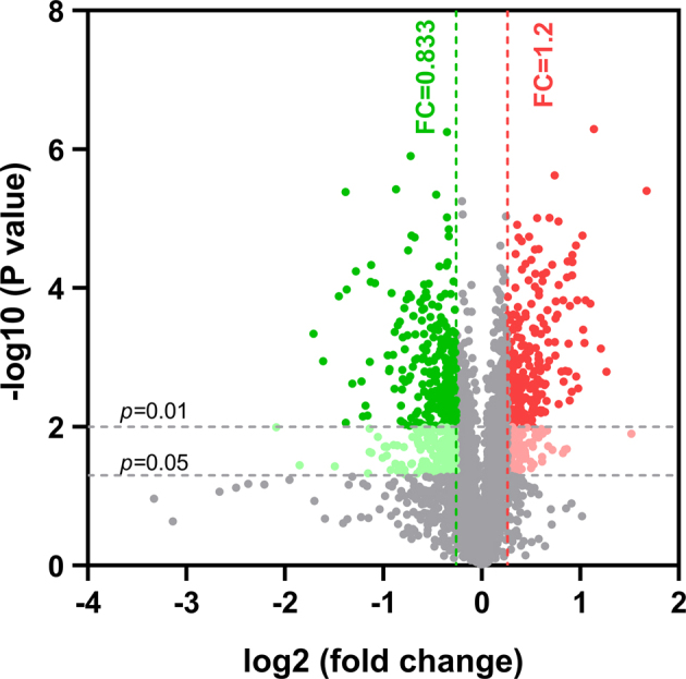
Volcano plot of differentially expressed proteins (DEPs) obtained from the labelled samples. The x -axis represents log2 expression fold-change (FC) in sudden death syndrome (SDS) vs. normal samples and the y -axis represents false discovery rate (FDR) (−log10). Up-regulated genes (FDR <0.01 and FC >1.2) are shown in red while down-regulated genes are shown in green (FDR <0.01 and FC <0.833).
Table 1.
Parts of the up-regulated proteins identified using isobaric tags for relative and absolute quantification (iTRAQ)-based quantitative proteomics.
| Accession | Description | Unique peptides | Ratio sudden death syndrome /Con | P value | Gene name |
|---|---|---|---|---|---|
| Q90765 | CASP protein | 1 | 2.864 | 0.013 | CUX1 |
| A0A1L1RYK7 | P2X purinoceptor | 1 | 1.958 | 0.000 | P2RX2 |
| F1NB57 | Serine/threonine-protein kinase | 1 | 1.942 | 0.002 | PLK2 |
| C6F1H2 | NADH-ubiquinone oxidoreductase chain 2 | 3 | 1.939 | 0.000 | MT-ND2 |
| Q2LK94 | Collectin-11 | 1 | 1.858 | 0.000 | COLEC11 |
| A0A1D5PR58 | Ensconsin | 1 | 1.830 | 0.000 | MAP7 |
| E1BS85 | Histone acetyltransferase | 1 | 1.771 | 0.000 | KAT6B |
| E1BWN6 | Coronin | 3 | 1.682 | 0.000 | CORO2A |
| Q76MS9 | Musculoskeletal embryonic nuclear protein 1 | 2 | 1.640 | 0.000 | MUSTN1 |
| F1NW02 | Alpha-amylase | 1 | 1.610 | 0.000 | AMY1A |
| A0A1D5PBV6 | Troponin T | 1 | 1.598 | 0.001 | TNNT2 |
| Q5ZHV4 | Phosphoglycerate mutase | 1 | 1.588 | 0.000 | PGAM1 |
| Q90Z42 | Roundabout 1 protein | 1 | 1.578 | 0.001 | ROBO1 |
| A0A1D5P4H0 | Tropomyosin beta chain | 1 | 1.533 | 0.000 | TPM2 |
Table 2.
Parts of the down-regulated proteins identified using isobaric tags for relative and absolute quantification (iTRAQ)-based quantitative proteomics.
| Accession | Description | Unique peptides | Ratio sudden death syndrome /Con | P value | Gene name |
|---|---|---|---|---|---|
| P04268 | Tropomyosin alpha-1 chain | 1 | 0.277 | 0.036 | TPM1 |
| Q8AY28 | Fast myosin heavy chain HCIII | 1 | 0.306 | 0.000 | MYH1A |
| Q5F3R7 | DDB1- and CUL4-associated factor 12 | 1 | 0.365 | 0.000 | DCAF12 |
| Q1JR86 | Growth hormone | 1 | 0.412 | 0.000 | GH |
| F1NZX1 | Aggrecan core protein | 1 | 0.429 | 0.002 | VCAN |
| F1NUI7 | Kinesin-like protein | 1 | 0.454 | 0.011 | KIF16B |
| F4ZCJ4 | MHC class II antigen | 1 | 0.481 | 0.014 | BMA2 |
| Q009U5 | KIT ligand form 2 | 1 | 0.529 | 0.000 | KITLG |
| P02565 | Myosin-1B | 12 | 0.547 | 0.000 | MYH1B |
| F1NLH9 | Inosine triphosphate pyrophosphatase | 2 | 0.573 | 0.002 | ITPA |
| F1NRB3 | Oxysterol-binding protein | 1 | 0.573 | 0.001 | OSBP2 |
| F1NG74 | Gamma-enolase | 2 | 0.581 | 0.001 | ENO2 |
| P02197 | Myoglobin | 15 | 0.589 | 0.000 | MB |
| Q90933 | Neuron-glia cell adhesion molecule | 4 | 0.666 | 0.003 | P35331 |
Annotation and Functional Enrichment
To gain global insights into the 186 DEPs, GO was used to analyze the dysregulated proteins to reveal the BP, cell components (CC), and molecular functions (MF) altered in the heart tissues of SDS broilers. A total of 2,541 BPs were enriched in this study, and 426 were statistically significant with P < 0.05. The top 10 significantly enriched terms sorted according to P- value are shown in Figure 4 A. The top 23 terms of GO annotation for BP showed that most of the dysregulated proteins participated in cellular process, single-organism process, metabolic regulation, cellular component organization or biogenesis, response to stimulus, multicellular organismal process, developmental process, and localization (Figure 4 D). For CC ontology, 364 components were enriched, and 109 were statically significant (P < 0.05). The top 10 significantly enriched CC terms are shown in Figure 4 B. The DEPs in CC were distributed in 16 GO terms, including cell, cell part, organelle, membrane, organelle part, macromolecular complex, membrane part, extracellular region, extracellular region part, and membrane-enclosed lumen (Figure 4 D). With insight into MF ontology, 490 MF terms were enriched and 107 were statically significant (P < 0.05). The top 10 significantly enriched MF terms are shown in Figure 4 C. The key functions of changed proteins were associated with binding, catalytic activity, and structural molecule activity (Figure 4 D). The predominant functions of DEPs were in BP, such as cellular process, metabolic regulation, and response to stimulus.
Figure 4.

Gene ontology analysis results of differentially expressed proteins in sudden death syndrome (SDS). Results of significantly enriched biological process classification (A), cell component (B) and molecular function enrichment (C) are illustrated. (D) Percentages of proteins involved in each of the biological process, cellular component and molecular function terms.
Pathway Analysis
Pathway analysis based on KEGG database was performed to collect information concerning protein functions in metabolic processes to discover the specific biological events of DEPs. A total of 31 KEGG pathways were enriched. Most of them are clustered into 3 sub-categories, including metabolism (14 pathways), cellular process (6 pathways), and environmental information processing (5 pathways) (Figure 5). TheP -values of 4 pathways were less than 0.05 (Figure 6). The most significant KEGG pathways were glycolysis/gluconeogenesis, biosynthesis of amino acids, cardiac muscle contraction, and ascorbate and aldarate metabolism.
Figure 5.

Class of enriched Kyoto Encyclopedia of Genes and Genomes (KEGG) pathways participated by the differentially expressed proteins (DEPs).
Figure 6.

Distribution of Kyoto Encyclopedia of Genes and Genomes (KEGG) pathways arranged in ascending order according to the P -value.
Protein–Protein Interaction Analysis
The protein–protein interaction network was analyzed using the publicly available program STRING, and several strong interactions were found among the DEPs related to cardiac muscle contraction (Figure 7). Troponin T (TNNT2), myoglobin, myosin-3 (MYH1), tropomyosin alpha-1 (TPM1), Gga.27138, Gga.27660, ACTB, and SRF had pivotal roles in the interaction network by regulating cardiac muscle contraction. The identification of the protein interaction network of DEPs related to glycolysis or biosynthesis of amino acids showed that ENO2, BPGM, MDH2, PGM2, and AMY1A had pivotal roles in the network. In addition, they may have important roles in regulating glycolysis and biosynthesis of amino acids in chicken.
Figure 7.

Protein–protein associations analyzed by the String software. The interaction network showing important proteins involved in the regulation of cardiac muscle contraction and glycolysis or biosynthesis of amino acids, respectively.
Confirmation of Selected DEPs Compared with iTRAQ Database
To validate the results obtained by iTRAQ LC-MS/MS analysis, DEPs were selected for confirmation by western blot analysis. Two DEPs (upregulated: PGAM1; downregulated: Myoglobin) which showed good species specificity and clear bands when detected by commercial antibodies were shown in this study.β -actin was used as the internal standard. As shown in Figure 8, the expression of PGAM1 protein was significantly increased (P < 0.05), whereas the expression of myoglobin was significantly decreased (P < 0.01) in the heart tissue of SDS broilers. These results agree with the findings of iTRAQ-based proteomic analysis (Table 1 and 2). The dysregulated proteins (PGAM1 and myoglobin) can significantly differentiate the SDS group from the healthy group, thereby indicating that they can be clinically used as biomarkers to obtain specimen from SDS broilers for further study.
Figure 8.
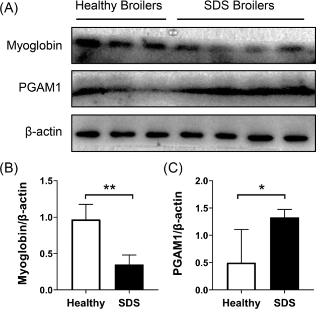
Validation of differentially expressed proteins (DEPs) using western blot. (A) Western blot result illustration. Group comparison based on gray value western blot result of myoglobin (B) and PGAM1 (C). β -actin was used as an internal standard. **: P < 0.01; *:P < 0.05.
DISCUSSION
SDS, a kind of nutritional metabolic disease related to cardiac dysfunction, is commonly observed in chickens selected for rapid growth. It has seriously affected breeding benefit in the broiler industry around the world for many years (Chung et al., 1993). Its pathogenesis in broilers is poorly understood, but more people point out that it may be related to structural damage of heart and cardiac arrhythmia in fast growing broilers (Itoh et al., 1997; Imaeda, 1999; Olkowski et al., 2008). Proteomic methods are often used in combination with bioinformatics techniques to screen various disease markers and reveal the physiological and pathological mechanisms of cells under various factors. To reveal the biological information of differential proteins in the myocardial tissue of SDS broilers and achieve potential biomarkers that play important roles in the pathogenesis of SDS, we focused on the DEPs in the left ventricle of SDS broilers. iTRAQ, a liquid-based technique in proteomics, labels the lysine residues and N terminus of all peptides with isotopes of identical masses. Thus, it exhibits better sensitivity and allows accurate quantitative comparison of proteins from multiple samples (Aggarwal and Yadav, 2016). To the best of our knowledge, this study is the first iTRAQ-based proteomic analysis for the left ventricle of broilers that suffered from SDS.
Consistent with previous reports, all chickens that died of SDS were in very good body condition and died suddenly without any discernible preexisting clinical signs (Olkowski, 2007; Olkowski et al., 2008). Histopathological examination showed the presence of focal cardiomyocyte degeneration and inflammatory response in the left ventricular myocardium, which showed no gross lesions, of SDS broilers. In addition, damaged mitochondria characterized by mitochondrial swelling, vacuolization, and crista fragmentation were found in the cardiomyocytes of SDS broilers, thereby demonstrating that mitochondrial damage was accompanied with myocardial injury in the hearts of SDS broilers. Heart-related mortalities are observed predominantly in fast-growing broiler chickens. Although structural damage, molecular changes, and biochemical factors were reported correlated with the progress of SDS (Imaeda, 1999; Olkowski et al., 2008), the pathogenesis of SDS is still poorly understood. In this study, comparative proteomic methods were used to study the DEPs in the left ventricular myocardium of SDS broilers, which has a certain theoretical basis and will improve our understanding of the molecular mechanisms associated with SDS.
The contraction and relaxation of the heart are important for the maintenance of cardiac function (Bers, 2002). In the present study, the proteins associated with the BP of cardiac muscle contraction, including TNNT2, TPM1, Fast myosin heavy chain HCIII (MYH1A), and myosin-1B (MYH1B) were identified among the top abundant proteins (Tables 1 and 2). Moreover, KEGG results showed that one of the most significant KEGG pathways was cardiac muscle contraction. The interaction network of cardiac muscle contraction-related proteins (Figure 7) showed that MYH1, TPM1, TNNT2, and MB are highly interrelated and play key roles in the entire network. Functional annotation results showed that these proteins were associated with GO terms for myofibril, contractile fiber part, and actin-binding cytoskeleton structures, consistent with the findings of a previous research (Perry, 1998; Oldfors et al., 2004; Wang et al., 2019). These results help clarify the relationship between cardiac dysfunction and the incidence of SDS from the perspective of proteomics, thereby further proving that the occurrence of SDS is closely related to cardiac dysfunction.
The heart is an organ with high activity and high energy expenditure. Both diastole and contraction require an adequate energy supply. An abnormal heart energy metabolism results in abnormal cardiac function and arrhythmia (Hessling, 2016; Wu et al., 2017). Due to the characteristics of genetic breeding and rapid growth of broilers, metabolic disorders, such as SDS, continue to occur (Julian, 2005). Broilers suffer from severe metabolic stress and are more prone to heart dysfunction and subclinical heart disease than other breeds of chickens (Wilson, Julian and Barker, 1988). SDS is confirmed to be associated with acute cardiac dysfunction induced by malignant arrhythmias, including ventricular flapping and ventricular fibrillation (Olkowski and Classen, 1997). Disturbance of energy metabolism balance and change of myocardial substrates promote atrial electromyography instability and arrhythmia via a variety of cellular pathways in myocardial cells (Barth and Tomaselli, 2009). There is a growing body of evidence proving that metabolic activity and arrhythmia are interdependent. An impaired cellular energetic state predisposes to atrial arrhythmias, and atrial rhythm disturbances influence metabolic activity. Life-threatening ventricular tachyarrhythmias are prevalent in every cardiovascular condition associated with structural and/or functional abnormalities (Brandenburg et al., 2016). In the present study, with a cutoff of 1.2-fold upregulation and 0.83-fold downregulation, we identified 186 significantly different proteins, of which 72 and 114 were upregulated and downregulated, respectively, in the SDS group. Among the identified DEPs, CASP, MT-ND2, alpha-amylase, PGAM1, ENO2, and myoglobin proteins were associated with the BP of energy metabolism and transport. The changes in expression levels of these metabolism-related enzymes may be related to the metabolic disorders of broilers (Olkowski and Classen, 1998). According to GO analysis, the DEPs identified using iTRAQ technique were involved in BP, including cellular process, single-organism process, and metabolic regulation. Moreover, KEGG analysis revealed that one of the most significant KEGG pathways was glycolysis/gluconeogenesis. Lactic acid, a product of glycolysis, causes a decrease in pH in cardiomyocytes, which are long-lived anaphase cells sensitive to changes in pH. Decreased pH can inhibit protein synthesis of cardiomyocytes, lead to a disturbance in the excitation-contraction coupling of the myocardium (Swietach et al., 2015), and directly damage the ultrastructure of cardiomyocytes, leading to organic lesions (Sorensen et al., 2015). Arrhythmia, an important cause of sudden cardiac death, is closely related to cardiomyocyte acidosis (Said et al., 2008; Godinjak et al., 2017). The blood lactic acid level of SDS broilers is significantly higher than that of live chickens. Arrhythmias and sudden death similar to SDS can occur in broilers by gavage or intravenous administration of lactic acid (Boulianne et al., 1993; Korte et al., 1999; Hassanzadeh et al., 2010). Thus, SDS broilers had metabolic disorders characterized by glycolytic enzyme activation and lactic acid production. The production of lactic acid during metabolic disorders damages cardiomyocytes and leads to arrhythmia, thereby eventually promoting the development of SDS.
CONCLUSIONS
Comprehensive proteome profiling was applied to identify and interpret the differences in protein expression during the progress of SDS. A highly efficient identification of cardiomyocyte proteome resulted in an observation of 186 DEPs, of which 7 were related to cardiac muscle contraction, and another seven were related to cardiac energy metabolism. TNNT, TPM1, TPM2, MYH1A, MYH1B, CORO2A, and MB may be involved in the regulation of cardiac muscle contraction. ENO2, PGAM1, CASP, MT-ND2, PLK2, MB, and AMY1A may affect cardiac energy metabolism or biosynthesis of amino acids. Our expression profiles provide useful information and new insights into SDS, thereby leading to better understanding of this condition.
ACKNOWLEDGMENTS
This work was supported by the Scientific and Technological Foundation of Henan Province in China (172102110026 and 192102110080) and Natural Science Foundation of Henan Province (182300410049).
Conflict of interest statement
The authors declare that they have no competing interests.
Footnotes
Supplementary data are available at Poultry Science online.
ps-19-0,9643-S001.xlsx.
Contributor Information
Dongfang Hu, Email: hdf@hist.edu.cn.
Yaming Ge, Email: ndgeyaming@126.com.
Supplementary Material
REFERENCES
- Aggarwal S., Yadav A.K. Dissecting the iTRAQ data analysis. Methods Mol. Biol. 2016;1362:277–291. doi: 10.1007/978-1-4939-3106-4_18. [DOI] [PubMed] [Google Scholar]
- Ai C., Kong L. CGPS: a machine learning-based approach integrating multiple gene set analysis tools for better prioritization of biologically relevant pathways. J. Genet. Genomics. 2018;45:489–504. doi: 10.1016/j.jgg.2018.08.002. [DOI] [PubMed] [Google Scholar]
- Barth A.S., Tomaselli G.F. Cardiac metabolism and arrhythmias. Circ. Arrhythm. Electrophysiol. 2009;2:327–335. doi: 10.1161/CIRCEP.108.817320. [DOI] [PMC free article] [PubMed] [Google Scholar]
- Basaki M., Asasi K., Tabandeh M.R., Aminlari M. Polymorphism identification and cardiac gene expression analysis of the calsequestrin 2 gene in broiler chickens with sudden death syndrome. Br. Poult. Sci. 2016;57:151–160. doi: 10.1080/00071668.2015.1099615. [DOI] [PubMed] [Google Scholar]
- Bers D.M. Cardiac excitation-contraction coupling. Nature. 2002;415:198–205. doi: 10.1038/415198a. [DOI] [PubMed] [Google Scholar]
- Boulianne M., Hunter D.B., Viel L., Physick-Sheard P.W., Julian R.J. Effect of exercise on the cardiovascular and respiratory systems of heavy turkeys and relevance to sudden death syndrome. Avian Dis. 1993;37:83–97. [PubMed] [Google Scholar]
- Brandenburg S., Arakel E.C., Schwappach B., Lehnart S.E. The molecular and functional identities of atrial cardiomyocytes in health and disease. Biochim. Biophys. Acta. 2016;1863:1882–1893. doi: 10.1016/j.bbamcr.2015.11.025. [DOI] [PubMed] [Google Scholar]
- Chung H.C., Guenter W., Rotter R.G., Crow G.H., Stanger N.E. Effects of dietary fat source on sudden death syndrome and cardiac sarcoplasmic reticular calcium transport in broiler chickens. Poult. Sci. 1993;72:310–316. doi: 10.3382/ps.0720310. [DOI] [PubMed] [Google Scholar]
- Godinjak A., Jusufovic S., Rama A., Iglica A., Zvizdic F., Kukuljac A., Tancica I., Rozajac S. Hyperlactatemia and the importance of repeated lactate measurements in critically ill patients. Med. Arch. 2017;71:404–407. doi: 10.5455/medarh.2017.71.404-407. [DOI] [PMC free article] [PubMed] [Google Scholar]
- Hassanzadeh M., Maddadi M.S., Mirzaie S., Assasie K., Moayyedian H. Partial pressure of carbon dioxide in the venous blood of young birds as a predictor of ascites susceptibility in broiler chickens. Acta. Vet. Hung. 2010;58:221–230. doi: 10.1556/AVet.58.2010.2.8. [DOI] [PubMed] [Google Scholar]
- Hessling G. [Arrhythmia in adults with congenital heart disease: acute and long-term management] Herzschrittmacherther. Elektrophysiol. 2016;27:81–87. doi: 10.1007/s00399-016-0430-x. [DOI] [PubMed] [Google Scholar]
- Huang da W., Sherman B.T., Lempicki R.A. Systematic and integrative analysis of large gene lists using DAVID bioinformatics resources. Nat. Protoc. 2009;4:44–57. doi: 10.1038/nprot.2008.211. [DOI] [PubMed] [Google Scholar]
- Imaeda N. Characterization of serum enzyme activities and electrolyte levels in broiler chickens after death from sudden death syndrome. Poult. Sci. 1999;78:66–69. doi: 10.1093/ps/78.1.66. [DOI] [PubMed] [Google Scholar]
- Itoh N., Moritsu Y., Taniyama H., Ichikawa S. Correlation between heart muscle damage and high-nutrient feed in broiler chickens. J. Vet. Med. Sci. 1997;59:209–211. doi: 10.1292/jvms.59.209. [DOI] [PubMed] [Google Scholar]
- Julian R.J. Production and growth related disorders and other metabolic diseases of poultry–a review. Vet. J. 2005;169:350–369. doi: 10.1016/j.tvjl.2004.04.015. [DOI] [PubMed] [Google Scholar]
- Karp N.A., Huber W., Sadowski P.G., Charles P.D., Hester S.V., Lilley K.S. Addressing accuracy and precision issues in iTRAQ quantitation. Mol. Cell. Proteomics. 2010;9:1885–1897. doi: 10.1074/mcp.M900628-MCP200. [DOI] [PMC free article] [PubMed] [Google Scholar]
- Korte S.M., Sgoifo A., Ruesink W., Kwakernaak C., van Voorst S., Scheele C.W., Blokhuis H.J. High carbon dioxide tension (PCO2) and the incidence of cardiac arrhythmias in rapidly growing broiler chickens. Vet. Rec. 1999;145:40–43. doi: 10.1136/vr.145.2.40. [DOI] [PubMed] [Google Scholar]
- Li H., Mao Y., Xiong Y., Zhao H.H., Shen F., Gao X., Yang P., Liu X., Fu D. A comprehensive proteome analysis of peripheral blood mononuclear cells (PBMCs) to identify candidate biomarkers of pancreatic cancer. Cancer Genomics Proteomics. 2019;16:81–89. doi: 10.21873/cgp.20114. [DOI] [PMC free article] [PubMed] [Google Scholar]
- Oldfors A., Tajsharghi H., Darin N., Lindberg C. Myopathies associated with myosin heavy chain mutations. Acta Myol. 2004;23:90–96. [PubMed] [Google Scholar]
- Olkowski A.A. Pathophysiology of heart failure in broiler chickens: structural, biochemical, and molecular characteristics. Poult. Sci. 2007;86:999–1005. doi: 10.1093/ps/86.5.999. [DOI] [PubMed] [Google Scholar]
- Olkowski A.A., Classen H.L. Malignant ventricular dysrhythmia in broiler chickens dying of sudden death syndrome. Vet. Rec. 1997;140:177–179. doi: 10.1136/vr.140.7.177. [DOI] [PubMed] [Google Scholar]
- Olkowski A.A., Classen H.L. High incidence of cardiac arrhythmias in broiler chickens. Zentralbl. Veterinarmed. A. 1998;45:83–91. doi: 10.1111/j.1439-0442.1998.tb00804.x. [DOI] [PubMed] [Google Scholar]
- Olkowski A.A., Wojnarowicz C., Nain S., Ling B., Alcorn J.M., Laarveld B. A study on pathogenesis of sudden death syndrome in broiler chickens. Res. Vet. Sci. 2008;85:131–140. doi: 10.1016/j.rvsc.2007.08.006. [DOI] [PubMed] [Google Scholar]
- Ouyang H., Wang Z., Chen X., Yu J., Li Z., Nie Q. Proteomic analysis of chicken skeletal muscle during embryonic development. Front Physiol. 2017;8:281. doi: 10.3389/fphys.2017.00281. [DOI] [PMC free article] [PubMed] [Google Scholar]
- Perry S.V. Troponin T: genetics, properties and function. J. Muscle Res. Cell Motil. 1998;19:575–602. doi: 10.1023/a:1005397501968. [DOI] [PubMed] [Google Scholar]
- Polansky O., Seidlerova Z., Faldynova M., Sisak F., Rychlik I. Protein expression in the liver and blood serum in chickens in response to Salmonella Enteritidis infection. Vet. Immunol. Immunopathol. 2018;205:10–16. doi: 10.1016/j.vetimm.2018.10.006. [DOI] [PubMed] [Google Scholar]
- Said M., Becerra R., Palomeque J., Rinaldi G., Kaetzel M.A., Diaz-Sylvester P.L., Copello J.A., Dedman J.R., Mundina-Weilenmann C., Vittone L., Mattiazzi A. Increased intracellular Ca2+ and SR Ca2+ load contribute to arrhythmias after acidosis in rat heart. Role of Ca2+/calmodulin-dependent protein kinase II. Am. J. Physiol. Heart Circ. Physiol. 2008;295:H1669–H1683. doi: 10.1152/ajpheart.00010.2008. [DOI] [PMC free article] [PubMed] [Google Scholar]
- Sorensen B.S., Busk M., Overgaard J., Horsman M.R., Alsner J. Simultaneous hypoxia and low extracellular pH suppress overall metabolic rate and protein synthesis in vitro. PLoS One. 2015;10 doi: 10.1371/journal.pone.0134955. [DOI] [PMC free article] [PubMed] [Google Scholar]
- Swietach P., Spitzer K.W., Vaughan-Jones R.D. Na(+) ions as spatial intracellular messengers for co-ordinating Ca(2)(+) signals during pH heterogeneity in cardiomyocytes. Cardiovasc. Res. 2015;105:171–181. doi: 10.1093/cvr/cvu251. [DOI] [PMC free article] [PubMed] [Google Scholar]
- Wang J., Tang C., Yang C., Zheng Q., Hou Y. Tropomyosin-1 functions as a tumor suppressor with respect to cell proliferation, angiogenesis and metastasis in renal cell carcinoma. J. Cancer. 2019;10:2220–2228. doi: 10.7150/jca.28261. [DOI] [PMC free article] [PubMed] [Google Scholar]
- Wang Z., Shang P., Li Q., Wang L., Chamba Y., Zhang B., Zhang H., Wu C. iTRAQ-based proteomic analysis reveals key proteins affecting muscle growth and lipid deposition in pigs. Sci. Rep. 2017;7 doi: 10.1038/srep46717. [DOI] [PMC free article] [PubMed] [Google Scholar]
- Wiese S., Reidegeld K.A., Meyer H.E., Warscheid B. Protein labeling by iTRAQ: a new tool for quantitative mass spectrometry in proteome research. Proteomics. 2007;7:340–350. doi: 10.1002/pmic.200600422. [DOI] [PubMed] [Google Scholar]
- Wilson J.B., Julian R.J., Barker I.K. Lesions of right heart failure and ascites in broiler chickens. Avian Dis. 1988;32:246–261. [PubMed] [Google Scholar]
- Wu Y., Li J., Xu L., Lin L., Chen Y.H. Mechanistic and therapeutic perspectives for cardiac arrhythmias: beyond ion channels. Sci. China. Life Sci. 2017;60:348–355. doi: 10.1007/s11427-016-9005-6. [DOI] [PubMed] [Google Scholar]
- Xing T., Wang C., Zhao X., Dai C., Zhou G., Xu X. Proteome analysis using isobaric tags for relative and absolute analysis quantitation (iTRAQ) reveals alterations in stress-induced dysfunctional chicken muscle. J. Agric. Food Chem. 2017;65:2913–2922. doi: 10.1021/acs.jafc.6b05835. [DOI] [PubMed] [Google Scholar]
- Zhang P., Zhu S., Li Y., Zhao M., Liu M., Gao J., Ding S., Li J. Quantitative proteomics analysis to identify diffuse axonal injury biomarkers in rats using iTRAQ coupled LC-MS/MS. J. Proteomics. 2016;133:93–99. doi: 10.1016/j.jprot.2015.12.014. [DOI] [PubMed] [Google Scholar]
- Zhao Z., Zhao Q., Zhu S., Huang B., Lv L., Chen T., Yan M., Han H., Dong H. iTRAQ-based comparative proteomic analysis of cells infected with Eimeria tenella sporozoites. Parasite. 2019;26:7. doi: 10.1051/parasite/2019009. [DOI] [PMC free article] [PubMed] [Google Scholar]
Associated Data
This section collects any data citations, data availability statements, or supplementary materials included in this article.