Abstract
Background
Clinical hypercoagulopathy in patients with COVID-19 has been anecdotally described, but there is lack of evidence due to the novelty of this disease. Our study reports the results of rotational thromboelastography (ROTEM) in relation to traditional laboratory coagulation tests and acute phase markers among a cohort of severely ill, mechanically ventilated patients with COVID-19.
Methods
Patients with COVID-19 (N=21) with respiratory failure requiring mechanical ventilation were included in this prospective case series. ROTEM was serially obtained for all patients on three different days during their intensive care unit (ICU) stay and analyzed using repeated measures analysis. Demographic variables, symptoms at the time of presentation, ROTEM values, laboratory values for traditionally measured coagulation profiles, and acute phase reactants were analyzed, in addition to the use of anticoagulation and clinical hypercoagulopathic complications.
Results
The average age of our cohort was 57.9 years old (SD=14.4) and 76.2% were male. The mortality rate was 14.3% (3 of 21). Two patients (12.5%) were identified to have new-onset deep vein thrombosis, two patients (12.5%) were found to have ≥3 episodes of central venous catheter thrombosis, and three patients (18.7%) had confirmed stroke. ROTEM demonstrated elevated EXTEM and INTEM clotting times, including elevated FIBTEM maximum clot firmness (MCFFIB). All patients treated with therapeutic anticoagulation still demonstrated hypercoagulopathy within the MCFFIB tests.
Discussion
Repeated measure ROTEMs were able to detect hypercoagulopathy in ICU patients with COVID-19 despite therapeutic anticoagulation with heparin.
Level of evidence
III.
Keywords: thrombelastography, fibrinolysis, blood coagulation, viruses
Introduction
The COVID-19 pandemic has posed distinct challenges. Reports of its characteristics are emerging daily, and although no organ system appears to be spared the immune system seems to be the most significantly affected. The illness initiates a surge of proinflammatory cytokines, which in turn hyperactivates the coagulation cascade. One of the consistent observations has been the presence of vasculitis and coagulopathies in patients stricken with COVID-19.1 Specifically, hypercoagulopathy has been described, as manifested by cerebral infarcts, pulmonary embolism, deep venous thrombosis, and clotting of indwelling central venous catheters.2–6 The upregulation of this immune-mediated cascade, left relatively unchecked, is postulated to be the cause of thromboembolic events.6
However, there is lack of data regarding specific coagulation pathways. Adjunctive laboratory tests such as prothrombin time (PT) and partial thromboplastin time (PTT) have been shown to be deranged in patients with COVID-19. Other markers such as elevated D-dimer levels have also been associated with increased risk of mortality.7 The clinical evaluation of coagulation has traditionally revolved around the measurement of conventional coagulation tests such as PT, PTT, platelet count, and fibrinogen. However, these tests, although they provide quantitative measurements of coagulation, fail to assess the dynamic and qualitative nature of clot formation, stability, and lysis. For this reason, point-of-care viscoelastic testing in the form of thromboelastography and rotational thromboelastometry has been widely used in trauma, perioperative surgical care, and critical care to evaluate coagulopathy.8 Derangements in rotational thromboelastography (ROTEM) values indicative of hypercoagulability among patients with COVID-19 were recently reported by Pavoni et al.9 The aim of our study was to report the results of ROTEM in relation to acute phase reactants and other markers of coagulopathy to further study disorders of coagulation among critically ill, mechanically ventilated patients with COVID-19. Given that anticoagulation is widely used in the setting of sepsis for venous thromboembolism (VTE) prophylaxis and/or therapeutically used to treat confirmed or suspected VTE and clinical coagulopathy, we further investigated the characteristics of ROTEM in relation to clinical manifestations of coagulopathy and the use of anticoagulation. Moreover, given that coagulopathy is a dynamic (not static) phenomenon, we sought to perform a repeated measure analysis to investigate the changes in ROTEM features over time during the clinical course of disease.
Methods
A prospective case series was conducted, after obtaining institutional review board approval, on 21 critically ill patients from our COVID-19 intensive care unit (ICU) from March 2020 to April 2020. Given limited resources and bed availability, our department of surgery provided daily care to 21 critically ill ICU patients during the study period. Our inclusion criteria included individuals who were COVID-19-positive and were mechanically ventilated. Patients who were on active extracorporeal membrane oxygenation during rotational thromboelastometry draws were excluded from the study. Thromboembolic events were recorded prospectively. These events included deep vein thrombosis (DVT), confirmed stroke, and three or more episodes of clotted central venous catheters. ROTEM was obtained from all 21 patients on three separate days while admitted to the ICU. The first ROTEM was drawn an average of 20.7 days from the date of admission (figure 1). The second ROTEM was drawn an average of 1.4 days from the first draw, and the third ROTEM was drawn an average of 3.2 days from the second ROTEM. With respect to patients with coagulopathic complications, the first ROTEM measurement was drawn an average of 3.4 days from the initial thromboembolic event (figure 1). The second ROTEM was drawn an average of 1.4 days from the first, and the third ROTEM was drawn on average of 3.2 days from the second.
Figure 1.
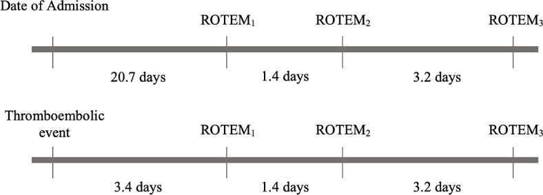
Rotational thromboelastography (ROTEM) and thromboembolic event timeline.
Given the repeated measures of this study, each ROTEM measurement was considered as a unique incident (n), resulting in 61 overall samples (discharge from the ICU or mortality disqualified patients from undergoing repeated measurements). ROTEM included the analysis of extrinsic clotting pathway defects (EXTEM: factors II, V, VII, X), defects of the intrinsic clotting cascade (INTEM: factors II, V, VIII, X, XI, XII), and a qualitative analysis on fibrinogen (FIBTEM: fibrin activity/contribution to clot firmness-extrinsic activation, platelet neutralization). Each section is further divided into various components that outline clot initiation, clot kinetics, clot strength, and fibrinolysis (table 1).
Table 1.
Rotational thromboelastography parameters
| Clot initiation | Clotting time |
| Clot kinetics | Clot formation time |
| Clot strength | Amplitude at 10 minutes Maximum clot firmness |
| Fibrinolysis | Maximum lysis |
Specifically, a prolonged clotting time (CT) intrinsic (CTIN) suggests heparin use or intrinsic pathway factor deficiency. Prolonged CT extrinsic (CTEX) suggests extrinsic pathway factor deficiency. A shortened clot formation time (CFT) suggests hypercoagulability. A reduced amplitude at 10 minutes (A10IN and EX) suggests inadequate clot firmness as a result of decreased platelets, fibrinogen, and/or factor VIII. Reduced INTEM and EXTEM maximum clot firmness (MCFIN and EX) suggests inadequate clot firmness as a result of decreased platelets, fibrinogen, and/or factor VIII. A reduced FIBTEM maximum clot firmness (MCFFIB) suggests inadequate fibrin, and finally maximum lysis (MLIN, EX, and FIB) greater than 15% suggests hyperfibrinolysis. A representative image of ROTEM parameters is included in online supplemental figure 1.10 A legend corresponding to the graphical representation of these ROTEM values is included in online supplemental figure 2.
tsaco-2020-000603supp001.pdf (914KB, pdf)
tsaco-2020-000603supp002.pdf (879.4KB, pdf)
DVT, confirmed strokes, and clotted central venous catheters were recorded into one variable labeled “hypercoagulopathic complications”. Patients with high clinical suspicion for stroke with concomitant radiographic findings of acute small vessel ischemic changes and diffuse cerebral effacement with loss of gray–white matter differentiation were included in our stroke population. Duplex studies were obtained on patients with clinical signs and symptoms of DVT. Patients were divided into two groups, those with hypercoagulopathic complications and those without. Another subgroup analysis was conducted examining patients with and without therapeutic heparin administration during their ICU stay. Typical coagulopathy profiles as well as acute phase markers were recorded and expressed as a function of their mean values: PT, international normalized ratio (INR), PTT, platelet count, C-reactive protein (CRP), ferritin, lactate dehydrogenase (LDH), fibrinogen, and D-dimer. Patient demographics including age, gender, and body mass index were included, as well as types of presenting symptoms prior to ICU admission. The use of therapeutic anticoagulation was also studied; therapeutic anticoagulation was defined as PTT values of 1.5 to 2 times the normal. Given the concern for end-organ damage seen in COVID-19-positive patients, anticoagulation was achieved with therapeutic doses of unfractionated heparin as opposed to low molecular weight heparin. All patients not receiving therapeutic anticoagulation were administered subcutaneous chemical VTE prophylaxis with 5000 units of unfractionated heparin three times daily or 40 mg low molecular weight heparin once daily for DVT prophylaxis. Statistical analysis was performed using IBM SPSS Statistics for Windows, V.26 (IBM, Armonk, NY, USA). Boxplots were created for graphical representation. Reference ranges were provided within both tables and graphs. Outliers were represented by a circle (mild outlier) or an asterisk (severe outlier).
Results
The average age of patients was 57.9 years old. There were 16 men and 5 women (table 2), and the average body mass index was 32.3 kg/m2. The mortality rate among these patients was 14.3% and occurred at a mean of 23.6 days (SD=7.0) after admission. The most common presenting symptom was fever (76.2% of patients), followed by shortness of breath and cough, which were observed in 71.4% of patients. Traditional coagulopathic markers such as PT, PTT, INR, and platelet counts were minimally elevated and suggestive of hypocoagulopathy. Acute phase markers, LDH, ferritin, fibrinogen, and D-dimer were all markedly elevated (table 3). Two patients were found to have DVT (treated with therapeutic anticoagulation), two additional patients had confirmed central venous catheter thrombosis, and three patients had confirmed cerebrovascular accidents. The overall rate of confirmed clinical thromboembolic events in this cohort was 33.3% (7 of 21) (table 4). In total, 71.4% of patients were treated with therapeutic anticoagulation at some point during their ICU course; patients without confirmed or suspected clinical coagulopathy were anticoagulated either empirically, or to prevent catheter-related thrombosis among those receiving hemodialysis. Seven patients (33.3%) received hemodialysis during their ICU course (table 4). All patients were mechanically ventilated at the time of ROTEM measurement. The first ROTEM measurements were taken an average of 20.8 days (STD=14.4) from the date of admission, the second –22.2 days (STD=14.4) and the third –25.4 days (STD=14.2).
Table 2.
Patient demographics, admission vital signs, and presenting symptoms
| Demographics, n (%) | |
| Age, mean (SD) | 57.9 (14.4) |
| Male | 16 (76.2) |
| BMI kg/m2, mean (SD) | 32.3 (9.2) |
| Mortality | 3 (14.3) |
| Admission vitals, mean (SD) | |
| Temperature, °F | 99.6 (2.1) |
| Pulse, beats per minute | 98.6 (25.1) |
| RR, breaths per minute | 25.3 (8.8) |
| O2 saturation (%) | 91.6 (6.3) |
| Systolic, mm Hg | 132.6 (19.6) |
| Diastolic, mm Hg | 75.6 (14.9) |
| Symptoms, n (%) | |
| Febrile | 16 (76.2) |
| Lethargy | 5 (23.8) |
| Shortness of breath | 15 (71.4) |
| Headache | 1 (4.8) |
| Anorexia | 0 (0) |
| Rhinorrhea | 0 (0) |
| Sputum | 3 (14.3) |
| Sore throat | 1 (4.8) |
| Cough | 15 (71.4) |
| Emesis | 1 (4.8) |
| Myalgia | 2 (9.5) |
| Diarrhea | 3 (14.3) |
| Recent travel | 0 (0) |
| Known sick contacts | 2 (9.5) |
| Length of symptoms, mean days (SD) | 5.2 (3.1) |
BMI, body mass index; RR, respiratory rate.
Table 3.
Acute phase reactants and coagulation markers
| Laboratory values, mean (SD) | Reference range | Mean (SD) | Peak, mean | No HC, mean (SD) n=14 |
HC, mean (SD) n=7 |
P value |
| Prothrombin time, seconds | 9.8–12.0 | 12.7 (1.9) | 18.7 | 12.6 (1.6) | 13.0 (2.5) | 0.6 |
| Partial thromboplastin time, seconds | 25.0–32.0 | 36.3 (7.7) | 53.3 | 35.5 (8.4) | 38.1 (6.5) | 0.5 |
| INR | 1.2 (0.2) | 1.85 | 1.1 (0.1) | 1.2 (0.3) | 0.4 | |
| Platelet count, k/mm3 | 160–410 | 181.2 (56.8) | 336.8 | 178.9 (45.6) | 185.7 (78.9) | 0.8 |
| D-dimer, mg/L | <0.59 | 10.8 (6.4) | 24.7 | 10.9 (5.6) | 10.4 (8.4) | 0.8 |
| Ferritin, µg/L | 18.0–370.0 | 1743.1 (1568.4) | 5268.9 | 1396.9 (1313.1) | 2435.5 (1904.1) | 0.1 |
| C-reactive protein, mg/dL | 0.00–0.50 | 13.4 (26.3) | 26.8 | 12.7 (6.5) | 14.7 (6.5) | 0.5 |
| LDH, U/L | 125–220 | 469.6 (175.1) | 874.9 | 440.3 (141.8) | 528.2 (229.4) | 0.3 |
| Fibrinogen, mg/dL | 180–400 | 406.3 (162.0) | 732.1 | 360.8 (126.3) | 474.5 (196.8) | 0.2 |
HC, hypercoagulopathic complications; INR, international normalized ratio; LDH, lactate dehydrogenase.
Table 4.
Description of hypercoagulopathic complications (confirmed DVT and catheter thrombosis (≥3 episodes), stroke, and use of therapeutic anticoagulation)
| Patient number | DVT | DVT location | Clotted central venous catheters | Confirmed stroke | Therapeutic anticoagulation | Anticoagulation use (days) | HD | Comorbidities |
| 1 | – | – | Y | – | Y | 4 | Y | MG |
| 2 | – | – | – | – | – | – | – | HLD, HTN |
| 3 | – | – | Y | – | Y | 24 | – | HTN, DM |
| 4 | – | – | – | – | Y | 6 | – | None |
| 5 | – | – | – | – | – | – | – | HTN, TIA |
| 6 | – | – | – | – | – | – | Y | HTN |
| 7 | Y | Brachial vein | – | Y | Y | 6 | – | None |
| 8 | – | – | – | – | Y | 2 | – | CP |
| 9 | – | – | – | – | Y | 3 | – | DA |
| 10 | – | – | – | – | – | – | – | CILI |
| 11 | Y | Internal jugular | – | – | Y | 7 | – | HTN, EtOH |
| 12 | – | – | – | – | Y | 23 | Y | HLD, PVD, SZ |
| 13 | – | – | – | – | Y | 3 | Y | HTN, CHF |
| 14 | – | – | – | – | – | – | – | HTN |
| 15 | – | – | – | – | Y | 9 | Y | HTN, DM, MG |
| 16 | – | – | – | – | – | – | – | CILI, EtOH |
| 17 | – | – | – | Y | Y | 14 | – | ARF, HTN, DM |
| 18 | – | – | – | – | Y | 15 | – | HTN, DM, TIA |
| 19 | – | – | – | – | Y | 11 | Y | HTN, DM |
| 20 | – | – | – | – | Y | 6 | – | HLD, CHF, HTN, COPD, CAD |
| 21 | – | – | – | Y | Y | 2 | Y | HLD, HTN, DM |
CAD, coronary artery disease; CHF, congestive heart failure; CILI, cirrhosis of the liver; COPD, chronic obstructive pulmonary disease; CP, cerebral palsy; DA, drug abuse; DM, diabetes; DVT, deep vein thrombosis; EtOH, alcohol use; HD, hemodialysis; HLD, hyperlipidemia; HTN, hypertension; MG, myasthenia gravis; PVD, peripheral vascular disease; SZ, seizure; TIA, transient ischemic attack; Y, yes.
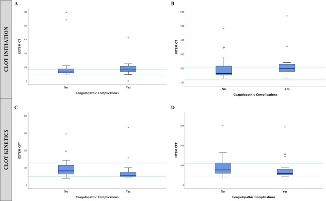
Although CFTEX and IN was within the reference range, patients with hypercoagulopathic complications were noted to have decreased values, suggesting a faster time to achieve clot formation compared with patients without hypercoagulopathic complications (figure 2C, D).
Figure 2.
(A and B) CTEX and IN and (C and D) CFTEX and IN for patients with and without coagulopathic complications. Outliers are represented by a circle (mild outlier) or an asterisk (severe outlier). CFT, clot formation time; CT, clotting time; EX, EXTEM; IN, INTEM.
Patients in the hypercoagulopathic group also exhibited an elevated A10EX and IN, which corresponds to overadequate clot formation and clot firmness (table 5).
Table 5.
ROTEM parameters and PTT by median and mean quartile of patients with hypercoagulopathy versus patients without coagulopathy
| HC (no=0, yes=1) | n | Reference range | Minimum | Q1 | Median | Q3 | Maximum | 95% median CI | |
| CTEX | 0 | 40 | 43–82 | 49 | 59 | 71 | 86.7 | 495 | 64.3 to 121.2 |
| 1 | 21 | 0 | 64.5 | 80 | 107 | 311 | 62.6 to 115.2 | ||
| CTIN | 0 | 40 | 122–208 | 123 | 150.2 | 162.5 | 215 | 482 | 168.2 to 213.4 |
| 1 | 21 | 123 | 172.5 | 209 | 228.5 | 572 | 176.2 to 261.1 | ||
| CFTEX | 0 | 40 | 48–127 | 39 | 61.7 | 81 | 115 | 295 | 76.3 to 106.6 |
| 1 | 21 | 42 | 48 | 56 | 74.5 | 331 | 47.2 to 105.3 | ||
| CFTIN | 0 | 40 | 45–110 | 35 | 58.2 | 76 | 111.5 | 301 | 72.5 to 101.4 |
| 1 | 21 | 44 | 51 | 58 | 80.5 | 294 | 52.5 to 104.9 | ||
| A10EX | 0 | 40 | 40–60 | 30 | 50.2 | 58.5 | 67.7 | 78 | 54.7 to 61.4 |
| 1 | 21 | 28 | 57.5 | 64 | 69 | 74 | 56.1 to 66.1 | ||
| A10IN | 0 | 40 | 40–60 | 29 | 47.5 | 56 | 63.7 | 74 | 53.6 to 59.7 |
| 1 | 21 | 29 | 54.5 | 63 | 65 | 72 | 52.8 to 63.2 | ||
| MCFEX | 0 | 40 | 52–70 | 43 | 59.2 | 66 | 72 | 83 | 63.1 to 68.5 |
| 1 | 21 | 39 | 67.5 | 70 | 73.5 | 79 | 65.2 to 72.8 | ||
| MCFIN | 0 | 40 | 51–72 | 39 | 58.2 | 64 | 71 | 79 | 61.1 to 66.6 |
| 1 | 21 | 38 | 63.5 | 67 | 72 | 77 | 61.8 to 70.1 | ||
| MCFFIB | 0 | 40 | 7–24 | 5 | 15 | 23 | 28 | 40 | 19.8 to 25.6 |
| 1 | 21 | 7 | 22 | 32 | 38 | 41 | 23.8 to 33.9 | ||
| PTT | 0 | 40 | 25–32 (seconds) | 19 | 26 | 29.3 | 35.5 | 77 | 29.5 to 36 |
| 1 | 21 | 25 | 32.7 | 41 | 48.6 | 63 | 36.8 to 46.9 |
A10, amplitude at 10 minutes; CFT, clot formation time; CT, clotting time; EX, EXTEM; FIB, FIBTEM; HC, hypercoagulopathic complications; IN, INTEM; MCF, maximum clot firmness; n, number of ROTEM test results; PTT, partial thromboplastin time; ROTEM, rotational thromboelastography.
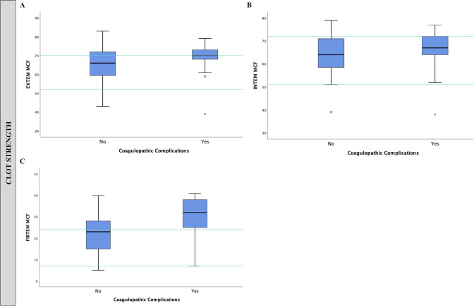
When comparing patients with and without coagulopathic complications, CTEX, CTIN, A10EX and IN, and MCFFIB were elevated in patients with hypercoagulopathic complications (tables 5 and 6). MCFFIB was also elevated above the reference range in patients with hypercoagulopathic complications (figure 3). Of note, an elevated MCF is indicative of high clot strength and is commonly used to detect hypercoagulability.11 12
Table 6.
ROTEM parameters and PTT by median and mean quartile of patients with and without therapeutic heparin administration
| Heparin drip use (no=0, yes=1) | n | Reference range | Minimum | Q1 | Median | Q3 | Maximum | 95% median CI | |
| CTEX | 0 | 18 | 43–82 | 49 | 57 | 68.5 | 76 | 89 | 61.3 to 74.1 |
| 1 | 43 | 0 | 62 | 79 | 107 | 495 | 72.9 to 129.8 | ||
| CTIN | 0 | 18 | 122–208 | 130 | 150.5 | 160 | 173.2 | 222 | 152.2 to 172.9 |
| 1 | 43 | 123 | 157 | 196 | 229 | 572 | 188.6 to 216.2 | ||
| CFTEX | 0 | 18 | 48–127 | 52 | 74 | 85 | 113 | 194 | 78.6 to 111.3 |
| 1 | 43 | 39 | 48 | 61 | 95 | 331 | 63.9 to 101.0 | ||
| CFTIN | 0 | 18 | 45–110 | 51 | 67.7 | 81 | 110 | 121 | 75.1 to 96.0 |
| 1 | 43 | 35 | 51 | 59 | 93 | 301 | 65.5 to 101.2 | ||
| A10EX | 0 | 18 | 40–60 | 41 | 50.7 | 55 | 59.5 | 70 | 51.4 to 58.8 |
| 1 | 43 | 28 | 55 | 63 | 70 | 78 | 57.3 to 64.3 | ||
| A10IN | 0 | 18 | 40–60 | 46 | 49.7 | 54.5 | 59.2 | 66 | 51.5 to 57.2 |
| 1 | 43 | 29 | 50 | 60 | 68 | 74 | 54.8 to 61.8 | ||
| MCFEX | 0 | 18 | 52–70 | 50 | 59.7 | 64.5 | 67.5 | 76 | 60.8 to 67.2 |
| 1 | 43 | 39 | 64 | 70 | 74 | 83 | 65.3 to 70.9 | ||
| MCFIN | 0 | 18 | 51–72 | 54 | 59.7 | 62.5 | 66.2 | 73 | 60.4 to 65.2 |
| 1 | 43 | 38 | 59 | 67 | 73 | 79 | 62.3 to 68.3 | ||
| MCFFIB | 0 | 18 | 7–24 | 11 | 12.7 | 18 | 25 | 37 | 15.6 to 22.9 |
| 1 | 43 | 5 | 21 | 29 | 36 | 41 | 24.1 to 30.3 | ||
| PTT | 0 | 18 | 25–32 (seconds) | 19 | 25.1 | 27 | 28.7 | 29 | 25.4 to 27.9 |
| 1 | 43 | 25 | 32.6 | 35.6 | 48.1 | 77 | 36.3 to 43.3 |
A10, amplitude at 10 minutes; CFT, clot formation time; CT, clotting time; EX, EXTEM; FIB, FIBTEM; IN, INTEM; MCF, maximum clot firmness; PTT, partial thromboplastin time; ROTEM, rotational thromboelastography.
Figure 3.
(A, B, and C) MCFEX, IN and FIB for patients with and without coagulopathic complications. Outliers are represented by a circle (mild outlier) or an asterisk (severe outlier). EX, EXTEM; FIB, FIBTEM; IN, INTEM; MCF, maximum clot firmness.
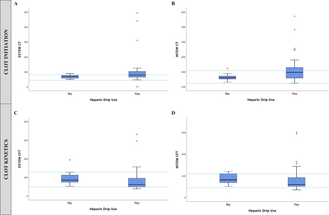
Patients without recorded coagulopathic complications during their hospital stay were placed on therapeutic heparinization due to extracorporeal membrane oxygenator (ECMO) (9.5%), prior DVT (4.7%), atrial fibrillation (4.7%), and clinical concern for coagulopathy (14.3%). Patients who were administered therapeutic heparinization also saw a similar increase or prolongation of all ROTEM components, except for CFT, when compared with patients not given therapeutic heparinization (figures 3 and 4). PTT was also elevated above the reference range in patients with hypercoagulopathic complications (tables 5 and 6). The PTT values reported in table 6 represent only one point in time, at the same time as ROTEM was drawn. For these same patients, PTT values for those receiving therapeutic heparinization were consistently noted to be therapeutic on repeated blood draws.
Figure 4.
(A and B) CTEX and IN and (C and D) CFTEX and IN for patients with and without heparin drip administration. Outliers are represented by a circle (mild outlier) or an asterisk (severe outlier). CFT, clot formation time; CT, clotting time; EX, EXTEM; IN, INTEM.
ROTEM did not capture any fibrinolytic activity in our cohort of ICU patients with COVID-19. MLEX, IN and FIB was not reported for this study given the medians for all cohorts were 0.
Discussion
The findings of this study support the hypothesis that many patients with COVID-19 are in a hypercoagulable state and this effect persisted despite heparin use. As acute phase reactants are typically non-specific (eg, D-dimer) and traditional parameters of coagulation (PT/PTT) may be normal even in the setting of coagulopathy or bleeding, we sought to examine the utility of ROTEM in identifying alterations of coagulation in patients with this disease. Prior studies examining thromboelastography in COVID-19-positive patients have noted derangements in K-angle and maximum amplitude, suggesting coagulopathy.13 Another study examining ROTEM values in critically ill patients with COVID-19 also noted hyperfibrinogenemia and increased fibrin polymerization.14 A trend toward hypercoagulability was also demonstrated by Pavoni et al9 as patients in their cohort tended to have accelerated clot formation (CFT) and higher clot strength (MCF).
In our cohort, the CTEX and IN in patients with hypercoagulopathic complications trended toward the upper limit of normal, and in some cases were elevated (figure 2). This can partially be explained by the effects of therapeutic anticoagulation where higher CTEX and IN was more pronounced in the subset of patients receiving heparin infusions.15–18 CTEX and IN prolongation has also been documented in studies using novel oral anticoagulants.19 In our study, seven (53.8%) patients without documented or suspected hypercoagulopathic complications were also anticoagulated.
When examining MCF, it is important to note that, although the majority of patients remained within their reference ranges, the median across most MCF values trended toward the upper limit of normal (figure 2C, D). This is especially true of patients with hypercoagulopathic complications, where the median was consistently elevated over patients without thromboembolic events and, as in the case of MCFFIB, exceeding the normal range. Given that higher MCF denotes a stronger clot,20 this further supports the hypothesis that patients with COVID-19 are in a hypercoagulable state. Although not graphically represented, the median A10 (a surrogate of clot strength) for patients with coagulopathic complications was also elevated above the normal range (table 5). However, as most values for MCFEX and IN remained in the reference range for patients with and without hypercoagulopathic complications, the clinical value of using ROTEM to predict thromboembolic events will need further elucidation. One area of promise may be focusing on MCFFIB. Our study shows that patients without hypercoagulopathic complications have a median value within the reference range, in stark contrast to patients with thromboembolic events where the median is above the upper limit of normal.
To investigate the effects of therapeutic anticoagulation on the analysis of ROTEM, we separately studied patients who did and who did not receive therapeutic anticoagulation during their ICU course. The use of anticoagulation has been shown to effect ROTEM clotting time to the extent that CT ROTEM values have been used in lieu of PTT to monitor treatment effectiveness of anticoagulation.21 The findings of prolonged CTIN and EX (figure 4A, B) are expected with patients under the effect of heparin anticoagulation. However, it is important to note that even under the influence of therapeutic heparinization (with appropriate PTT values), A10EX and IN still remained elevated (tables 5 and 6). Typically, A10 is used as a surrogate for clot strength, with elevated values suggestive of fibrinogen and/or platelet hyperfunctionality. It is equally important to note that CFTEX and IN, although within the reference range, was surprisingly lower in patients treated with therapeutic anticoagulation (heparin infusion). Shortened CFT is also typically seen in hypercoagulopathic ROTEM profiles.22 This would suggest that patients with COVID-19, despite adequate anticoagulation, are able to form clots faster than those without anticoagulation.
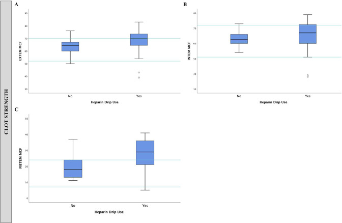
Although the majority of patients on therapeutic heparinization had MCFIN values within the reference range, both MCFEX and MCFFIB exhibited medians that were borderline and highly deranged, respectively (figure 5). As such, we demonstrate in this cohort that, even in the presence of adequately heparinized patients, critically ill patients with COVID-19 still remain hypercoagulable, as demonstrated by their decreased CFTEX and IN as well as elevated A10EX and IN and MCFEX/MCFFIB. PTT levels were also recorded during the same day as ROTEM was drawn, in addition to standard interval monitoring based on institutional protocols. When examining the median and 75th percentile levels of patients with hypercoagulopathic complications, the majority of patients were shown to have PTT levels approximately 1.5 times the reference range (table 4). In addition, patients receiving therapeutic heparinization were protocolized to have the infusion titrated to maintain therapeutic PTT (1.5–2× greater than the baseline reference range). This further suggests that despite proper anticoagulation, critically ill patients with COVID-19 remain at risk of clotting. A representative figure is also included in supplemental online supplemental figure 3 for graphical representation.
Figure 5.
(A, B, and C) MCFEX, IN and FIB for patients with and without heparin drip administration. Outliers are represented by a circle (mild outlier) or an asterisk (severe outlier). EX, EXTEM; FIB, FIBTEM; IN, INTEM; MCF, maximum clot firmness.
tsaco-2020-000603supp003.pdf (942.6KB, pdf)
Our additional findings of elevated PT, PTT, and D-dimer as well as inflammatory markers (LDH, ferritin, and CRP) are consistent with other studies in the COVID-19 literature, but do not fully explain the thrombogenic tendencies of the COVID-19 illness.23 24 ROTEM alone may not be sufficient to predict or fully capture the degree and nature of hypercoagulability among patients with COVID-19. As an adjunct, however, ROTEM may help to identify those patients with increased propensity toward hypercoagulopathic complications, even under the influence of therapeutic anticoagulation. Historically, point-of-care viscoelastic testing of coagulation has been used, particularly in trauma and surgical critical care, to understand the nuances of the bleeding patient to the extent of replacing factors in an evidence-based fashion. Amidst the COVID-19 pandemic and based on clinical reports of clinical hypercoagulopathy, many have advocated for empiric anticoagulation. As our study suggests, ROTEM may be used in settings such as COVID-19 critical illness to not only identify hypercoagulopathic tendencies, but also to guide and/or develop efficacious anticoagulation, as heparin alone, in our study, does not appear to reverse the hypercoagulable tendencies of patients with COVID-19, as measured by ROTEM. Other anticoagulants that use other pathways may need to be investigated to treat hypercoagulopathy.
Limitations
Our study has several limitations. First, ROTEMs collected on these patients were drawn at various times during their ICU course and three separate samples (on different days) were obtained for each patient. Second, our sample size was small and potentially underpowered to draw conclusions regarding statistical significance. The results, therefore, may not be representative of all critically ill patients with COVID-19. However, this analysis reveals both the utility and limitations of ROTEM in the clinical evaluation of hypercoagulopathy in patients with COVID-19.
Conclusion
Our analysis of ROTEM supports prior retrospective studies showing the higher incidence of thromboembolic events associated with COVID-19. Critically ill, mechanically ventilated patients with COVID-19 with known or suspected clinical thromboembolic events demonstrate deranged ROTEM values that reveal a tendency toward hypercoagulability. Given that these derangements were present even in the presence of therapeutic heparinization, our study suggests that anticoagulation with antithrombin inhibitor (heparin) may not be a comprehensive approach to treating COVID-19 coagulopathies. The coagulopathy observed among patients during this pandemic requires further investigation with respect to etiology, physiology, and the appropriate laboratory methods for screening and measurement.
Acknowledgments
We thank the critical care nurses, residents, and infectious disease colleagues who have worked tirelessly to care for our patients with COVID-19. Our results would not have been possible without their hard work and courage.
Footnotes
Contributors: All persons who meet the authorship criteria are listed as authors, and all authors certify that they have participated sufficiently in the work to take public responsibility for the content, including participation in the concept, design, analysis, writing, or revision of the article. Furthermore, each author certifies that this material or similar material has not been and will not be submitted to or published in any other publication. Acquisition of data: JKC, KP, RL, AS, JK, GL, PR. Analysis and/or interpretation of data: JKC, KP, RL, AS, PR. Revising the article critically for important intellectual content: JKC, KP, RL, AS, JK, GL, PR. Approval of the version of the article to be published: JKC, KP, RL, AS, JK, GL, PR.
Funding: The authors have not declared a specific grant for this research from any funding agency in the public, commercial or not-for-profit sectors.
Disclaimer: All authors listed on this article do not have other relationships or activities, including financial interests, which would have influenced the results of this study.
Competing interests: None declared.
Provenance and peer review: Not commissioned; externally peer reviewed.
Supplemental material: This content has been supplied by the author(s). It has not been vetted by BMJ Publishing Group Limited (BMJ) and may not have been peer-reviewed. Any opinions or recommendations discussed are solely those of the author(s) and are not endorsed by BMJ. BMJ disclaims all liability and responsibility arising from any reliance placed on the content. Where the content includes any translated material, BMJ does not warrant the accuracy and reliability of the translations (including but not limited to local regulations, clinical guidelines, terminology, drug names and drug dosages), and is not responsible for any error and/or omissions arising from translation and adaptation or otherwise.
Data availability statement
Data are available upon reasonable request. All data relevant to the study are included in the article or uploaded as supplementary information. All of the de-identified individual participant data reported in this article will be made available, including the study protocol and statistical analysis plan, beginning 9 months and ending 36 months after article publication. Data will be shared with investigators whose proposed use of data has been approved by an independent review committee identified for this purpose. Proposals may be submitted up to 36 months after article publication.
Ethics statements
Patient consent for publication
Next of kin consent obtained.
Ethics approval
This research received ethics approval from the Westchester Medical Center - New York Medical College Institutional Review Board (IRB ID: 14217). Informed consent was obtained prior to participation.
References
- 1.Varga Z, Flammer AJ, Steiger P, Haberecker M, Andermatt R, Zinkernagel AS, Mehra MR, Schuepbach RA, Ruschitzka F, Moch H. Endothelial cell infection and endotheliitis in COVID-19. Lancet 2020;395:1417–8. 10.1016/S0140-6736(20)30937-5 [DOI] [PMC free article] [PubMed] [Google Scholar]
- 2.Zhou F, Yu T, Du R, Fan G, Liu Y, Liu Z, Xiang J, Wang Y, Song B, Gu X, et al. Clinical course and risk factors for mortality of adult inpatients with COVID-19 in Wuhan, China: a retrospective cohort study. Lancet 2020;395:1054–62. 10.1016/S0140-6736(20)30566-3 [DOI] [PMC free article] [PubMed] [Google Scholar]
- 3.Léonard-Lorant I, Delabranche X, Séverac F, Helms J, Pauzet C, Collange O, Schneider F, Labani A, Bilbault P, Molière S, et al. Acute pulmonary embolism in patients with COVID-19 at CT angiography and relationship to D-dimer levels. Radiology 2020;296:E189–91. 10.1148/radiol.2020201561 [DOI] [PMC free article] [PubMed] [Google Scholar]
- 4.Connors JM, Levy JH. COVID-19 and its implications for thrombosis and anticoagulation. Blood 2020;135:2033–40. 10.1182/blood.2020006000 [DOI] [PMC free article] [PubMed] [Google Scholar]
- 5.Helms J, Tacquard C, Severac F, Leonard-Lorant I, Ohana M, Delabranche X, Merdji H, Clere-Jehl R, Schenck M, Fagot Gandet F, et al. High risk of thrombosis in patients with severe SARS-CoV-2 infection: a multicenter prospective cohort study. Intensive Care Med 2020;46:1089–98. 10.1007/s00134-020-06062-x [DOI] [PMC free article] [PubMed] [Google Scholar]
- 6.Jose RJ, Manuel A. COVID-19 cytokine storm: the interplay between inflammation and coagulation. Lancet Respir Med 2020;8:e46–7. 10.1016/S2213-2600(20)30216-2 [DOI] [PMC free article] [PubMed] [Google Scholar]
- 7.Zhang L, Yan X, Fan Q, Liu H, Liu X, Liu Z, Zhang Z. D-Dimer levels on admission to predict in-hospital mortality in patients with Covid-19. J Thromb Haemost 2020;18:1324–9. 10.1111/jth.14859 [DOI] [PMC free article] [PubMed] [Google Scholar]
- 8.Drumheller BC, Stein DM, Moore LJ, Rizoli SB, Cohen MJ. Thromboelastography and rotational thromboelastometry for the surgical intensivist: a narrative review. J Trauma Acute Care Surg 2019;86:710–21. 10.1097/TA.0000000000002206 [DOI] [PMC free article] [PubMed] [Google Scholar]
- 9.Pavoni V, Gianesello L, Pazzi M, Stera C, Meconi T, Frigieri FC. Evaluation of coagulation function by rotation thromboelastometry in critically ill patients with severe COVID-19 pneumonia. J Thromb Thrombolysis 2020;50:281–6. 10.1007/s11239-020-02130-7 [DOI] [PMC free article] [PubMed] [Google Scholar]
- 10.Anderson L, Quasim I, Steven M, Moise SF, Shelley B, Schraag S, Sinclair A. Interoperator and intraoperator variability of whole blood coagulation assays: a comparison of thromboelastography and rotational thromboelastometry. J Cardiothorac Vasc Anesth 2014;28:1550–7. 10.1053/j.jvca.2014.05.023 [DOI] [PubMed] [Google Scholar]
- 11.Hincker A, Feit J, Sladen RN, Wagener G. Rotational thromboelastometry predicts thromboembolic complications after major non-cardiac surgery. Crit Care 2014;18:549. 10.1186/s13054-014-0549-2 [DOI] [PMC free article] [PubMed] [Google Scholar]
- 12.Akay OM, Ustuner Z, Canturk Z, Mutlu FS, Gulbas Z. Laboratory investigation of hypercoagulability in cancer patients using rotation thrombelastography. Med Oncol 2009;26:358–64. 10.1007/s12032-008-9129-0 [DOI] [PubMed] [Google Scholar]
- 13.Panigada M, Bottino N, Tagliabue P, Grasselli G, Novembrino C, Chantarangkul V, Pesenti A, Peyvandi F, Tripodi A. Hypercoagulability of COVID-19 patients in intensive care unit: a report of thromboelastography findings and other parameters of hemostasis. J Thromb Haemost 2020;18:1738–42. 10.1111/jth.14850 [DOI] [PMC free article] [PubMed] [Google Scholar]
- 14.Spiezia L, Boscolo A, Poletto F, Cerruti L, Tiberio I, Campello E, Navalesi P, Simioni P. COVID-19-Related severe hypercoagulability in patients admitted to intensive care unit for acute respiratory failure. Thromb Haemost 2020;120:998–1000. 10.1055/s-0040-1710018 [DOI] [PMC free article] [PubMed] [Google Scholar]
- 15.Schöchl H, Maegele M, Solomon C, Görlinger K, Voelckel W. Early and individualized goal-directed therapy for trauma-induced coagulopathy. Scand J Trauma Resusc Emerg Med 2012;20:15. 10.1186/1757-7241-20-15 [DOI] [PMC free article] [PubMed] [Google Scholar]
- 16.Görlinger K, Dirkmann D, Solomon C, Hanke AA. Fast interpretation of thromboelastometry in non-cardiac surgery: reliability in patients with hypo-, normo-, and hypercoagulability. Br J Anaesth 2013;110:222–30. 10.1093/bja/aes374 [DOI] [PubMed] [Google Scholar]
- 17.Lier H, Vorweg M, Hanke A, Görlinger K. Thromboelastometry guided therapy of severe bleeding. Essener Runde algorithm. Hamostaseologie 2013;33:51–61. 10.5482/HAMO-12-05-0011 [DOI] [PubMed] [Google Scholar]
- 18.Dirkmann D, Görlinger K, Dusse F, Kottenberg E, Peters J. Early thromboelastometric variables reliably predict maximum clot firmness in patients undergoing cardiac surgery: a step towards earlier decision making. Acta Anaesthesiol Scand 2013;57:594–603. 10.1111/aas.12040 [DOI] [PubMed] [Google Scholar]
- 19.Chojnowski K, Górski T, Robak M, Treliński J. Effects of rivaroxaban therapy on ROTEM coagulation parameters in patients with venous thromboembolism. Adv Clin Exp Med 2015;24:995–1000. 10.17219/acem/42147 [DOI] [PubMed] [Google Scholar]
- 20.Anıl H, Kılıç Yıldırım G, Harmancı K, Bozkurt Turhan A, Akay OM, Bör Özcan, Aydoğdu S, Kocak A. Thromboelastogram as a tool to predict hypercoagulability in children with cystic fibrosis. Clin Appl Thromb Hemost 2018;24:348–52. 10.1177/1076029616683045 [DOI] [PMC free article] [PubMed] [Google Scholar]
- 21.Najafi A, Nikeish M, Etezadi F, Pourfakhr P, Imani F, Khajavi MR, Shariat Moharari R. Comparison of aPTT and CT parameter of the ROTEM test to monitor heparin anti-coagulation effect in ICU patients: an observational study. Acta Med Iran 2015;53:643–6. [PubMed] [Google Scholar]
- 22.Giaccherini C, Verzeroli C, Marchetti M, Gamba S, Piras F, Russo L, Tessarolo S, Vignoli A, Finazzi G, Rambaldi A, et al. PO-26 - Whole blood rotational thromboelastometry (ROTEM) to detect hypercoagulability in patients with myeloproliferative neoplasms (MPN). Thromb Res 2016;140 Suppl 1:S185–6. 10.1016/S0049-3848(16)30159-1 [DOI] [PubMed] [Google Scholar]
- 23.Tang N, Li D, Wang X, Sun Z. Abnormal coagulation parameters are associated with poor prognosis in patients with novel coronavirus pneumonia. J Thromb Haemost 2020;18:844–7. 10.1111/jth.14768 [DOI] [PMC free article] [PubMed] [Google Scholar]
- 24.Brady L, Stein MD. Coagulopathy associated with COVID-19. NEJM Journal Watch 2020;2020. 10.1056/nejm-jw.NA51254 [DOI] [Google Scholar]
Associated Data
This section collects any data citations, data availability statements, or supplementary materials included in this article.
Supplementary Materials
tsaco-2020-000603supp001.pdf (914KB, pdf)
tsaco-2020-000603supp002.pdf (879.4KB, pdf)
tsaco-2020-000603supp003.pdf (942.6KB, pdf)
Data Availability Statement
Data are available upon reasonable request. All data relevant to the study are included in the article or uploaded as supplementary information. All of the de-identified individual participant data reported in this article will be made available, including the study protocol and statistical analysis plan, beginning 9 months and ending 36 months after article publication. Data will be shared with investigators whose proposed use of data has been approved by an independent review committee identified for this purpose. Proposals may be submitted up to 36 months after article publication.


