Description
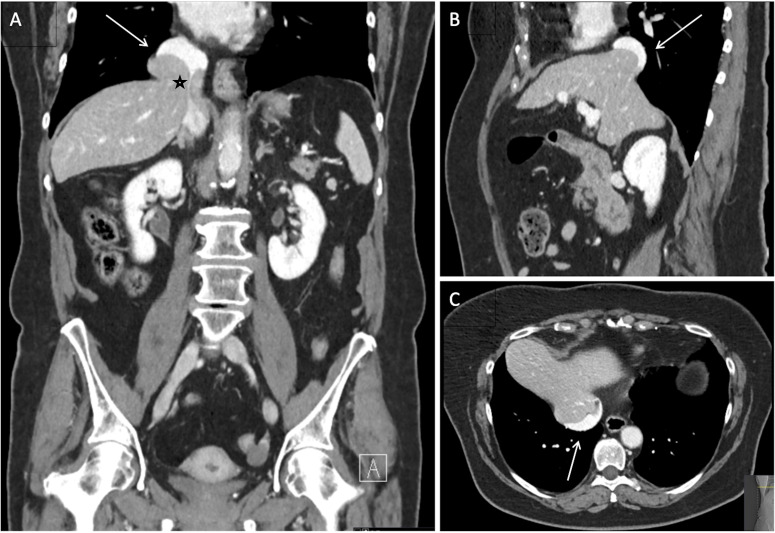
A woman in her 60s presented with long-standing persistent right hypochondrial pain. The patient demonstrated elevated liver function tests (ALT: 47 U/L (normal range: 10–35 U/L); AST: 40 U/L (normal range: 8–36 U/L)) and was otherwise healthy with no history of trauma. Given the chronicity of the complaints, a CT scan (figure 1) was conducted and revealed the herniation of the caudate lobe of the liver protruding through the right diaphragmatic crura, probably through the inferior vena cava foramen, into the thoracic cavity. The patient refused any surgical intervention.
Figure 1.
Coronal (A) sagittal (B) and axial (C) contrast-enhanced CT images of the abdomen showing herniation (A-B-C-arrow) of the caudate lobe of the liver parenchyma protruding through the right diaphragmatic crura into the thoracic cavity. Note also secondary significant constriction of the inferior vena cava within the herniated caudate lobe (A-black star).
The majority of diaphragmatic hernias comprise the herniation of abdominal visceral organs such as the stomach, colon and the small bowels.1 The isolated herniation of the caudate lobe of the liver into the thoracic cavity, in the absence of a history of trauma or spontaneous rupture, is extremely rare.2 This type of herniation is believed to be related to congenital structural defects. This condition has almost always been misdiagnosed on chest roentgenograms as hydatid cyst, pulmonary sequestration or pulmonary tumour, which were then followed by unnecessary surgical interventions.3 Treatment of a liver herniation associated with trauma or acute spontaneous diaphragmatic rupture will usually require urgent surgery. Uncomplicated cases, however, depending on the extent of the herniation may be followed conservatively or may eventually be scheduled for elective surgery. The cause of distorted liver function tests in our patient was probably due to the impaired drainage of the caudate lobe. On 3-month follow-up, the patient had similar liver function tests (ALT: 40 U/L (normal range: 10–35 U/L); AST: 38 U/L (normal range: 8–36 U/L)). Advanced imaging studies such as CT will usually clearly demonstrate the pathology. Therefore, a high level of suspicion is the key to a timely and accurate diagnosis and appropriate treatment plan.
Learning points.
The isolated herniation of the caudate lobe of the liver into the thoracic cavity, in the absence of a history of trauma or spontaneous rupture, is extremely rare.
Misdiagnosis is frequent and results in unnecessary surgical interventions.
A high level of suspicion is the key to a timely and accurate diagnosis and appropriate treatment plan.
Footnotes
Contributors: MM and FVR contributed to the conception and design of the study. SG additionally conducted the literature research and provided the references. FVR additionally contributed in the design and presenting of the case report. KDS participated in reviewing and revising the manuscript. KDS provided important intellectual contribution.
Funding: The authors have not declared a specific grant for this research from any funding agency in the public, commercial or not-for-profit sectors.
Case reports provide a valuable learning resource for the scientific community and can indicate areas of interest for future research. They should not be used in isolation to guide treatment choices or public health policy.
Competing interests: None declared.
Provenance and peer review: Not commissioned; externally peer reviewed.
Ethics statements
Patient consent for publication
Consent obtained directly from patient(s).
References
- 1.Loumiotis I, Kinkhabwala MM, Bhargava A. Acute trans-diaphragmatic herniation of the caudate lobe of the liver. Ann Thorac Surg 2018;105:e5–6. 10.1016/j.athoracsur.2017.08.031 [DOI] [PubMed] [Google Scholar]
- 2.Chen Y-Y, Huang T-W, Chang H, et al. Intrathoracic caudate lobe of the liver: a case report and literature review. World J Gastroenterol 2014;20:5147–52. 10.3748/wjg.v20.i17.5147 [DOI] [PMC free article] [PubMed] [Google Scholar]
- 3.Sharma OP, Duffy B. Transdiaphragmatic intercostal hernia: review of the world literature and presentation of a case. J Trauma 2001;50:1140–3. 10.1097/00005373-200106000-00026 [DOI] [PubMed] [Google Scholar]


