Abstract
Objective:
To evaluate the long-term effects of successful slow maxillary expansion without fixed appliances or retainers in the mixed dentition on patients with unilateral crossbites, using Haas-type, hyrax, or quad helix appliances.
Materials and Methods:
Serial dental casts of 110 patients were evaluated at three time points: preexpansion (T1) (mean age 7 years/7 months), postexpansion (T2) (mean age 8 years/8 months), and approximately 4 years later in the permanent dentition (T3) (mean age 12 years/9 months). Maxillary and mandibular intercanine and intermolar widths, arch length, and perimeter and molar angulation were measured at all three time intervals with the Michigan published growth norms serving as a control.
Results:
Successful treatment by slow maxillary expansion (SME) produced similarly favorable expansion by all three expanders in all measurements for both arches. Maxillary arch widths were narrower than controls pretreatment (T1) and wider than controls immediately post treatment (T2). Long-term (T3) maxillary intermolar width was the same as controls, with intercanine width significantly wider than controls. Maxillary intercanine and intermolar width increased from T1 to T3, by 4.5 mm and 3.5 mm, respectively, with 98% of intercanine and 80% of intermolar expansion remaining at T3. Maxillary arch circumference increased by 1 mm from T1 to T3. Mandibular width did not change significantly.
Conclusion:
Maxillary arch dimensions in early mixed dentition in patients with unilateral posterior crossbite showed good stability 4 years post treatment in the permanent dentition.
Keywords: Maxillary expansion, Unilateral crossbite, Arch dimension changes
INTRODUCTION
Posterior crossbite (PXB) exhibits an abnormal transverse relationship in centric occlusion, often due to maxillary constriction.1 The prevalence of PXB ranges from 8% to 23% in the primary and mixed dentition.2,3 A functional shift of the mandible results in a unilateral presentation (UPXB) in 90% of cases, with maxillary expansion the most common treatment.3–6 The primary goal is to widen the constricted palate and eliminate the shift. Therefore, UPXB is usually an indication for early orthodontic treatment.7
Palatal expansion can be obtained by either rapid maxillary expansion (RME) or slow maxillary expansion (SME).8–11 Animal studies suggest that SME maintains sutural integrity during expansion, producing a more stable result than RME.12,13 Clinical studies have supported this, but used small sample sizes in the short term.14–16
Lagravere et al.17 stated that most crossbite studies suffer from problems such as small sample size, bias, confounding variables, deficient statistical methods, lack of method analysis and long-term data, blinding in measurements, and deficient controls.7,14–21 Since RME studies used fixed appliances post expansion, the effects of pure expansion alone on arch dimensions remain unanswered.18–21
Huynh et al.22 demonstrated 84% long-term success in treating early mixed dentition UPXB and bilateral posterior crossbite with their data referring successful and unsuccessful cases. There are no data on arch dimensions after successful early treatment of UPXB. Our purpose was to conduct a retrospective clinical study to evaluate short- and long-term effects of SME on arch dimensions in early mixed dentition patients exhibiting a UPXB.
MATERIALS AND METHODS
Subjects
Methodology was approved by the Institutional Review Board of the University of Southern California. All patients were treated in the private orthodontic practices of one of the authors in Vancouver and Richmond, British Columbia, Canada between July 1981 and June 2000. A total of 4800 models were surveyed to yield 330 unilateral PXB cases which were then screened for inclusion in this retrospective nonrandomized study using the following criteria:
patients with a UPXB of at least two consecutive posterior teeth with a functional pretreatment mandibular shift that was clearly noted in the chart at the initial exam;
patients no older than 10 years at pretreatment (T1) with first permanent molars present;
pretreatment (T1), posttreatment (T2), and early permanent dentition models (T3) and notes available with a minimum of 2 years between T2 and T3;
slow maxillary expansion with only one appliance and no other treatment involved that would affect the SME effects, either during or after the expansion period; and
successful UPXB correction at T2 and T3.
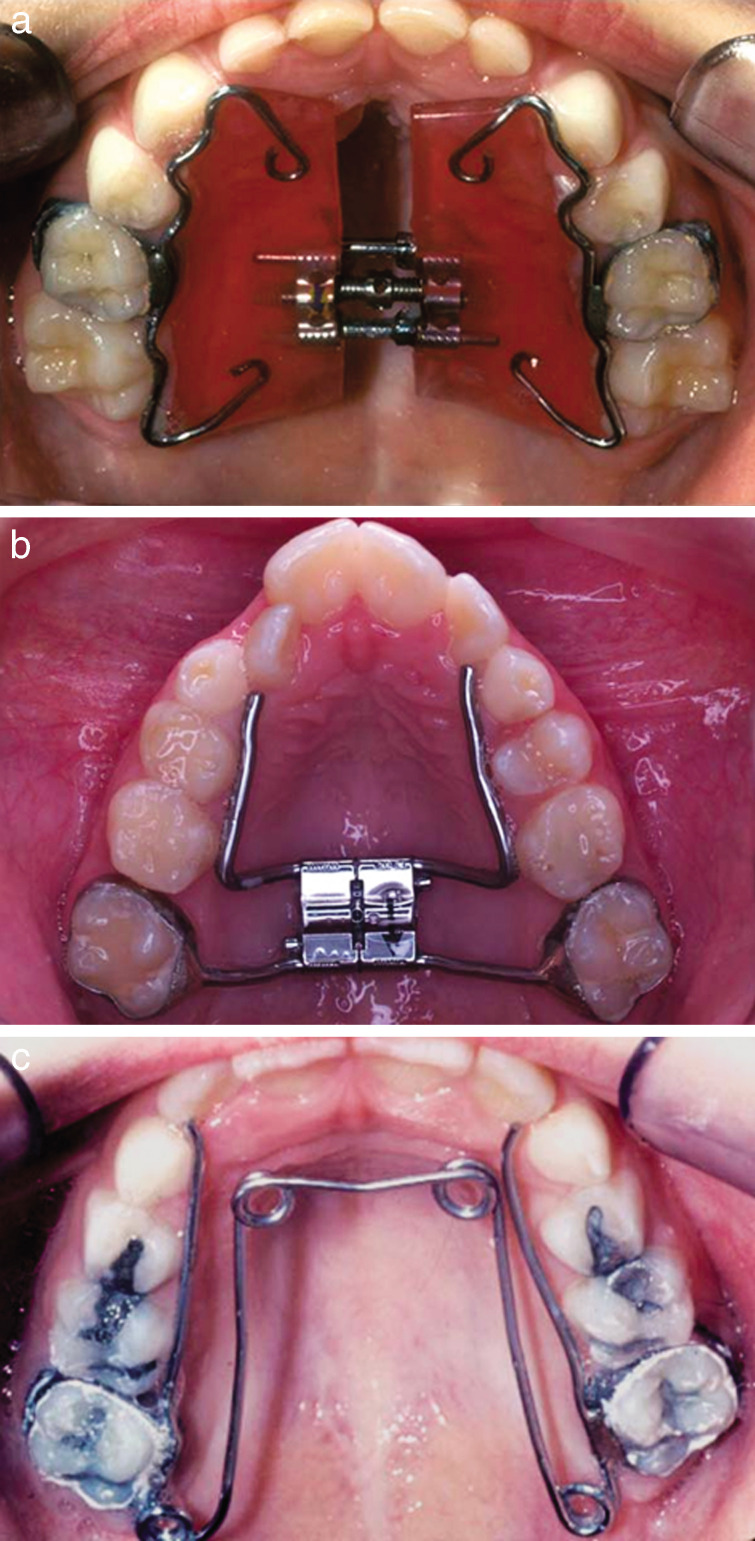
Of the 330 PXB cases screened, 110 met the criteria. All were treated by SME using one of the three types of fixed expanders: a modified Haas-type acrylic appliance with two bands (18 boys and 38 girls), hyrax (11 boys and 15 girls), or quad helix appliance (13 boys and 15 girls) (Figure 1a through c). Mean patient ages and ranges at the three time intervals are as follows: T1 - 7 years and 7 months; T2 - 8 years and 8 months; and T3 - 12 years and 9 months.
Figure 1.
(a) Haas-type acrylic coverage modified appliance. (b) Hyrax. (c) Quad helix.
The expanders were arbitrarily chosen, regardless of the severity of the PXB. However, the quad helix was used if rotation of permanent first molars was needed, and the acrylic coverage Haas-type appliance was used when a digit habit correction crib was required.
The treatment protocol involved one turn (0.25 mm) every 2 days for the Haas-type acrylic coverage or hyrax appliance and one activation every 4 to 6 weeks for the quad helix. The appliances were over expanded approximately 1 mm each side, and left intra-orally (without activation) for a minimum of 3 months of retention. On removal, a new set of study models was taken (T2). T3 records were taken at least 2 years post expansion in the permanent dentition.
Measurements
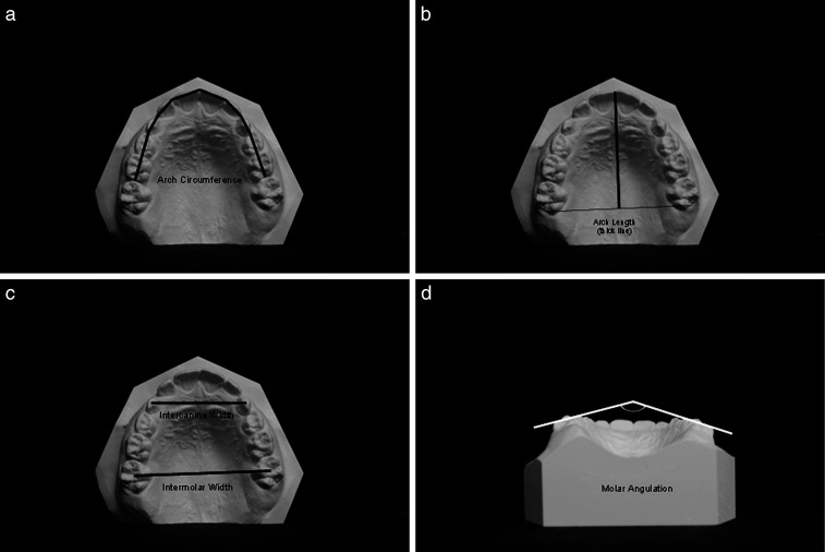
All models were photocopied face-down and parallel to the surface onto 8 × 11-inch white paper with a clear ruler for measurement calibration allowing measurements at a secondary location. Measurement techniques are described below and are similar to those described by McNamara et al.,20 except that in our study, a digital caliper was used to the nearest 1/10 of a millimeter, with each set of casts being independently calibrated. Measurements for arch circumference, intercanine and intermolar width (centroid), arch length, and molar angulation are diagrammed in Figure 2a through d.
Figure 2.
(a) Arch circumference. (b) Arch length. (c) Intercanine and intermolar width. (d) Intermolar angulation.
Predicted changes for each measurement due to normal growth (University of Michigan untreated control group [CG]) were calculated via a quadratic interpolation using published norms matched to gender and chronologic age of study subjects.23 Intermolar angulation measurements were not available for this CG.
Statistical Analysis
All statistical analyses were conducted using SPSS version 16, Chicago, Ill. Utilizing a generalized linear model for repeated measures mixed between-within subjects effects, a multivariate analysis of variance (MANOVA) was conducted to test for differences in the means of the dependent variables on each of the independent variables, and for significant interaction effects between the independent variables. A one-way analysis of variance (ANOVA) with Scheffe's and Dunnett T-3 post hoc analyses was performed to assess significant pair-wise differences for each time group on each of the dependent variables, and for the differences in time intervals for each of the time-dependent variables. An experiment-wide level of significance level of at least P < .05 was used throughout the analysis. Intrarater reliability was determined by measuring and remeasuring 10 cases on two different occasions, 1 week apart. Test-retest reliability was determined by utilizing an intraclass correlation coefficient (ICC) according to the method of Denegar.24
RESULTS
Utilizing a two-way random effect model (absolute agreement) intraclass correlation coefficient (ICC), measurement error showed a reliability range of 0.833 to 0.999, with an average measure ICC of 0.964.
MANOVA
The omnibus MANOVA showed significant main effects (within-subjects) for the pooled dependent variables on each of the independent variables (trial, gender, and type of expander) with a significant interaction effect between gender and type of expander. Significant between-subjects effects on multiple dependent variables were noted for the independent variables trial and gender. Of note is that, with respect to the independent variable expander, significant differences were noted only for maxillary molar angulation, mandibular arch circumference, mandibular arch length, and mandibular molar angulation.
Given the observed significant within group and between group effects, it was deemed appropriate to investigate further using pair-wise comparisons25 the nature of the relationships among the independent and dependent variables.
Pair-wise Univariate Comparisons Comparison of Pretreatment Arch Measurements (T1)
Both maxillary and mandibular dental arch measurements showed no significant differences (P > .001) between expander groups at T1. The treatment subgroups were then pooled for comparison to the control group (Table 1). Maxillary arches of the treatment group (TG) were mildly, but significantly narrower than the CG by approximately 1 mm (P < .001) for both the canines and molars (Table 1). Maxillary arch circumference was shorter by approximately 4.6 mm (P < .001), while maxillary arch length was approximately equal to the CG. For the mandibular arch, the TG arches were narrower by approximately 2 mm (P < .01) between the canines but similar to the CG between the molars (P not significant). Mandibular arch circumference was similar, but TG arch length was close to 2 mm shorter than the CG.
Table 1.
Comparison of Pooled Treated Groups (TG) and the Control Group (CG) at T1 (Preexpansion)a
Evaluation of Initial Treatment Effects (T1–T2)
All three expanders showed similar amounts of expansion, therefore the data were pooled for comparison with the control group. Active expansion produced intercanine and intermolar increases respectively of 4.5 mm (P < .001) and 4.3 mm (P < .001), and a 3.27-mm increase in arch circumference (P < .001; Table 2). Maxillary arch length remained stable while 4 degrees of total buccal crown tipping took place between the molars.
Table 2.
Comparison of the Changes T2–T1 Between the Pooled Treated Group (TG) and the Control Group (CG)a
Normal growth accounted for all changes between T1 and T2 for the CG with mean intercanine and intermolar increases of 0.57 mm to 0.70 mm, respectively (Table 2). The T2 arch maxillary width for the TG was wider than CG by 3.4 mm at the canines and by 2.5 mm at the molars (Table 3). Maxillary arch circumference remained shorter by 2.5 mm (P < .001), while arch length remained the same when compared with the CG (Table 3).
Table 3.
Comparison of the Pooled Treated Group (TG) and the Control Group (CG) at T2 (Postexpansion)a
No significant changes took place for all mandibular measurements for both the TG and the CG from T1–T2, although 4 mm of total buccal tipping occurred between the molars (P not significant) in the treatment group (Table 2). Consequently, the relationship for all variables between the TG and CG is similar to that seen at T1.
Evaluation of Posttreatment Changes (T3–T2)
The T2 to T3 change measurements for the three treatment groups were pooled for comparison with the controls (Table 4). There was minimal maxillary intercanine width relapse of 0.09 mm (0.2%) and 0.67 mm (20%) of intermolar width relapse (Table 4). Maxillary arch circumference was reduced by 2 mm (P < .001), while 9 degrees of total uprighting occurred at the molars (Table 4). Significant reductions were seen for mandibular arch circumference (−4.5 mm, P < .001) and arch length (−1.5 mm, P < .001), whereas arch width and molar angulations remained stable (Table 4).
Table 4.
Comparison of the Changes T2–T3 Between the Treated (TG) and Control (CG) Groupa
Evaluation of Overall Treatment Changes (T1–T3)
Treated groups measurements were pooled to compare with controls (Table 5). Maxillary intercanine width increased 4.47 mm at T3, which reflects both treatment and normal growth which averaged 2.47 mm in the CG. Maxillary intermolar width increased 3.65 mm in the TG compared with 2.39 mm in the CG (Table 5). Notable changes over this 5-year period include a significant increase in maxillary arch width by 1 mm to 2 mm (P < .001), and a reduction in mandibular arch circumference and arch length (−2 mm to −3 mm; P < .001) (Table 5). The mandibular arch width remained stable throughout treatment. Maxillary molars tipped to the lingual 5 degrees (P < .001), while mandibular molars tipped to the buccal 3.5 degrees (Table 5).
Table 5.
Comparison of the Changes T3–T1 Between the Treated (TG) and Control (CG) Groupa
Comparison of Final Arch Measurements (T3)
Treated groups T3 data were pooled for comparison with controls (Table 6). Maxillary arch transverse measurements were similar between the TG and the norms (CG) with the exception of maxillary intercanine width, which was greater by 4 mm in the TG (Table 6). Arch circumference and mandibular arch length was shorter in the TG than in the CG, whereas the two groups were similar in mandibular arch width (Table 6).
Table 6.
Comparison of Pooled Test Groups (TG) and the Control Group (CG) at T3 (Permanent Dentition)a
DISCUSSION
A distinctive feature of this study was that fixed appliances or retainers were not placed after SME therapy. This is one of the few studies that has examined the pure effects of successful expansion alone on the upper and lower arches short and long term.
Pretreatment Comparisons (T1)
At all time intervals, the three expanders were similar (P not significant) in all measurements in both arches. Consequently, the expansion effects throughout treatment could be measured against one another without initial selection bias between the treatment groups (TG), despite this being a retrospective nonrandomized study. Prior to treatment, mild but significant differences compared with the CG were seen when comparing maxillary arch width. All TG subjects at T1 had a constricted maxillary arch of approximately 1 mm between the canines (28.32 mm vs 29.36 mm) and molars (41.58 mm vs 42.75 mm) compared with the CG. This was expected since all treated patients exhibited UPXB requiring maxillary expansion. The 3.5-mm shorter maxillary arch circumference of the TG compared with that of the CG (75.19 vs 79.85 mm) results from a reduced arch circumference associated with narrow arches. With maxillary expansion treatment, arch circumference increases and space is gained.19
Active Expansion: T1–T2 Changes
All three expanders produced similar amounts of change (P < .001) for all measurements in both arches and were equally effective at correcting UPXB in early mixed dentition aged children, even though a small difference was seen between boys and girls. Maxillary intercanine width increased 4.56 mm (P < .001) and an intermolar width increased 4.32 mm (P < .001) during active expansion (Table 2). This intermolar expansion is less than that reported by Huynh et al.22 because our sample included only UPXB cases, not UPXB and bilateral PXB cases together. These results are comparable to those reported by Bell and LeCompte11 and Boysen et al.,26 who both used methodologies similar to this study, treating 8-year-old patients with the quad helix appliance and SME. These findings are similar to ours as the selection of the type of appliance did not produce any bias in the outcomes. This maxillary intercanine width increase may have significant clinical value in children with insufficient room for their permanent maxillary lateral incisors.
Intercanine and Intermolar Width Stability (T2–T3 Changes)
Ninety-eight percent of maxillary intercanine expansion (ie, 4.47 of 4.56 mm) and 84% of intermolar expansion (ie, 3.65 of 4.32 mm) was maintained over the 4-year period from T2 to T3 without the use of retainers or fixed appliances (Table 4). Children with crowded maxillary lateral incisors may benefit from this maxillary intercanine width stability. Unfortunately, it is difficult to compare our findings with other studies that used RME followed by fixed appliances and retention after the active expansion phase. The SME study by Boysen et al.26 was the only study similar to ours where 87% of intercanine and 73% of intermolar width was maintained after the postretention period. The 3-month post expansion time in the study by Boysen et al.26 was much shorter than the 4 years of post expansion experienced by our TG. Our apparent high percentage of stability could also be due to growth effects, since 1.5 mm to 2 mm of width increase was expected over the 4-year period from ages 9 to 13 years. The skeletal classification was not considered because the Michigan control group was not structured in this fashion.
Arch Circumference Changes (T2–T3)
From T2 to T3, significant decreases in arch perimeter occurred when compared with the control for both the maxillary and mandibular arches. One possible reason for this could be that the control group was comprised of people of Northern European decent, whereas our sample was comprised of a mixed population reflecting different tooth sizes.27,28 Thus, there was a greater loss of leeway space, resulting in a greater decrease in arch circumference and arch length when compared with the White norm.27,28 The Michigan control group represents the gold standard for this type of study and has been used by many authors in the past.20,21
Molar Angulation
Buccal crown tipping of approximately 4 degrees for both arches during the expansion phase (T1 to T2) was similar to the findings of McNamara et al.,20 after RME and fixed appliances (Table 2). From T2 to T3, McNamara et al.20 showed little if any changes in molar angulation (<1 degree) for both arches, whereas we obtained maxillary lingual crown tipping of approximately 9 degrees (P < .001; Table 6) and no change in mandibular molar angulation (P not significant). A probable reason for this is that no appliances were placed in our study to hold and stabilize the molars after expansion, while other studies used fixed appliances and removable retainers after expansion.20
CONCLUSIONS
All appliances produced similar maxillary arch expansion short and long term.
UPXB patients had narrower maxillary widths than controls prior to expansion. Post expansion, maxillary intercanine and intermolar widths were significantly greater than controls.
In the early permanent dentition, maxillary intermolar width was the same as controls, but intercanine width remained significantly larger.
From pretreatment to early permanent dentition, the maxillary intermolar width increased 3.5 mm and intercanine width increased by 4.5 mm representing 80% and 98% stability, respectively, of the initial expansion.
REFERENCES
- 1.Proffit W, Sarver D. Contemporary Orthodontics. Philadelphia, Pa: Mosby; 2007. [Google Scholar]
- 2.Egermark-Eriksson I, Carlsson G. E, Magnusson T, Thilander B. A longitudinal study on malocclusion in relation to signs and symptoms of cranio-mandibular disorders in children and adolescents. Eur J Orthod. 1990;12:399–407. doi: 10.1093/ejo/12.4.399. [DOI] [PubMed] [Google Scholar]
- 3.Thilander B, Myrberg N. The prevalence of malocclusion in Swedish schoolchildren. Scand J Dent Res. 1973;81:12–21. doi: 10.1111/j.1600-0722.1973.tb01489.x. [DOI] [PubMed] [Google Scholar]
- 4.Hesse K. L, Årtun J, Joondeph D. R, Kennedy D. B. Changes in condylar position and occlusion associated with maxillary expansion for correction of functional unilateral posterior crossbite. Am J Orthod Dentofacial Orthop. 1997;111:410–418. doi: 10.1016/s0889-5406(97)80023-6. [DOI] [PubMed] [Google Scholar]
- 5.Myers D. R, Barenie J. T, Bell R. A, Williamson E. H. Condylar position in children with functional posterior crossbites: before and after crossbite correction. Pediatr Dent. 1980;2:190–194. [PubMed] [Google Scholar]
- 6.Bell R. A. A review of maxillary expansion in relation to rate of expansion and patient's age. Am J Orthod. 1982;81:32–37. doi: 10.1016/0002-9416(82)90285-8. [DOI] [PubMed] [Google Scholar]
- 7.Harrison J. E, Ashby D. Orthodontic treatment for posterior crossbites. Cochrane Database Syst Rev. 2001:CD000979. doi: 10.1002/14651858.CD000979. [DOI] [PubMed] [Google Scholar]
- 8.Barber A. F, Sims M. R. Rapid maxillary expansion and external root resorption in man: a scanning electron microscope study. Am J Orthod. 1981;79:630–652. doi: 10.1016/0002-9416(81)90356-0. [DOI] [PubMed] [Google Scholar]
- 9.Linder-Aronson S, Lindgren J. The skeletal and dental effects of rapid maxillary expansion. Br J Orthod. 1979;6:25–29. doi: 10.1179/bjo.6.1.25. [DOI] [PubMed] [Google Scholar]
- 10.Hicks E. P. Slow maxillary expansion. A clinical study of the skeletal versus dental response to low-magnitude force. Am J Orthod. 1978;73:121–141. doi: 10.1016/0002-9416(78)90183-5. [DOI] [PubMed] [Google Scholar]
- 11.Bell R. A, LeCompte E. J. The effects of maxillary expansion using a quad-helix appliance during the deciduous and mixed dentitions. Am J Orthod. 1981;79:152–161. doi: 10.1016/0002-9416(81)90313-4. [DOI] [PubMed] [Google Scholar]
- 12.Cotton L. A. Slow maxillary expansion: skeletal versus dental response to low magnitude force in Macaca mulatta. Am J Orthod. 1978;73:1–23. doi: 10.1016/0002-9416(78)90098-2. [DOI] [PubMed] [Google Scholar]
- 13.Storey E. Tissue response to the movement of bones. Am J Orthod. 1973;64:229–247. doi: 10.1016/0002-9416(73)90017-1. [DOI] [PubMed] [Google Scholar]
- 14.Lagravere M. O, Major P. W, Flores-Mir C. Skeletal and dental changes with fixed slow maxillary expansion treatment: a systematic review. J Am Dent Assoc. 2005;136:194–199. doi: 10.14219/jada.archive.2005.0141. [DOI] [PubMed] [Google Scholar]
- 15.Mossaz-Joelson K, Mossaz C. F. Slow maxillary expansion: a comparison between banded and bonded appliances. Eur J Orthod. 1989;11:67–76. doi: 10.1093/oxfordjournals.ejo.a035967. [DOI] [PubMed] [Google Scholar]
- 16.Mew J. Relapse following maxillary expansion. A study of twenty-five consecutive cases. Am J Orthod. 1983;83:56–61. doi: 10.1016/0002-9416(83)90272-5. [DOI] [PubMed] [Google Scholar]
- 17.Lagravere M. O, Major P. W, Flores-Mir C. Long-term skeletal changes with rapid maxillary expansion: a systematic review. Angle Orthod. 2005;75:1046–1052. doi: 10.1043/0003-3219(2005)75[1046:LSCWRM]2.0.CO;2. [DOI] [PubMed] [Google Scholar]
- 18.Lagravere M. O, Major P. W, Flores-Mir C. Long-term dental arch changes after rapid maxillary expansion treatment: a systematic review. Angle Orthod. 2005;75:155–161. doi: 10.1043/0003-3219(2005)075<0151:LDACAR>2.0.CO;2. [DOI] [PubMed] [Google Scholar]
- 19.Adkins M. D, Nanda R. S, Currier G. F. Arch perimeter changes on rapid palatal expansion. Am J Orthod Dentofacial Orthop. 1990;97:194–199. doi: 10.1016/S0889-5406(05)80051-4. [DOI] [PubMed] [Google Scholar]
- 20.McNamara J. A, Jr, Baccetti T, Franchi L, Herberger T. A. Rapid maxillary expansion followed by fixed appliances: a long-term evaluation of changes in arch dimensions. Angle Orthod. 2003;73:344–353. doi: 10.1043/0003-3219(2003)073<0344:RMEFBF>2.0.CO;2. [DOI] [PubMed] [Google Scholar]
- 21.McNamara J. A., Jr Long-term adaptations to changes in the transverse dimension in children and adolescents: an overview. Am J Orthod Dentofacial Orthop. 2006;129:S71–S74. doi: 10.1016/j.ajodo.2005.09.020. [DOI] [PubMed] [Google Scholar]
- 22.Huynh T, Kennedy D. B, Joondeph D. R, Bollen A. M. Treatment response and stability of slow maxillary expansion using Haas, hyrax, and quad-helix appliances: a retrospective study. Am J Orthod Dentofacial Orthop. 2009;136:331–339. doi: 10.1016/j.ajodo.2007.08.026. [DOI] [PubMed] [Google Scholar]
- 23.Moyers R. E, Van der Linden F. P. G. M, Riolo M. L, McNamara J. A., Jr . Standards of Human Occlusal Development Monograph 5—Craniofacial Growth Series Center for Human Growth and Development. Ann Arbor, MI: University of Michigan; 1976. [Google Scholar]
- 24.Denegar C. R. Assessing reliability and precision of measurement: an introduction to intraclass correlation and standard error of measurement. J Sport Rehabil. 1993;2:35–42. [Google Scholar]
- 25.Tabachnick B. G, Fidell L. S. Using Multivariate Statistics 4th ed. Boston, MA: Allyn & Bacon; 2001. [Google Scholar]
- 26.Boysen B, La Cour K, Athanasiou A. E, Gjessing P. E. Three-dimensional evaluation of dentoskeletal changes after posterior cross-bite correction by quad-helix or removable appliances. Br J Orthod. 1992;19:97–107. doi: 10.1179/bjo.19.2.97. [DOI] [PubMed] [Google Scholar]
- 27.Lavelle C. L. Secular trends in different racial groups. Angle Orthod. 1972;42:19–25. doi: 10.1043/0003-3219(1972)042<0019:STIDRG>2.0.CO;2. [DOI] [PubMed] [Google Scholar]
- 28.Lavelle C. L. Maxillary and mandibular tooth size in different racial groups and in different occlusal categories. Am J Orthod. 1972;61:29–37. doi: 10.1016/0002-9416(72)90173-x. [DOI] [PubMed] [Google Scholar]


