Abstract
Objective:
To use an accurate method of tooth visualization in magnetic resonance imaging (MRI) movie for the observation of spatio-temporal relationships among articulators.
Materials and Methods:
The subjects were two volunteers. Each subject repeated a vowel-consonant-vowel syllable (ie, /asa/; /ata/), and the run was measured using a gradient echo sequence. A custom-made clear retainer filled with the jelly form of ferric ammonium citrate was then fit onto the dental arch, and a T1-weighted turbo-spin-echo sequence was taken. Landmarks were used for superimposition of the incisor boundary onto sequential images of MRI movie. Tracings were conducted to observe the spatio-temporal relationships among articulators.
Results:
The incisor boundary was clearly visible in the magnetic resonance images. After superimposition, the contact distance of the tongue to palate/incisor was found to be longer during /t/-articulation than during /s/-articulation. There were prominent differences in images with and without tooth superimposition in the front oral cavity.
Conclusions:
The method could distinctly extract a tooth boundary in MRI. Detailed configurational relationships between the tongue and tooth were observed during the production of a fricative and a plosive in MRI movie using this method.
Keywords: MRI movie, Articulation, Fricative, Plosive
INTRODUCTION
Magnetic resonance imaging (MRI) has been used in speech research to observe the shape of articulatory organs and to measure the vocal tract area with no known harmful effects.1–3 Recently, MRI movie has been recognized as a new method for visualizing and quantifying the spatio-temporal details of speech production.4,5 MRI movie is a type of cine-loop MRI, which was modified from a technique originally developed for cardiac examinations to be applied to imaging of articulatory movement with the subject repeating certain syllables in a synchronized manner. In MRI movie, motion of soft-tissue organs can be captured, but calcified structures such as teeth are almost invisible because they have little mobile hydrogen.6 Teeth are important structures for articulation, especially in the production of dental-lingual consonants such as /s/ and /t/.7 In the evaluation of these dental-lingual articulations, similar pixel-intensity values in the oral cavity and on the tooth make it difficult to extract contours in the front cavity.3
Various methods have been used for tooth visualization in MRI. Previous studies8,9 have scanned dental casts in water using MRI to obtain the shape of the teeth to determine the boundaries of the oral cavity; but it has been reported that foam produced on the dental casts causes an artifact in the shape of the teeth on magnetic resonance (MR) image.10 Wakumoto et al.11 used double plastic plates separated by an oil layer to image tooth boundaries. However, as reported in a previous study,10 plastic plates could not be visualized in MR image and thus it was difficult to extract accurate tooth boundaries from MRI data.11
A few different approaches have also been attempted. Takemoto et al.6 used blueberry juice and Olt and Jakob12 used water as the oral contrast medium. However, the subject had to remain in a prone position during the scan and had to hold blueberry juice or water inside his or her mouth for a few minutes. This condition is not comfortable for the subject, and artifacts may arise from the motion or flow of the contrast liquid. Furthermore, a previous study reported that, while in the prone position, the subject might move his or her head to breathe, which can cause motion artifacts.10 Although other researchers have used different methods for tooth visualization, these also had inevitable drawbacks.3,13,14
Thus, the aim of our study was to use a jelly form of an effective and easily obtained contrast medium to accurately visualize a tooth in MRI movie for analyzing the spatio-temporal relationships between the incisors, tongue, lips, and palate during the articulation of a fricative /s/ and a plosive /t/.
MATERIALS AND METHODS
Phantom Experiment
In our study, we performed a T1-weighted turbo-spin-echo (TSE-T1) sequence to have a clearer image of soft-tissue anatomy within the oral cavity; 4 mg/mL of ferric ammonium citrate (FAC) was reported to provide good signal intensity on T1-weighted image in MRI.15 However, the pH for 4 mg/mL of FAC was 5.2. To avoid the possibility of tooth decalcification, we added 1.4 mg/mL of sodium bicarbonate, which is used in daily food ingredients for neutralization, to make the pH 7.1. Blueberry juice has also been reported to be an effective oral contrast agent for MRI,16 and prune juice was found to have a relatively high signal intensity on T1-weighted image in MRI.17 In our phantom experiment, we chose prune extract rather than prune juice for comparison because prune extract has a high viscosity, which resists flow, so it would not be necessary to add agar if prune extract were used for tooth imaging in MRI. Furthermore, prune extract is commercially available, and concentration in extract should be higher than in juice. It was thought that higher concentration would provide a better signal intensity in MRI. Therefore in this experiment, we compared 4 mg/mL of FAC (Wako Pure Chemical Industries, Ltd, Osaka, Japan) with commercially available concentrated blueberry juice (mySmoothie, Haruna Ecology Co, Ltd, Tokyo, Japan)16 and prune extract (California Prune Extract, Minami Healthy Foods Co, Ltd, Saitama, Japan). A 1.5T MRI apparatus (Magnetom Vision, Siemens AG, Erlangen, Germany) was used in this experiment. MRI was performed using a TSE-T1 sequence (TR = 600 milliseconds; TE = 15 milliseconds).
MRI Experiment
Two healthy volunteers, a 32-year-old man and a 29-year-old woman, participated in this study. They had a skeletal Class I molar relationship with no malocclusions. Both subjects were in good health and exhibited no obvious speech difficulties as judged by the experimenter. All of the experimental procedures complied with the Code of Ethics of the World Medical Association (Declaration of Helsinki) and were approved by the institutional ethics committee (No. 472). Written informed consent was obtained from both subjects after the experimental procedures were fully explained.
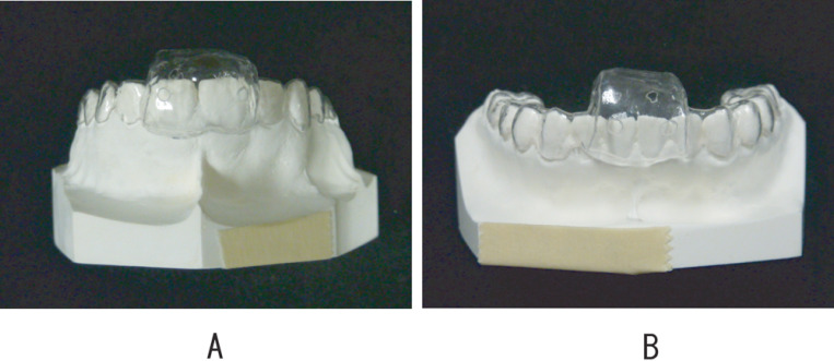
To visualize central incisors on the midsagittal plane in MR image, a customized clear Biostar (3171 IMPRELON S, SCHEU Dental Technology, Iserlohn, Germany) retainer with space around the central incisors was made on dental casts for the upper and lower dental arches (Figure 1). Small holes were opened on the labial and lingual sides for drainage of extra contrast medium in a jelly form. A mixture of 40 mg of FAC, 14 mg of sodium bicarbonate, and 950 mg of agar powder in 10 mL of boiled water was used as the contrast medium. Sugar (1100 mg) was also added to improve the taste. The mixture was then cooled in the refrigerator.
Figure 1.
Clear retainer with space around the central incisors of the upper (A) and lower (B) dental arches.
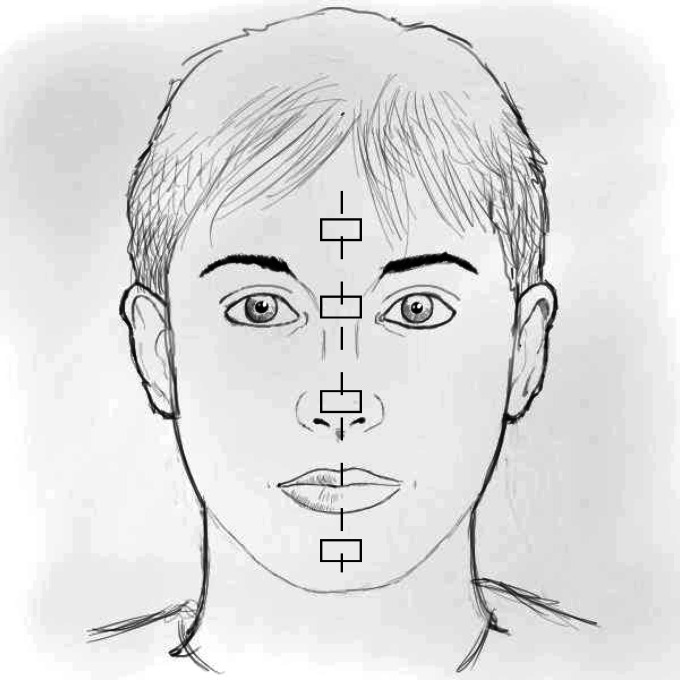
Four plastic tube landmarks, filled with 4 mg/mL of FAC, were taped along the midline of the face at the forehead, nasal root, nasal tip, and chin (Figure 2).
Figure 2.
Schematic illustration of tubes filled with 4 mg/mL of FAC that were taped along the midline of the face on the forehead, nasal root, nasal tip, and chin.
Custom-made circuitry was connected to the MRI apparatus, which was equipped with a head coil, to enable an external trigger pulse to control the timing of the scanning sequence and to provide an auditory cue for synchronization of the subject's utterance.4,5,18 Two target sounds (ie, a fricative /s/ and a plosive /t/) were chosen because teeth are important articulators for these sounds. All data were taken in the midsagittal plane. Each image had a 238 × 380 mm field of view with a pixel size of 1.88 × 1.48 mm (slice thickness = 5 mm); the matrix size was 126 × 256. The subject was in a supine position and repeatedly uttered a vowel-consonant-vowel syllable (ie, /asa/, /ata/) in a synchronized manner in response to the auditory cue. Each run was measured using a gradient echo (GRE) sequence for a cardiac cine (repetition time [TR] = 30 milliseconds; echo time [TE] = 4.8 milliseconds; flip angle = 30°). During data acquisition, an external trigger pulse was fed to the MRI scanner 42 times. The time required for data acquisition was approximately 1 minute. Because wearing the retainer would affect speech production, the subject was first required to repeat each vowel-consonant-vowel syllable 42 times without wearing the retainer.
The prepared jelly form of the contrast medium was then used to fill the space of the retainer, which was fit onto the upper and lower dental arches separately. Extra contrast medium that overflowed from the holes on the retainer was wiped away. The subject was required to remain still during data acquisition. Static MR imaging was performed using a TSE-T1 sequence (TR = 600 milliseconds; TE = 15 milliseconds), which produced a higher-resolution image. Each image had a 238 × 380 mm field of view with a pixel size of 0.94 × 0.74 mm (slice thickness = 5 mm); the matrix size was 252 × 512. The height and width of the pixels for both GRE and TSE-T1 sequences were set at an exact ratio of 2∶1 to eliminate error from linear interpolation during superimposition. For superimposition, both TSE-T1 and GRE sequences were also taken in the rest position, while the subject did not wear the retainer.
Superimposition of the Incisor Boundary
After the tooth boundary was extracted, the upper and lower incisors were colored before superimposition. Superimposition was conducted using customized software (Image Rugle© 2009, Medic Engineering Inc, Kyoto, Japan).
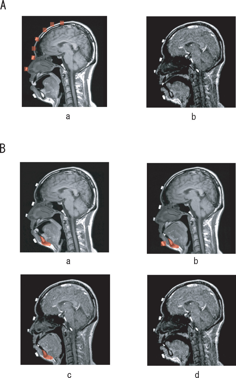
For superimposition of the upper incisor, three landmarks on the forehead, nasal root, and nasal tip were used in conjunction with the contour of the cranium (Figure 3Aa). Together with the upper incisor, the hard palate was also superimposed because it was not clearly visible on images with the GRE sequence. One of the MRI movie images obtained from this superimposition is shown in Figure 3Ab. For superimposition of the lower incisor, three procedures were carefully performed by using one landmark on the chin and symphysis:
Figure 3.
(A) Superimposition of the upper incisor and hard palate. Landmarks on the nasal tip, nasal root, forehead, and points along the cranium (Aa) were used for superimposition. (Ab) One of the MRI movie images obtained from this superimposition. (B) Superimposition of the lower incisor. First, the landmark at the symphysis (Ba) was used to superimpose the lower incisor onto the image taken at rest with a TSE-T1 sequence. The landmarks on the chin and symphysis (Bb) were then used to superimpose the lower incisor onto the image taken at rest with a GRE sequence. Finally, the landmark at the symphysis (Bc) was used to superimpose the lower incisor onto an MRI movie image. (Bd) The image obtained after superimposition.
First, the lower incisor boundary obtained (Figure 3Ba) was superimposed onto the image at rest taken with a TSE-T1 sequence by using the contour of the symphysis as a landmark.
Next, based on the landmarks of the tube on the chin and symphysis (Figure 3Bb), the lower incisor was superimposed onto the image at rest taken with a GRE sequence. Both images were taken in the rest position, and thus the landmark of the tube on the chin was considered to be stable. This step was performed to transfer the image of the lower incisor onto an image that had the same resolution as the MRI movie images.
The obtained image (Figure 3Bc) was then used for superimposing the lower incisor onto each sequential image of MRI movie by using the contour of the symphysis as a landmark.
RESULTS
Phantom Experiment
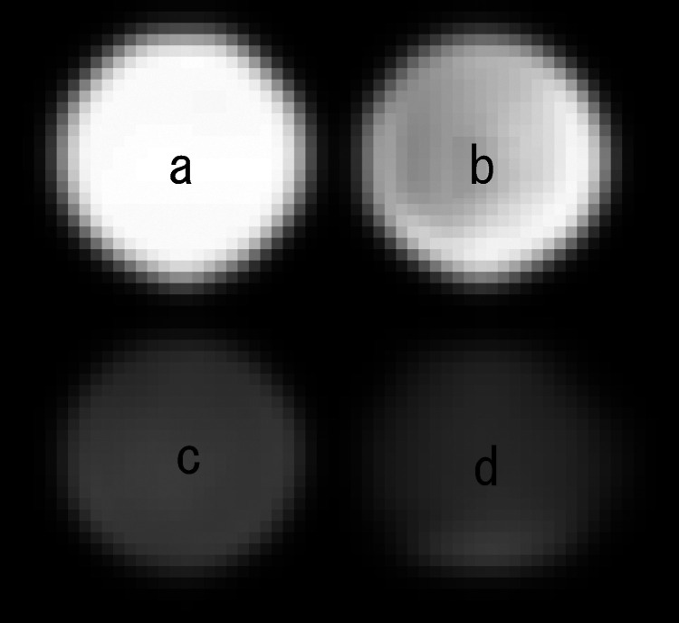
In the phantom experiment, FAC showed the highest signal intensity among the four contrast media (Figure 4). Thus, we suggest that FAC has the highest potential to be an effective oral contrast medium for tooth imaging using a TSE-T1 sequence (TR = 600 milliseconds; TE = 15 milliseconds) in MRI.
Figure 4.
Comparison of the signal intensity among contrast media using a TSE-T1 sequence (TR = 600 milliseconds; TE = 15 milliseconds) in MRI. (a) 4 mg/mL of FAC (along with 1.4 mg/mL of sodium bicarbonate). (b) Blueberry juice. (c) Water. (d) Prune extract.
Tooth Superimposition
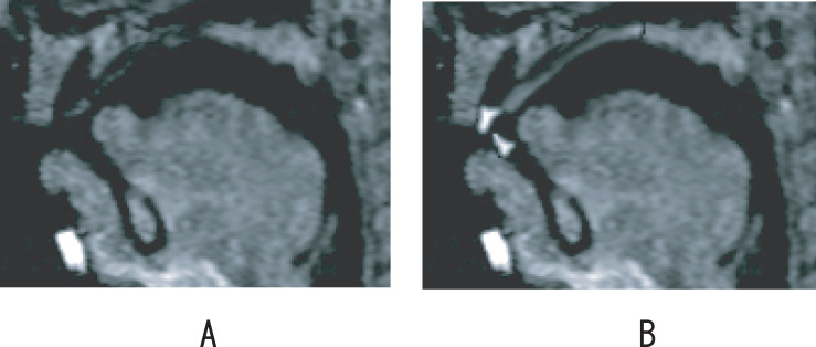
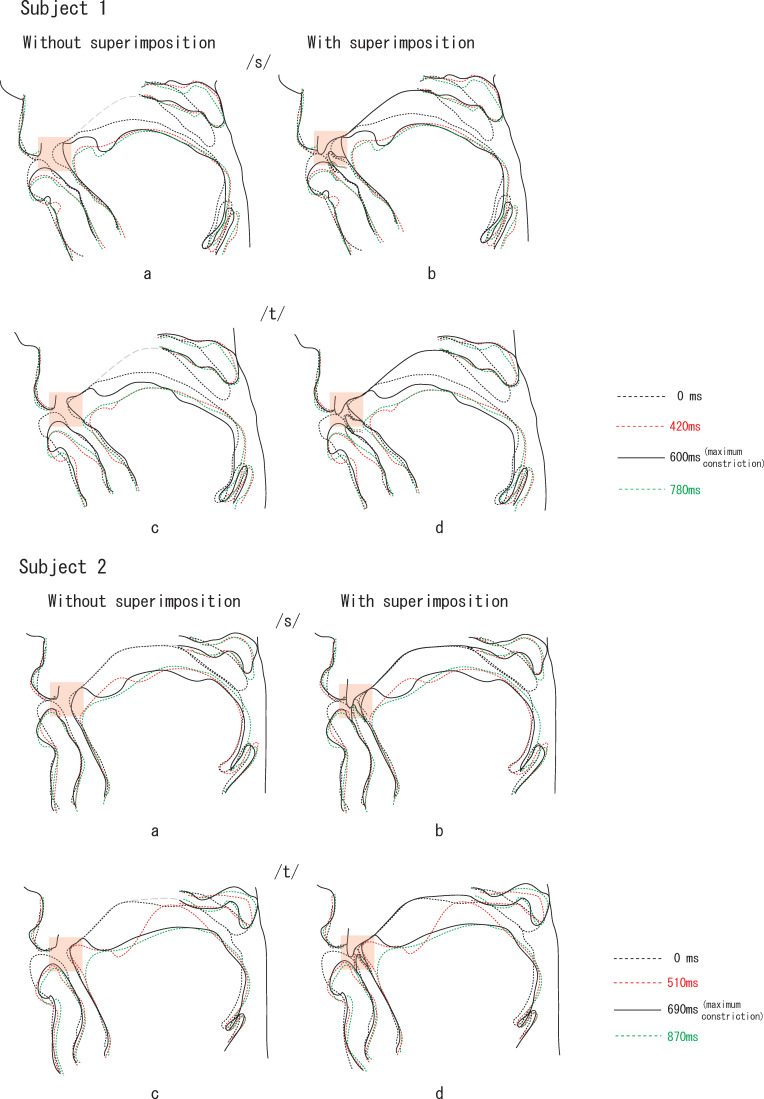
Images of incisors were successfully superimposed on MR images (Figure 5). Sequential tracings of oral structures without and with tooth superimposition during the articulation of /asa/ and /ata/ in the two subjects are shown in Figure 6. There was a prominent difference in images with and without tooth superimposition in the front oral cavity. Without tooth superimposition, the contours of the front oral cavity could not be determined and were overestimated (Figure 6). The pattern of the front oral cavity could be clearly viewed with the superimposition of incisors. The vague line of the hard palate in movie images could also be clearly observed after superimposition.
Figure 5.
Comparison of MRI movie images before (A) and after (B) superimposition.
Figure 6.
Comparison of images without and with tooth superimposition during the articulation of /asa/ and /ata/ in the two subjects (subject 1 and subject 2). Tracings were made before, during, and after maximum constriction (at 600 milliseconds for subject 1 and 690 milliseconds for subject 2) for the articulation of /s/ in /asa/ (a, b) and /t/ in /ata/ (c, d), without (a, c) and with (b, d) tooth superimposition, respectively. The time interval was set at 180 milliseconds. Clear contact/separation among the tip of the tongue, lips, and palate (shaded areas) can be observed with tooth superimposition in both subjects.
DISCUSSION
In this study, we applied the proposed method of using agar-based contrast medium14 for the purpose of observing articulatory movement in MRI movie. This method has been successfully used to clearly extract a tooth boundary in MR image. Compared with methods described in some studies,3,6,8–10,12 this method does not cause artifacts. The contrast medium is made into a jelly form by adding agar powder14 to fill the space in the retainer. Because the contrast medium is in direct contact with the tooth surface, the actual shape and size of tooth could be visualized in MR image. Indeed, previous researchers have reported that the soft tissues surrounding teeth and saliva retained in the mouth allow for indirect visualization of tooth crowns.13,14 However, these studies have been done for canines and molars. Thus, future study should examine the feasibility of the method for the central incisor in the midsagittal dimension.
With regard to the contrast medium in MRI, the paramagnets of Gd3+, Fe3+, and Mn2+ have a positive contrast-enhancing effect.15,16,19 FAC, which includes Fe3+, has been shown to have high signal intensity in MRI.15 However, FAC dissolved in water is acidic, has an unpleasant iron-metallic taste, and is in liquid form. Our method enabled us to neutralize FAC, improve its flavor, and eliminate the need for the subject to hold the liquid form of FAC inside their mouth for tooth visualization, which would otherwise cause undesired tissue reactions and be unpleasant for the subject. Furthermore, all of the low-cost materials in the contrast medium are used in daily food ingredients, and their safety is ensured by the concerned authorities. These advantages suggest that this method may also be effectively and easily applied in other scientific fields.
Landmarks on the forehead, nasal root, nasal tip, and points along the cranium that were used for superimposition of the upper incisor were all stable because they did not move together with the lower jaw during speech. Furthermore, the upper incisor is embedded in the upper jaw and anatomically connected to the frontal and nasal bone in the cranium. Therefore, the position of the upper incisor was in a direct relationship to those landmarks. On the other hand, the soft tissue around the mouth moves together with the lower jaw during speech. Thus, the landmarks on the soft tissue around the mouth could not be used for superimposition, but the symphysis was used in the final step for superimposition of the lower incisor onto the image.
Tooth visualization and superimposition in this preliminary study were used to observe articulatory movement during the articulation of a fricative /s/ and a plosive /t/. The fricative /s/ is produced by forcing air through a narrow channel in the vocal tract toward the edge of the incisor. The incisor acts as an obstacle to airflow, and thereby plays an essential role in the generation of turbulence in fricatives.20 For the plosive /t/, it has been reported that complete closure across the anterior palate is attained.21 Our study also revealed that there was a significant amount of tongue to palate/incisor contact in the front region during maximum constriction in /t/. Moreover, we found that /t/ was associated with a narrower front oral cavity than /s/, which is probably due to the closure needed to produce /t/. The present finding was consistent with findings in previous studies and further demonstrated the meticulous spatio-temporal relationship among articulators during the production of /asa/ and /ata/.
CONCLUSIONS
A clear incisor boundary can be extracted in MR image by using a retainer with space filled with a jelly form of FAC.
The superimposition of an incisor onto an MRI movie image enabled a more detailed observation of the front oral cavity during the production of a fricative and a plosive.
This technique may be useful in further studies to evaluate spatio-temporal relationships among articulators during speech in subjects with malocclusion, such as open bite.
Acknowledgments
This study was supported by Grants-in-Aid for Scientific Research Project (#20659322 and #20390521) from the Japan Society for the Promotion of Science, and by a grant from the Japanese Ministry of Education, Global Center of Excellence Program, International Research Center for Molecular Science in Tooth and Bone Diseases.
REFERENCES
- 1.Shinagawa H, Ono T, Honda E, et al. Dynamic analysis of articulatory movement using magnetic resonance imaging movies: methods and implications in cleft lip and palate. Cleft Palate Craniofac J. 2005;42:225–230. doi: 10.1597/03-007.1. [DOI] [PubMed] [Google Scholar]
- 2.Narayanan S. An approach to real-time magnetic resonance imaging for speech production. J Acoust Soc Am. 2004;115:1771–1776. doi: 10.1121/1.1652588. [DOI] [PubMed] [Google Scholar]
- 3.Ventura S. R, Freitas D. R, Tavares J. M. Application of MRI and biomedical engineering in speech production study. Comput Methods Biomech Biomed Eng. 2009;12:671–681. doi: 10.1080/10255840902865633. [DOI] [PubMed] [Google Scholar]
- 4.Inoue M. S, Ono T, Honda E, Kurabayashi T, Ohyama K. Application of magnetic resonance imaging movie to assess articulatory movement. Orthod Craniofac Res. 2006;9:157–162. doi: 10.1111/j.1601-6343.2006.00366.x. [DOI] [PubMed] [Google Scholar]
- 5.Inoue M. S, Ono T, Honda E, Kurabayashi T. Characteristics of movement of the lips, tongue and velum during a bilabial plosive: a noninvasive study using a magnetic resonance imaging movie. Angle Orthod. 2007;77:612–618. doi: 10.2319/071706-298.1. [DOI] [PubMed] [Google Scholar]
- 6.Takemoto H, Kitamura T, Nishimoto H, Honda K. A method of tooth superimposition on MRI data for accurate measurement of vocal tract shape and dimensions. Acoust Sci Tech. 2004;25:468–474. [Google Scholar]
- 7.Mohri D, Shimazu K. Articulation disorders caused by abnormal dentition. J Osaka Dent Univ. 2006;40:99–111. [Google Scholar]
- 8.Yang C. An accurate method to measure the shape and length of the vocal tract for the five Japanese vowels by MRI. Jpn J Logop Phoniatr. 1994;35:317–321. [Google Scholar]
- 9.Hasegawa-Johnson M, Pizza S, Alwan A, Cha J. S, Haker K. Vowel category dependence of the relationship between palate height, tongue height, and oral area. J Speech Lang Hear Res. 2003;46:738–753. doi: 10.1044/1092-4388(2003/059). [DOI] [PubMed] [Google Scholar]
- 10.Kitamura T, Hirata H, Honda K, et al. A method for measuring tooth shape by magnetic resonance imaging using a thermoplastic elastomer dental mouthpiece. IEICE Tech Rep. 2007;107:7–10. [Google Scholar]
- 11.Wakumoto M. Visualization of dental crown shape in an MRI-based speech production study. Int J Oral Maxillofac Surg. 1997;26:189–190. [Google Scholar]
- 12.Olt S, Jakob P. M. Contrast-enhanced dental MRI for visualization of the teeth and jaw. Magn Reson Med. 2004;52:174–176. doi: 10.1002/mrm.20125. [DOI] [PubMed] [Google Scholar]
- 13.Tutton L. M, Goddard P. R. MRI of the teeth. Br J Radiol. 2002;75:552–562. doi: 10.1259/bjr.75.894.750552. [DOI] [PubMed] [Google Scholar]
- 14.Tymofiyeva O, Rottner K, Gareis D, et al. In vivo MRI-based dental impression using an intraoral RF receiver coil. Concepts Magn Reson Pt B Magn Reson Engin. 2008;33B:244–251. [Google Scholar]
- 15.Patten R. M, Lo S. K, Phillips J. J, et al. Positive bowel contrast agent for MR imaging of the abdomen: phase II and III clinical trials. Radiology. 1993;189:277–283. doi: 10.1148/radiology.189.1.8372205. [DOI] [PubMed] [Google Scholar]
- 16.Hiraishi K, Narabayashi I, Fujita O, et al. Blueberry juice: preliminary evaluation as an oral contrast agent in gastrointestinal MR imaging. Radiology. 1995;194:119–123. doi: 10.1148/radiology.194.1.7997537. [DOI] [PubMed] [Google Scholar]
- 17.Riordan R. D, Khonsari M, Jeffries J, Maskell G. F, Cook P. G. Pineapple juice as a negative oral contrast agent in magnetic resonance cholangiopancreatography: a preliminary evaluation. Br J Radiol. 2004;77:991–999. doi: 10.1259/bjr/36674326. [DOI] [PubMed] [Google Scholar]
- 18.Inoue-Arai M. S, Ono T, Honda E, Kurabayashi T, Moriyama K. Motor coordination of articulators depends on the place of articulation. Behav Brain Res. 2009;199:307–316. doi: 10.1016/j.bbr.2008.12.008. [DOI] [PubMed] [Google Scholar]
- 19.Gries H, Miklautz H. Some physicochemical properties of the gadolinium-DTPA complex, a contrast agent for MRI. Physiol Chem Phys Med NMR. 1984;16:105–112. [PubMed] [Google Scholar]
- 20.Narayanan S. An articulatory study of fricative consonants using magnetic resonance imaging. J Acoust Soc Am. 1995;98:1325–1347. [Google Scholar]
- 21.Cheng H. Y. Electropalatographic assessment of tongue-to-palate contact patterns and variability in children, adolescents, and adults. J Speech Lang Hear Res. 2007;50:375–392. doi: 10.1044/1092-4388(2007/027). [DOI] [PubMed] [Google Scholar]


