Abstract
Primary tracheal schwannoma is a rare disease with no specific symptoms. At the molecular level, neurofibromatosis type 2 (NF2) gene mutation of Schwann cells is the major tumorigenic element. Herein, we present the case of a 54-year-old man with refractory shortness of breath and dry cough, which was resistant to bronchodilator treatment. Computed tomography revealed a transmural mass in the dorsolateral trachea. The tumor was surgically resected, and the diagnosis of schwannoma was confirmed by pathological examination. Furthermore, for this case, we performed whole-exome sequencing and identified several novel mutated schwannoma genes. The specific roles of these mutations need further confirmation.
Keywords: tracheal tumor, schwannoma, surgical treatment, whole-exome sequencing, mutation
Introduction
Primary tracheal tumor is an uncommon disease, with an estimated incidence of one case per million.1 The majority of these neoplasms are malignancies and mostly consists of squamous cell carcinoma and adenoid cystic carcinoma.2 Primary tracheal neurogenic tumors are extremely rare, constituting less than 0.5% of primary tracheal tumors.3,4 This type of tumor arises from nerves located inside the tracheal wall, including schwannomas and neurofibromas, and are usually benign.5 Schwannomas originate from tumorigenic Schwann cells, mainly attributed to the loss of function mutations of the neurofibromatosis type 2 (NF2) tumor suppressor gene.6 However, to our knowledge, no case has yet been described with molecular features of primary tracheal schwannomas. We herein present a rare case of a transmural schwannoma in the trachea with whole-exome sequencing.
Case Presentation
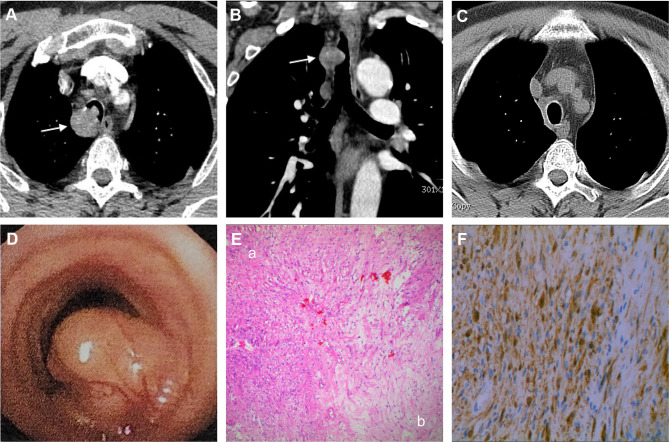
A 54-year-old man had a 1-month history of progressive shortness of breath and dry cough. He was a non-smoker with no history of pulmonary disease. His symptoms were exacerbated in the supine position, with little relief after bronchodilator treatment. These symptoms developed without apparent cause and in the absence of problems such as chest pain, fever, nausea, or vomiting. Upon admission, physical examination revealed a temperature of 36.6°C, a heart rate of 85 to 90 beats/min, a blood pressure of 126/71 mmHg, a respiratory rate of 17 to 21 breaths/min and a transcutaneous oxygen saturation 97% on ambient air. Lung auscultation indicated a prolonged expiratory phase with wheezing in the thoracic part of the trachea. Respiratory function studies demonstrated a severe obstructive ventilation defect with a forced expiratory volume in 1 s (FEV1) of 0.97 L and FEV1% of 28.1, which did not respond to aerosolized bronchodilators. There were no clinically significant changes in other physical examinations and biochemical tests. A computed tomography (CT) enhancement scan of his chest showed a smooth-edged, homogenous, intra-tracheal mass measuring approximately 27×25 × 20 mm, with extratracheal extension (Figure 1). Fiberoptic bronchoscopy revealed a smooth-edged, intact capsule mass obstructing about 70% of the tracheal lumen (Figure 1). The margin of the mass was between the 8th to 11th trachea cartilage ring, with abundant blood vessels on the surface. Given the potential for bleeding and airway obstruction, no biopsies were taken.
Figure 1.
(A) CT showing a transmural mass in the tracheal (arrow). (B) Coronal reconstruction of the tumor (arrow). (C) Six months after tracheal segmental resection and anastomosis. (D) Bronchoscopy revealed a wide-based mass on the dorsolateral tracheal wall, with abundant blood vessels on the surface. (E) Histopathology of the tumor, HE staining (magnification, ×100); (a) Antoni A tissue, densely packed cells with elongated nuclei; (b) Antoni B tissue, loose myxoid stroma with microcystic cellular organization. (F) S-100 was positive on immunohistochemical examination (magnification, ×400).
The patient immediately underwent surgical treatment. Thoracotomy was performed with the patient in the left lateral position under general anesthesia. Upon adequate exposure of the trachea, a transmural tracheal mass was found at 5 cm from the carina. The tumor was completely resected by circumferential tracheal excision with end-to-end anastomosis. Macroscopically, the tumor had a homogeneous brown color with clear borders, about 27×25 × 15 mm in size. Morphologically, hematoxylin and eosin (HE) staining showed that the cytoplasm of tumor cells were abundant, with tightly packed spindle cells with elongated nuclei, arranged in a palisading pattern (Figure 1). The mitotic rate was low, ranging from 0 to 1 mitoses/10 high-power fields. On immunohistochemical analysis (Figure 1), the tumor was found to be positive for S-100, SOX-10, BCL-2 and negative for SATA6, CD117, Dog-1, CD34, SMA, Desmin, and ALK; the diagnosis of schwannoma was confirmed. The patient recovered successfully without complications, and there was no evidence of recurrence after six months follow-up by chest CT (Figure 1).
To pursue a molecular diagnosis, whole-exome sequencing was performed in tumor tissue specimens and matched normal tissue samples. After ruling out genetic variations, 19 tumor-related somatic mutations were confirmed (shown in Table 1). The mutations included FLNB, CLCA2, MICALCL, MUC16, DCXR, SREK1, PCGF5, BEX3, MYH6, KHNYN, NANOG, RFC3, CARD8, PHACTR3, KATNA1, AIM2, EIF2B3, CPEB3, and CLMN. There has been no prior report of these mutations in schwannomas.
Table 1.
Somatic Variants Detected in the Patient
| Gene | Transcript | Nucleotide Change | Amino Acid Change | Frequency |
|---|---|---|---|---|
| FLNB | NM_001457.4 | c.7159G>T | p.V2387L | 16.6% |
| CLCA2 | NM_006536.7 | c.448A>G | p.N150D | 16.6% |
| MICALCL | NM_032867.3 | c.894A>T | p.Q298H | 15.0% |
| MUC16 | NM_024690.2 | c.25472C>T | p.A8491V | 14.0% |
| DCXR | NM_016286.4 | c.686T>G | p.M229R | 13.6% |
| SREK1 | NM_001077199.3 | c.299A>G | p.K100R | 12.5% |
| PCGF5 | NM_032373.5 | c.242A>T | p.K81M | 11.0% |
| BEX3 | NM_001282674.1 | c.233dup | p.N78Kfs*31 | 10.2% |
| MYH6 | NM_002471.3 | c.4010C>T | p.S1337L | 10.0% |
| KHNYN | NM_001290256.1 | c.2131_2134del | p.S711Rfs*31 | 9.5% |
| NANOG | NM_024865.4 | c.576C>A | p.N192K | 8.3% |
| RFC3 | NM_002915.4 | c.244del | p.I82Lfs*25 | 7.9% |
| CARD8 | NM_001351782.2 | c.1093A>G | p.M365V | 7.6% |
| PHACTR3 | NM_080672.5 | c.146G>A | p.R49H | 6.6% |
| KATNA1 | NM_007044.4 | c.1248_1268del | p.S417_N423del | 5.7% |
| AIM2 | NM_004833.3 | c.1027del | p.T343Hfs*14 | 4.6% |
| EIF2B3 | NM_020365.5 | c.450dup | p.A151Sfs*6 | 3.0% |
| CPEB3 | NM_014912.5 | c.188del | p.P63Rfs*19 | 2.9% |
| CLMN | NM_024734.4 | c.2594dup | p.E866Gfs*25 | 2.7% |
Discussion
Primary tracheal schwannoma is a rare disease that accounts for less than 0.5% of tracheal tumors.3,4 Straus et al first described this tumor in 1951.7 Arising from Schwann cells of nerve sheaths, schwannomas in the respiratory system were mostly been found in the lungs and bronchi but not tracheal. Primary tracheal schwannoma is a benign tumor that usually presents as a single, well-defined, slow growing mass. The symptoms of this disease are not obviously specific in the early stage, and are easily confused with respiratory diseases such as asthma, which may lead to delayed diagnosis.8 In the present case, the primary symptoms were shortness of breath and dry cough, with little relief after bronchodilator treatment.
Pulmonary function tests suggesting an upper airway narrowing may be of significant diagnostic value. Standard chest radiographs are of limited diagnostic utility because superimposed soft tissues and the spine may interfere with tumor imaging. CT could evaluate the tumor’s location, size, presence or absence of transmural involvement, and relationship with the surrounding tissues. Bronchoscopy was used not only for direct observation of the tumor but equally for the biopsy and excision given the large airway. However, for patients with tumors that almost completely obstruct the airway, bronchoscope placement may cause airway blockage. The definitive diagnosis of a schwannoma requires histopathological examination. The internal structure of the tumor consists of an Antoni A area (densely packed cells with elongated nuclei) and an Antoni B area (loose myxoid stroma with microcystic cellular organization). In the process of tumor growth, Antoni A type tissue may transform into Antoni B tissue.6 S-100 protein expression is consistently positive in immunohistochemical examination.9
The optimal treatment of primary tracheal schwannomas has not yet been established. Surgical resection, which includes tracheal resection and end-to-end anastomosis, was most adopted in the treatment of tracheal schwannomas. However, it is not suitable for long segmental tracheal resection, owing to the lack of an ideal material for a tracheal substitute.1 Alternative treatment consists of a complete endoscopic approach, especially in patients whose tumors were pedunculated-type and intraluminal.10 Of note, there were some cases of tumor local recurrence after endoscopic resection,5,8,11 whereas no relapse was recorded in surgical resection. Therefore, selecting the proper approach for the appropriate patient and adequate long-term follow-up is essential. At present, there are no approved pharmacological treatments for schwannoma. Several targeted therapies are under development as potential therapeutics.12 In the present case, a patient with transmural tumor underwent segmental tracheal resection and reconstruction, and no recurrence was observed during the six-month follow-up period.
At the molecular level, loss of NF2 most frequently occurs in sporadic schwannomas. Sameer Agnihotri et al13 precisely mapped the genomic landscape of schwannomas. They found that the most common mutation was NF2, accounting for 77% of cases, followed by ARID1 (including ARID1A and ARAD1B), accounting for 29% of cases. Other recurrent but low-frequency variants include DDR1, TAB3, ALPK2, CAST, TSC1 and TSC2, and were first described in schwannomas. However, there was no tracheal schwannoma in their study. The variety of mutations may considerably vary in schwannomas of different origins. For example, NF2 mutations in vestibular schwannomas were significantly higher than those in spinal schwannomas.13 Our finding showed that FLNB had the highest gene mutation abundance (16.62%), followed by CLCA2 (16.57%) and MICALCL (14.97%). These mutated genes were novel in schwannomas. Till date, there is no relevant report on molecular features in tracheal schwannomas, except for the present report. We were unable to explain the likely roles of these variants in tracheal schwannomas. Our finding needs further confirmation.
In conclusion, primary tracheal schwannoma is a rare disease with no specific symptoms that may further lead to delayed diagnosis. Pulmonary function, CT, and bronchoscopy can help in early diagnosis. Current treatment options, including Surgery and endoscopic treatment, depend on the tumor type and substantial status of the patients. Despite usually being a benign disease, there is a possibility of recurrence; therefore, long-term follow-up is necessary. We found several novel mutated genes in schwannomas, and its mechanism needs to be studied further.
Acknowledgments
We thank the patient in this report and his family. This research was partially supported by grants from scientific research program of Tianjin Education Commission (2020KJ151). The funders had no role in study design, data collection and analysis, decision to publish, or in the preparation of the manuscript.
Ethics Statement
The study was approved by institutional ethics committee board of Tianjin Medical University General Hospital. Written informed consent was obtained from the patient for publication of this case report and any accompanying images.
Author Contributions
All authors made a significant contribution to the work reported, whether that is in the conception, study design, execution, acquisition of data, analysis and interpretation, or in all these areas; took part in drafting, revising or critically reviewing the article; gave final approval of the version to be published; have agreed on the journal to which the article has been submitted; and agree to be accountable for all aspects of the work.
Disclosure
The authors declare that the research was conducted in the absence of any commercial or financial relationships that could be construed as a potential conflict of interest.
References
- 1.Hao ZR, Yao ZH, Zhao JQ, et al. Clinical efficacy of treatment for primary tracheal tumors by flexible bronchoscopy: airway stenosis recanalization and quality of life. Exp Ther Med. 2020;20(3):2099–2105. doi: 10.3892/etm.2020.8900 [DOI] [PMC free article] [PubMed] [Google Scholar]
- 2.Grillo HC, Mathisen DJ. Primary tracheal tumors: treatment and results. Ann Thorac Surg. 1990;49(1):69–77. doi: 10.1016/0003-4975(90)90358-D [DOI] [PubMed] [Google Scholar]
- 3.Han DP, Xiang J, Ye ZQ, et al. Primary tracheal schwannoma treated by surgical resection: a case report. J Thorac Dis. 2017;9(3):E249–E252. doi: 10.21037/jtd.2017.02.85 [DOI] [PMC free article] [PubMed] [Google Scholar]
- 4.Hamdan AL, Moukarbel RV, Tawil A, El-Khatib M, Hadi U. Tracheal schwannoma: a misleading entity. Middle East J Anaesthesiol. 2010;20(4):611–613. [PubMed] [Google Scholar]
- 5.Righini CA, Lequeux T, Laverierre MH, Reyt E. Primary tracheal schwannoma: one case report and a literature review. Eur Arch Otorhinolaryngol. 2005;262(2):157–160. doi: 10.1007/s00405-004-0778-0 [DOI] [PubMed] [Google Scholar]
- 6.Helbing DL, Schulz A, Morrison H. Pathomechanisms in schwannoma development and progression. Oncogene. 2020;39(32):5421–5429. doi: 10.1038/s41388-020-1374-5 [DOI] [PMC free article] [PubMed] [Google Scholar]
- 7.Straus GD, Guckien JL. Schwannoma of the tracheobronchial tree. A case report. Ann Otol Rhinol Laryngol. 1951;60(1):242–246. doi: 10.1177/000348945106000122 [DOI] [PubMed] [Google Scholar]
- 8.Ge X, Han F, Guan W, Sun J, Guo X. Optimal treatment for primary benign intratracheal schwannoma: a case report and review of the literature. Oncol Lett. 2015;10(4):2273–2276. doi: 10.3892/ol.2015.3521 [DOI] [PMC free article] [PubMed] [Google Scholar]
- 9.Dorfman J, Jamison BM, Morin JE. Primary tracheal schwannoma. Ann Thorac Surg. 2000;69(1):280–281. doi: 10.1016/S0003-4975(99)01195-9 [DOI] [PubMed] [Google Scholar]
- 10.Kasahara K, Fukuoka K, Konishi M, et al. Two cases of endobronchial neurilemmoma and review of the literature in Japan. Intern Med. 2003;42(12):1215–1218. doi: 10.2169/internalmedicine.42.1215 [DOI] [PubMed] [Google Scholar]
- 11.Chen H, Zhang K, Bai M, et al. Recurrent transmural tracheal schwannoma resected by video-assisted thoracoscopic window resection: a case report. Medicine. 2019;98(51):e18180. doi: 10.1097/MD.0000000000018180 [DOI] [PMC free article] [PubMed] [Google Scholar]
- 12.Ahmed SG, Abdelnabi A, Maguire CA, et al. Gene therapy with apoptosis-associated speck-like protein, a newly described schwannoma tumor suppressor, inhibits schwannoma growth in vivo. Neuro Oncol. 2019;21(7):854–866. doi: 10.1093/neuonc/noz065 [DOI] [PMC free article] [PubMed] [Google Scholar]
- 13.Agnihotri S, Jalali S, Wilson MR, et al. The genomic landscape of schwannoma. Nat Genet. 2016;48(11):1339–1348. doi: 10.1038/ng.3688 [DOI] [PubMed] [Google Scholar]


