Abstract
Objectives:
To compare the clinical efficacy of unilateral and bilateral puncture PKP in the treatment of OVCFs and explored whether there is a difference in the efficacy of unilateral and bilateral puncture PKP after surgery.
Methods:
A total of 98 patients with OVCFs treated by PKP from August 2016 to June 2018 were selected. There were 62 cases in the unilateral puncture group and 36 cases in the bilateral puncture group. The operation time, the amount of bone cement injection, the height of the anterior edge of the vertebral body and the visual analog scale (Visual Analog Scale, VAS) scores before and after the operation were analyzed, and whether the differences between the 2 groups were statistically significant was analyzed.
Results:
All patients were followed up completely. The operation time and the number of X-ray fluoroscopies of the unilateral puncture group were significantly reduced compared to those of the bilateral group, and the difference was statistically significant (p<0.05). In terms of the bone cement injection volume, the average injection volume of the bilateral group was greater than that of the unilateral group, and the difference was statistically significant (p<0.05); the postoperative VAS scores of the 2 groups of patients were significantly improved, and the difference was statistically significant compared with that before surgery (p<0.05) but that of the unilateral group was not statistically significant compared with that of the bilateral group (p>0.05). The height of the anterior edge of the vertebral body in both groups was significantly improved compared with that before the operation, and the difference was statistically significant (p<0.05).
Conclusion:
Unilateral and bilateral puncture PKP can achieve good clinical efficacy in the treatment of osteoporotic vertebral compression fractures, but unilateral PKP has the advantages of short operation time and low X-ray exposure.
Osteoporosis (OP) is caused by a decrease in bone mass for a variety of reasons, especially a decrease in the amount of cancellous bone in the vertebral body and damage to the microstructure of bone tissue, bone mineral composition and bone matrix per unit volume. Osteoporosis is one of the diseases with high morbidity and mortality in the world and has become an important disease that endangers the health of middle-aged and elderly people.1 Osteoporotic vertebral compression fractures (OVCFs) are one of the major complications of osteoporosis, which often cause stubborn waist and back aches. Severe thoracolumbar osteoporotic vertebral body compression fractures may lead to cardiopulmonary and other multisystem dysfunctions, seriously affecting the patient’s quality of life.2
For the treatment of OVCFs, the current recommendations are conservative treatment and surgical treatment. Conservative treatment may cause various complications due to long-term bed rest, including bedsores, delayed fracture healing, deformity healing or nonunion, respiratory and urinary tract infections, and lower extremity venous thrombosis, which can threaten the life of the patient.3,4 Therefore, patients with OVCFs who have early out-of-bed activity requirements and surgical indications are more likely to undergo surgical treatment.
The traditional surgical treatment for OVCFs is posterior laminectomy and decompression pedicle screw internal fixation, but due to the higher degree of osteoporosis in older patients, the long-term screw internal fixation effect is poor, and surgical trauma has a greater impact on patients; thus, the long-term efficacy is not ideal.5 In recent years, with the improvement of minimally invasive spine technology, percutaneous vertebralplasty (PVP) and percutaneous balloon dilatation kyphoplasty (Percutaneous kyphoplasty, PKP) have achieved satisfactory results in the treatment of OVCF. Compared with PVP, PKP uses a balloon or other expansion system to expand the compressed vertebral body to form a relatively low-pressure vertebral body space, followed by low-pressure injection of bone cement, which can better correct kyphosis and reduce the penetration of bone cement leakage.6,7
The PKP surgical puncture consists of a bilateral pedicle approach or a unilateral pedicle approach. While the advantages of the transdermal bilateral pedicle approach include better diffusion of bone cement and reduced risk of puncture, there are shortcomings, such as long operation time, large radiation exposure and high hospitalization costs.8 At present, there is no unified conclusion as to which PKP approach is better for use to treat OVCFs. Therefore, it is of great clinical significance to clarify the difference between unilateral and bilateral PKP in the treatment of OVCFs.
The OVCFs are one of the common diseases that cause lumbago and kyphosis in the elderly. At present, PKP is one of the common methods for the treatment of OVCFs. Bilateral puncture of the pedicle approach is the classic operation method of PKP, but some scholars believe that unilateral puncture bone cement injection can achieve the same surgical effect. This record-based case–control study retrospectively analyzed patients with OVCFs treated in our hospital from August 2016 to June 2018, performed an in-depth analysis and comparison of the unilateral and bilateral PKP treatment of OVCFs, and provided a reference for the clinical approach to PKP treatment of OVCFs.
Methods
A retrospective analysis of the clinical data of 98 patients with OVCFs admitted to our hospital from August 2016 to June 2018 for PKP was performed. Inclusion criteria were: (1) age ≥65 years, with typical clinical symptoms, including thoracolumbar and back pain, turning over, and difficulty moving; (2) MRI or ECT diagnosed with fresh vertebral compression fracture (MRI: T1WI is low signal, T2WI is high signal or equal signal, and the fat suppression sequence STIR is high signal), the course of disease is less than 2 weeks; (3) dual energy X-ray bone densitometry to check the bone mineral density (BMD) diagnosis of osteoporosis; (4) observers who can cooperate to complete clinical follow-up; (5) no symptoms of spinal cord nerve damage; and (6) patients undergoing PKP surgery and using percutaneous unilateral or bilateral puncture; (7) all patients were single vertebral compression fractures, and no adjacent or distant fractures. The exclusion criteria were: (1) the patient had poor basic physical condition and could not tolerate the surgery; (2) the course of disease was more than 2 weeks; (3) the injured vertebral body had an incomplete posterior wall with symptoms of spinal cord or nerve root compression; 4) clinical imaging and other data were incomplete; and (5) those who were treated by surgical methods other than PKP. The study was approved by the hospital ethics committee.
Among the 98 patients, 32 were male and 66 were female, and the age range was 65-87 years. The distribution of thoracic vertebrae (T6~10) was 20 segments, thoracolumbar (T11~L2) 62 segments, and lumbar spine (L3~5) 16 segments. All patients underwent PKP surgery. Among them, 36 patients underwent bilateral pedicle puncture as the bilateral group, and 62 patients underwent unilateral pedicle puncture as the unilateral group. There was no statistically significant difference in general data, such as age, gender, and fracture segment distribution, between the 2 groups (p>0.05) (Table 1).
Table 1.
- Comparison of general information of the 2 groups (Mean±SD).
| Groups | Unilateral | Bilateral |
|---|---|---|
| Total | 62 | 36 |
| Gender (male/female) | 20/42 | 12/24 |
| Age | 77.3±8.6 | 75.6±9.5 |
| There was no statistically significant difference in gender and age between the 2 groups (p<0.05) | ||
The patient was placed in a prone position, with the abdomen suspended. C-arm X-ray fluoroscopy was used to determine the location of the diseased vertebral body and pedicle and accurately locate and mark it, and 1% lidocaine was used for anesthesia with full-thickness soft tissue infiltration from the skin of the puncture point to the pedicle.
Unilateral group
Take the upper part of the pedicle shadow (11 points on the left side of the pedicle projection and 2 points on the right side) as the puncture point. Slowly penetrate and enter the vertebral body through the pedicle and advance into the vertebral body after l/3, withdraw the puncture needle core, use the dilator to expand the tunnel in front of the vertebral body to reach the middle of the vertebral body. A balloon is inserted into the lower puncture needle sleeve, and contrast medium is injected for expansion and reduction. Withdraw the balloon and inject bone cement in the drawing stage under full fluoroscopy. If leakage occurs, stop the injection of bone cement immediately and observe the patient’s lower limb sensation and movement.
Bilateral group
The puncture method is the same as before; one side of the pedicle is punctured, and then the balloon is expanded, and the same method is followed on the other side of the pedicle, with the same steps of puncture and balloon expansion. Under continuous fluoroscopy, both sides are simultaneously cemented by injecting the cement into the vertebral body.
The operation time, the number of X-ray exposures, the amount of bone cement injection, and the VAS scores of the patient before and after operation at 3 days after surgery were recorded, and the changes of the anterior edge height of the fractured vertebral body before and after operation were measured.
The SPSS 16.0 software was used for statistical processing. Before operation, after operation, and between groups, the measurement data of each observation index were evaluated by t test, and p<0.05 was considered statistically significant.
Results
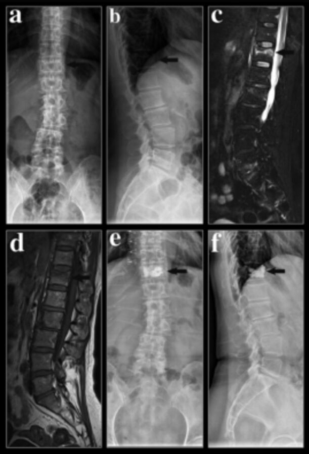
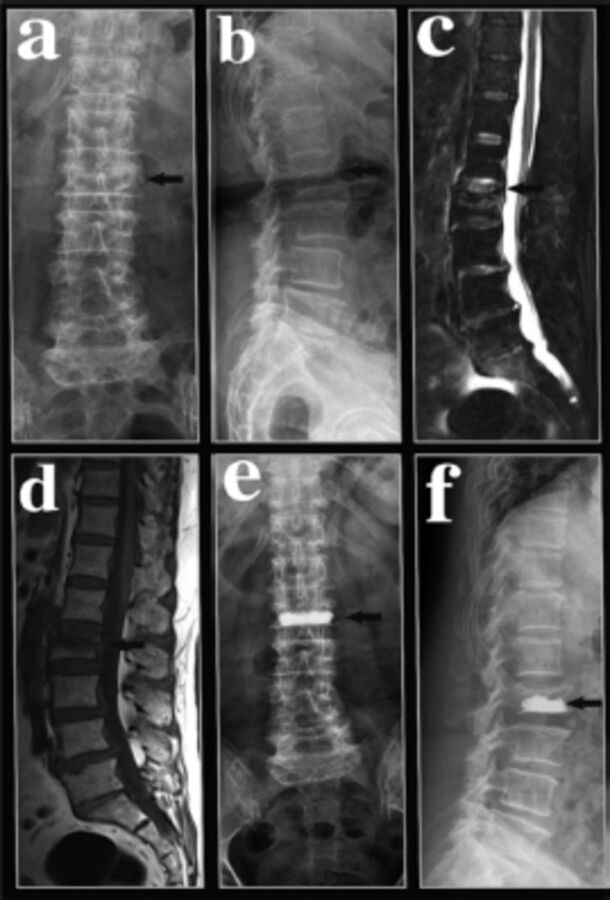
All patients had a successful operation and completed preoperative and postoperative imaging examinations and clinical symptom scoring. The operation time of (53.6±8.7) min, the bone cement injection volume of (5.6±1.3) ml and the X-ray irradiation frequency of (21.5±4.3) in the bilateral puncture group were higher than those in the unilateral puncture group of (35.7±6.2) min, (3.5±0.9) ml and (12.6±3.2), respectively; the differences were statistically significant (p<0.05). The anterior and postoperative vertebral body anterior margin heights were (19.2±6.1) mm and (23.7±5.3) mm in the unilateral puncture group, and the VAS scores were (8.2±2.1) and (2.2±1.6), respectively. In the bilateral puncture group, the anterior and posterior vertebral body heights were (19.2±6.1) mm and (22.9±5.1) mm, respectively, and the VAS scores were (8.5±2.5) and (2.2±1.3) points, respectively. There were statistically significant differences between the anterior edge height and the VAS score after surgery (3 days after surgery) and before surgery (p<0.05), but there was no significant difference between the unilateral puncture group and the bilateral puncture group in the recovery of vertebral body anterior height and postoperative symptom improvement (p>0.05) (Table 2 & 3). Typical cases are shown in Figure 1 a-f (unilateral puncture) and Figure 2 a-f (bilateral puncture).
Table 2.
- Comparison of operation time, bone cement dosage and X-ray perspective times between the 2 groups (Mean±SD).
| Groups | Unilateral | Bilateral | P-value |
|---|---|---|---|
| Operation time (min) | 35.7±6.2a | 53.6±8.7 | 0.032 |
| Bone cement dosage | 3.5±0.9a | 5.6±1.3 | 0.041 |
| X-ray perspective times | 12.6±3.2a | 21.5±4.3 | 0.038 |
Compared with bilateral group, the difference was statistically significant (p<0.05)
Table 3.
- Comparison of anterior height of vertebral body and VAS score between the 2 groups before and after operation (Mean±SD).
| Groups | Anterior height of vertebral body | VAS score | ||
|---|---|---|---|---|
| Preoperative | Postoperative | Preoperative | Postoperative | |
| Unilateral | 19.6±6.5 | 23.7±5.3 a | 8.2±2.1 | 2.2±1.6 a |
| Bilateral | 19.2±6.1 | 22.9±5.1ab | 8.5±2.5 | 2.2±1.3 ab |
The corresponding p-values of a1, a2, a3 and a4 are 0.036, 0.037, 0.002 and 0.001; The corresponding P-values of b1 and b2 are 0.108 and 0.065.
Compared with preoperatively, the difference was statistically significant (p<0.05);
compared with the unilateral group, the difference was not statistically significant (p>0.05)
Figure 1.
- A 60-year-old female who suffered a back injury and suffered back pain accompanied by restricted motion of the lumbar spine was admitted to the hospital for 2 days. a-b) Preoperative lumbar spine lateral X-ray film prompts: T12 vertebral wedge shape change. c-b) -Preoperative MRI shows T12 vertebral bone marrow edema as a fresh fracture. e-f) The T12 vertebral body was treated with PVP, unilateral pedicle puncture, and the injection of 3.9 ml of PMMA bone cement. Postoperative lumbar spine X-ray film showed that T12 had changed postoperatively, and the bone cement was well distributed.
Figure 2.
- A 68-year-old woman was admitted to the hospital after a week of backache due to a fall. a-b) Preoperative lumbar spine lateral X-ray film findings: L2 vertebral body wedge changes. c-d) Preoperative MRI showed L2 vertebral bone marrow edema as a fresh fracture. e-f) PVP treatment of L2 vertebral body, bilateral pedicle puncture, and injection of 5.0 ml of PMMA bone cement. Postoperative lumbar spine X-ray film showed that L2 had changed postoperatively, and the bone cement was well distributed.
Discussion
The OVCFs caused by osteoporosis are a common and frequently occurring disease in the elderly. The clinical manifestations are low back pain, spinal deformity, and motor dysfunction, which seriously affect the quality of life of patients. The PKP treatment of OVCFs features less trauma, simple operation, good safety and effectiveness, and rapid relief of low back pain and restoration of injured vertebral body morphology as its main clinical features. In this study, the clinical symptoms of the 2 groups of patients were significantly improved after surgery, indicating that both puncture methods could quickly and effectively relieve pain and stabilize the maintenance effect. Possible causes: (1) Mechanical reasons. Bone cement injection quickly enhances the stability of the injured vertebrae, reduces the fretting friction at the fractured end of the injured vertebrae, and prevents further compression of the vertebral body, reducing pain. (2) Thermal effects and chemistry. Bone cement powder liquid heat blocking and toxic paralysis during the polymerization process cause degeneration and necrosis of surrounding tissues, destroy nerve endings at the fracture site, and reduce pain. (3) It has been reported in the literature that PKP can remove blood and leakage in injured vertebrae, reduce the pressure in the injured vertebrae, reduce the stimulation of nerve endings around the fractured end, and thus reduce pain 9.
At present, the commonly used PKP procedures are mainly unilateral and bilateral vertebral kyphoplasty. Generally, the best bone cement distribution in cancellous bone should be obtained by bilateral puncture, but the choice of unilateral or bilateral approaches is still controversial, and evidence-based medicine and other relevant evidence are still lacking. A study found that through a three-dimensional finite element model study, unilateral injection will cause uneven distribution of bone cement in the vertebral body, which causes the vertebral body to bear the fracture and can lead to spinal instability.10 It is easy to flex laterally to the opposite side of the perfusion under constant load. As a result, the vertebral body is compressed and deformed, but Steinmann et al.11 found that a fracture model made on fresh cadavers can restore the strength and rigidity of the fractured vertebral body with single and bilateral puncture. Side operation does not present a greater risk of side compression. Bilateral pedicle puncture is a common method of PKP. The study found that the bilateral pedicle approach is better than the unilateral pedicle approach because of the better diffusion of bone cement, which can fully stabilize the microfractures of the diseased vertebral body, and the degree of symptom improvement is more obvious. Garfin S et al12 compared the clinical efficacy of single and bilateral puncture PKP treatment of OVCFs and showed that the bilateral puncture group is superior to the single point in terms of the recovery of the anterior edge height of the injured vertebrae, kyphosis, Cobb’s angle, and early VAS score improvement. For the side puncture group, Chen et al13 also showed that bilateral puncture is more advantageous for vertebral body height recovery. However, there have been an increasing number of reports on unilateral pedicle puncture in recent years, and the literature reports that there is no significant difference between the unilateral and bilateral pedicle approaches for pain relief after PKP. Kim et al14 confirmed through clinical studies36 that unilateral pedicle puncture can achieve the same therapeutic effect as bilateral pedicle puncture; thus, bilateral pedicle puncture is not recommended. In this study, the statistical analysis of the operation time, fluoroscopy time, and bone cement injection index of single and double puncture PKP surgery initially confirmed that single puncture PKP surgery can achieve the same satisfactory clinical efficacy as bilateral puncture PKP surgery and has a shorter operation time, lower radiation exposure, less tissue trauma, and less bone cement injection for patients and medical staff. More importantly, unilateral and bilateral approaches to PKP can obviously restore the height of the compressed vertebral body. The high degree of recovery can effectively relieve patients’ pain symptoms. Of course, if the patient’s general condition is poor or affected by a variety of medical diseases, or if the patient cannot tolerate the long time in prone position required for the bilateral surgery, to reduce the patient’s pain and shorten the operation time, unilateral pedicle puncture is a good choice. Of course, everything is not absolute, and the patient’s health should be considered in all aspects according to the patient’s age, general health, fracture type, number of responsible vertebral bodies, and various emergencies and precautions that may be encountered during the operation. The situation ultimately determines whether to use unilateral or bilateral pedicle puncture.
Studies have shown that the recovery of compressed vertebral body height is related to the height of the vertebral body before surgery, the patient’s bone density, the type of fracture, and the course of disease. The PKP technology uses a specific device to inject bone cement into the compressed vertebral body to repair bone defects, correct the degree of vertebral compression and kyphosis, and restore the strength and rigidity of the vertebral body to avoid further collapse of the injured vertebral body.15 The restoration of compressed vertebral body height is beneficial to restore the balance of the sagittal plane of the patient’s spine and correct kyphosis.16 Therefore, clinically effective restoration of the height of the diseased vertebral body in patients with osteoporotic vertebral compression fractures and subsequent correction of convex deformity are necessary. Studies have shown that changes in vertebral body height caused by vertebral compression fractures can advance the load-bearing line of the spine. This change can not only increase the continued collapse of the compressed vertebral body but also increase the risk of fractures of adjacent vertebral bodies. PKP can change the load-bearing force line of the vertebral body, moving it back to a near-normal level, thereby restoring the stability of the spine and alleviating the pain of the patient.17 This study found that unilateral and bilateral approaches to PKP can significantly restore the height of the compressed vertebral body. Postoperative height restoration of the vertebral body can effectively relieve the patient’s pain symptoms.
Theoretically, the more bone cement injected, the higher the biomechanical strength and the better the clinical effect. However, recent studies have found that there is no direct relationship between the amount of bone cement injected and the clinical effect after surgery. Injecting a small amount of bone cement into the diseased vertebral body can achieve the purpose of relieving pain.18 The VAS scores of all patients in this study were significantly improved after surgery, and the difference was statistically significant compared with that before surgery (p<0.05).
This study has several limitations that should be considered. First, the number of subjects was relatively small and the follow-up period relatively short, and a larger study sample and longer follow-up period may be needed for statistical assessment in the future. Second, selective bias might have been introduced, which may lead to errors in the results. Although these limitations are important, this study contributes to the comparison of clinical efficacy of unilateral and bilateral puncture PKP in the treatment of OVCFs.
In conclusion, unilateral and bilateral PKP treatment of osteoporotic vertebral compression fractures can quickly relieve pain in patients, and there is no significant difference in the clinical efficacy between the 2. However, the former has the advantages of a short operation time and low X-ray exposure, while the bilateral approach is more complicated, and the potential complications are greater. We believe that unilateral percutaneous kyphoplasty can be used as a treatment for osteoporotic fractures and is the preferred method for lumbar compressive fractures.
Acknowledgements
The authors would like to thank Dr. Bing Zhang for her help with radiological evaluation.
Footnotes
References
- 1. McCarthy J, Davis A.. Diagnosis and Management of Vertebral Compression Fractures. Am Fam Physician 2016; 94: 44–50. [PubMed] [Google Scholar]
- 2. Musbahi O, Ali AM, Hassany H, Mobasheri R.. Vertebral compression fractures. Br J Hosp Med 2018; 79: 36–40. [DOI] [PubMed] [Google Scholar]
- 3. Goldstein CL, Chutkan NB, Choma TJ, Orr RD.. Management of the Elderly With Vertebral Compression Fractures. Neurosurgery 2015; 77: S33–S45. [DOI] [PubMed] [Google Scholar]
- 4. Richmond BJ. Vertebral Augmentation for Osteoporotic Compression Fractures. J Clin Densitom 2016; 19: 89–96. [DOI] [PubMed] [Google Scholar]
- 5. Martikos K, Greggi T, Faldini C, Vommaro F, Scarale A.. Osteoporotic thoracolumbar compression fractures: long-term retrospective comparison between vertebroplasty and conservative treatment. Eur Spine J 2018; 27: 244–247. [DOI] [PubMed] [Google Scholar]
- 6. Cheng Y, Liu Y.. Percutaneous curved vertebroplasty in the treatment of thoracolumbar osteoporotic vertebral compression fractures. J Int Med Res 2019; 47: 2424–2433. [DOI] [PMC free article] [PubMed] [Google Scholar]
- 7. He S, Zhang Y, Lv N, Wang S, Wang Y, Wu S, et al. The effect of bone cement distribution on clinical efficacy after percutaneous kyphoplasty for osteoporotic vertebral compression fractures. Medicine (Baltimore) 2019; 98: e18217. [DOI] [PMC free article] [PubMed] [Google Scholar]
- 8. Tang J, Guo W-c, Hu J-f, Yu L.. Unilateral and Bilateral Percutaneous Kyphoplasty for Thoracolumbar Osteoporotic Compression Fractures. J Coll Physicians Surg Pak 2019; 29: 946–950. [DOI] [PubMed] [Google Scholar]
- 9. Yang S, Chen C, Wang H, Wu Z, Liu L.. A systematic review of unilateral versus bilateral percutaneous vertebroplasty/percutaneous kyphoplasty for osteoporotic vertebral compression fractures. Acta Orthop Traumatol Turc 2017; 51: 290–297. [DOI] [PMC free article] [PubMed] [Google Scholar]
- 10. Liebschner MAK, Rosenberg WS, Keaveny TM.. Effects of bone cement volume and distribution on vertebral stiffness after vertebroplasty. Spine (Phila Pa 1976) 2001; 26: 1547–1554. [DOI] [PubMed] [Google Scholar]
- 11. Steinmann J, Tingey CT, Cruz G, Dai Q.. Biomechanical comparison of unipedicular versus bipedicular kyphoplasty. Spine (Phila Pa 1976) 2005; 30: 201–205. [DOI] [PubMed] [Google Scholar]
- 12. Wong W, Reiley MA, Garfin S.. Vertebroplasty/kyphoplasty. J Womens Imaging 2000; 2: 117–124. [Google Scholar]
- 13. Chen C, Chen L, Gu Y, Xu Y, Liu Y, Bai XL, et al. Kyphoplasty for chronic painful osteoporotic vertebral compression fractures via unipedicular versus bipedicular approachment: A comparative study in early stage. Injury 2010; 41: 356–359. [DOI] [PubMed] [Google Scholar]
- 14. Kim AK, Jensen ME, Dion JE, Schweickert PA, Kaufmann TJ, Kallmes DF.. Unilateral transpedicular percutaneous vertebroplasty: Initial experience. Radiology 2002; 222: 737–741. [DOI] [PubMed] [Google Scholar]
- 15. Wang C, Zhang X, Liu J, Shan Z, Li S, Zhao F.. Percutaneous kyphoplasty: Risk Factors for Recollapse of Cemented Vertebrae. World Neurosurg 2019; 130: e307–e315. [DOI] [PubMed] [Google Scholar]
- 16. Robinson Y, Heyde CE, Forsth P, Olerud C.. Kyphoplasty in osteoporotic vertebral compression fractures - Guidelines and technical considerations. J Orthop Surg Res 2011; 6: 43. [DOI] [PMC free article] [PubMed] [Google Scholar]
- 17. Liu Q, Cao J, Kong JJ.. Clinical effect of balloon kyphoplasty in elderly patients with multiple osteoporotic vertebral fracture. Niger J Clin Pract 2019; 22: 289–292. [DOI] [PubMed] [Google Scholar]
- 18. He X, Li H, Meng Y, Huang Y, Hao DJ, Wu Q, et al. Percutaneous Kyphoplasty Evaluated by Cement Volume and Distribution: An Analysis of Clinical Data. Pain Physician 2016; 19: 495–506. [PubMed] [Google Scholar]


