Abstract
Objective:
To describe the age-related changes of the soft tissue facial profile from the second to fourth decades of life.
Materials and Methods:
Cephalograms from the same subjects in their 20s, 30s, and 40s were analyzed. A coordinate system analysis based on stable landmarks is used. A line connecting Walker's point (W) and sphenoethmoidal (SE) created the x-axis. Walker's point was origin. Depending on data distribution, landmark displacements from T1 to T2, from T2 to T3, and from T1 to T3 were analyzed using the paired t-test or the Wilcoxon test for zero expected change versus a two-sided alternative. For each landmark the mean, standard deviation, P value, and lower and higher 95% confidence intervals were calculated.
Results:
During T2–T1, for males, the whole profile was displaced anteriorly and slightly superiorly, and for females, the lower facial profile was displaced in a posterior and inferior direction. Greater changes occurred in the female profile than the male profile. During T3–T2, the female profile changed slightly while the male profile underwent great changes: the upper facial profile was displaced anteriorly, and the lower profile was displaced posteriorly. The whole profile was displaced in the inferior direction.
Conclusions:
Significant changes occurred in the soft tissue facial profile from the second to fourth decades. Aging of the male facial profile began 10 years later than for females; however, when the changes did occur, they were of greater magnitude. The upper facial profile was displaced in the anterior direction and the whole profile was displaced inferiorly for both sexes.
Keywords: Coordinate system, Soft tissue, Age-related changes, Facial profile
INTRODUCTION
The standard method and its variations are most commonly used methods to analyze lateral cephalograms. They are based on the structural method developed by Björk1 in 1963. When studying changes over time with the standard method, cephalograms from the time periods of interest are superimposed on, for example, the fairly stable structures of the anterior cranial base. Then a line chosen on, for example, the first cephalogram is transferred to all the others while the cephalograms are held in place.
A limitation of the standard method (and its variations) is that an individual landmark cannot be studied on its own. All information is based on relations to other landmarks, either through angles or distances. Another common problem is how to superimpose lateral cephalograms and then how to register the changes found. Several different methods are used: (1) best fit of anterior cranial base anatomy; (2) superimposition on the SN line, registered at S, (3) superimposition on registration point R with Bolton-nasion planes parallel; and (4) superimposition on Basion-nasion (Ricketts), registered at point CC or point N.2 One common method has been to use SNL as the reference line. Nasion has been shown not to be stable.3 Methods to correct for this have been tried.4 Even if this superposition is perfect, how does one mathematically and scientifically register these points, lines, and even curves and the changes they undergo? Coordinate system–based analysis techniques have been developed to solve these issues.
In this article a coordinate system–based method to analyze lateral cephalograms is used. In literature two main types of coordinate systems are used for cephalometric analysis. The x-axis chosen for the different coordinate systems is either (1) the sella-nasion line5,6 or (2) the sella-nasion line followed by a 7° (the angle can vary) posterior rotation.7,8 Once the x-axis is established a landmark is chosen in order to draw the y-axis. The landmarks that define the x and y landmarks of these methods are often not completely stable themselves.3 Any movement of the landmarks defining the different coordinate systems also displaces the coordinates of all the landmarks being studied. This creates systematic errors in all longitudinal measurements. The landmarks used to construct the coordinate system for this study are based on the same landmarks found stable by Björk's implant studies1,9–13 and Melsen's14 histologic studies on human biopsy material. These landmarks have been also previously used for coordinate system construction.15
The age-related changes of the soft tissue facial profile have not only been of interest to the medical professional but also to laypeople. The development of the soft tissue profile is a result of complex changes within the hard and soft tissue structures of the face. The anterior cranial base lengthens until the end of normal growth via bone apposition at the nasion. This elongates the cranial base. The nasion influences the sagittal maxillary relationships. The face and dentition develop along the nasion-gonion and sella-gnathion distance. During growth, the viscerocranium increases in height.16 Even after the growth period the soft tissue visibly changes; a man's face in his 20s looks different than it will when he is in his 40s. The modern orthodontist looks beyond occlusion and is putting more emphasis on the influence of the treatment on the facial profile. Therefore it can be an advantage for the orthodontist to know what the natural age-related changes of the soft tissue profile are, as it might influence treatment planning.
In this study we examined how the facial profile changes from the second to the fourth decade of life in horizontal and vertical directions. There can be big differences between individuals. The aim of this study was to examine the overall common age-related changes irrespective of such factors as occlusion, mandibular plane angle, lip position or thickness, and so on. The field of interest was from glabella to the soft tissue menton.
MATERIALS AND METHODS
Longitudinal lateral head cephalograms of 56 white subjects with northern European ancestry, 25 of whom were men, were selected from the archives of orthodontist and associated professor Olav Bondevik at the University of Oslo. The subjects were third-year Norwegian dental students at the University of Oslo, Norway, from 1972 to 1989 (T1). All the subjects had lateral cephalograms taken by the same x-ray machine; Lumex B Cephalostat (Siemens Norge A/S, Oslo, Norway) and sat in the same chair which was nailed to the ground. The focus-median plane was 180 cm, and the film-median plane was 10 cm. The cephalograms were checked to make sure they had the same linear expansion coefficient. The cephalograms were taken of the same people in their second (T1), third (T2), and fourth decade (T3) of life. Detailed information regarding the time periods can be found in Table 1. A letter of exemption for this study has been received from the Regional Ethical Committee.
Table 1.
Mean Ages for T1, T2, and T3 and Mean Years Elapsed T2–T1, T3–T2
All of the cephalometric radiographs were scanned using the Epson Expression 1680 Pro scanner (Epson, Long Beach, Calif) with the help of Adobe Photoshop 6 (Adobe Systems Inc, San Jose, Calif). At the same time, the program corrected for the 5.6% enlargement of the cephalograms that occurred during the scanning process.
The following criteria were used to select the sample.
None of the subjects received orthodontic treatment or oral surgery during the observation period.
Three radiographs were taken at the second, third, and fourth decade of life.
A soft tissue demarcation was visible on all radiographs.
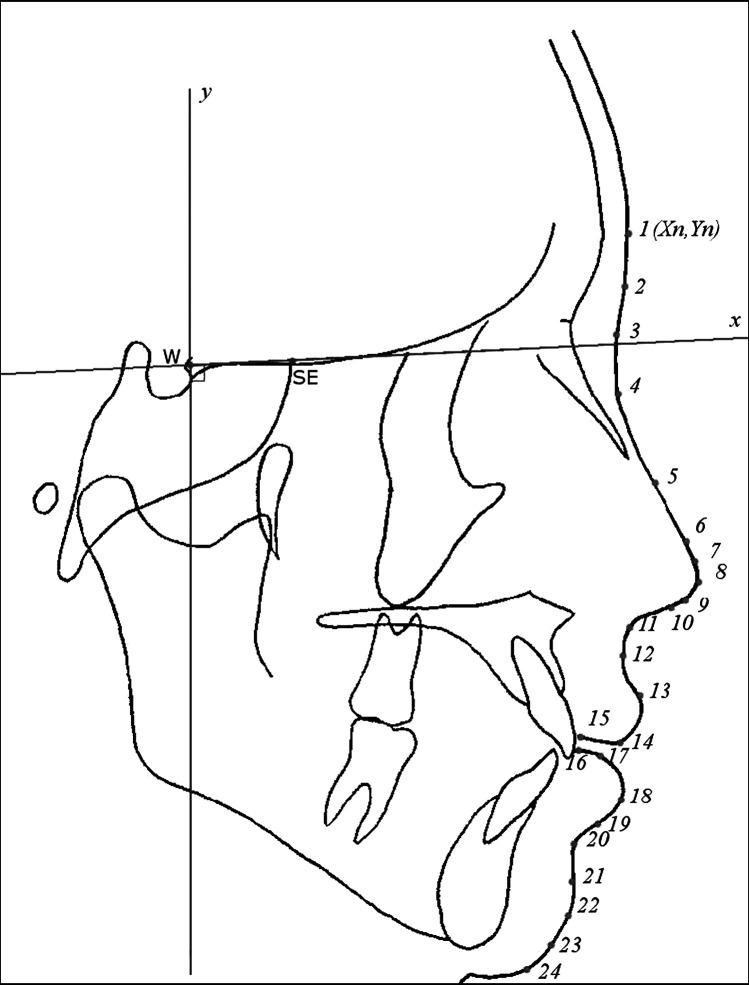
The 24 soft tissue landmarks based on the Rickett's factor analysis, Walker's point [W] (A. Bjõrk) and sphenoethmoidal [SE] are shown and listed in Figure 1. These two landmarks are also known as tuberculum sella (T) and wing (W), respectively.
Figure 1.
Coordinate system applied to lateral cephalogram with 24 soft tissue cephalometric landmarks: 1 = glabella, 2 = halfway between glabella and soft tissue nasion, 3 = soft tissue nasion, 4 = at junction of inferior limit of the concavity overlying the naso-frontal suture, 5 = nasal sorsum (halfway from nasion to pronasale), 6 = at junction of the dorsom and the tip of the nose, 7 = superior nasal tip, 8 = pronasale, 9 = inferior nasal tip, 10 = columella, 11 = subnasale, 12 = superior labial sulcus, 13 = labrale superius, 14 = midway between labre superius and stomion superius, 15 = stomion superius, 16 = stomion inferius, 17 = midway between stomion inferius and labre inferius, 18 = labrale inferius, 19 = midway between labre inferius and labiomental fold, 20 = labiomental fold, 21 = midway between labiomental fold and soft tissue pogonion, 22 = soft tissue pogonion, 23 = soft tissue gnathion, and 24 = soft tissue menton.
The use of the sphenoethmoidal as a stable reference point was recommended by others.17,18 Walker's point was found to be stable after the age of 5.16 Arat and coworkers15 also found that the length of the mid-cranial base (W-SE) remains unchanged in all periods of pubertal growth.
A line was drawn connecting Walker's point [W] and the sphenoethmoidal [SE], creating the x-axis. Thereafter a second line was drawn 90° to the first line creating the y-axis. Walker's point was chosen as origin. The coordinate system is illustrated in Figure 1.
The program Facad (Ilexis AB, Linköping, Sweden) was used to digitally place the landmarks and analyze the cephalograms. After statistical analysis figures were constructed using the mathematical software Graph (IES National Center for Educational Statistics, Washington, DC) to allow for a visual analysis of the data.19
Error of Method
Random error was assessed by statistically analyzing the difference between double measurements taken two weeks apart on 15 cephalograms. Measurements were carried out by the same operator. No systematic difference in locating the landmarks was found between the first and the second measurements of these 15 cephalograms. Random error was calculated using Dahlberg's formula:  to be 0.35 mm or less for the x-axis and 0.58 mm or less for the y-axis.
Because the coordinate system rotates with the head in the anterior-posterior direction, different natural head positions that might occur while taking cephalograms do not affect the measurements. Coordinate system–based cephalometric analysis techniques cannot be traced by hand, unlike the standard method; however, Sayinsu and colleagues20 demonstrated that the use of computer software for cephalometric analysis carried out on scanned images does not increase measurement error compared with hand tracing.
Statistical Method
The raw data were transferred to the statistical program. Evaluation of the data distribution was performed by means of the Shapiro-Wilk test. For the data found to be normally distributed, analysis of changes for men and women from T1 to T2, from T2 to T3, and from T1 to T3 was performed by the paired t-test to test the null hypothesis of zero expected change, versus a two-sided alternative. For the data for which the Shapiro-Wilk test was rejected, that is, for which the distribution was not normal, we used the nonparametric Wilcoxon test. Using the appropriate test, the displacement data were calculated in terms of the mean, median, standard deviation, P value, and lower and higher 95% confidence intervals for each landmark at all three time periods.
RESULTS
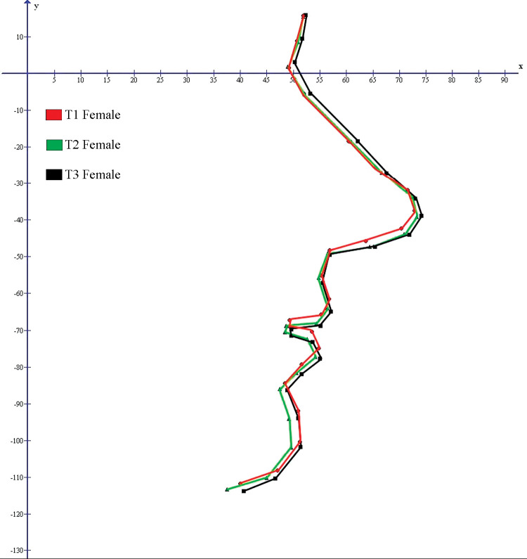
Figure 2 illustrates mean female profiles at T1, T2, and T3. Figure 2 shows the upper facial profile with slight horizontal growth in the area of the frontal sinus from T2 to T3. The dorsum of the nose experiences horizontal growth. The tip of the nose grows outward and downward. The subnasal landmark is fairly stable throughout the whole observational period. The lower facial profile shows downward movement of both lips, especially from T1 to T2. The chin area from T1 to T2 shows a posterior and inferior movement. From T2 to T3 the area shows anterior movement.
Figure 2.
Mean female profile at T1, T2, and T3.
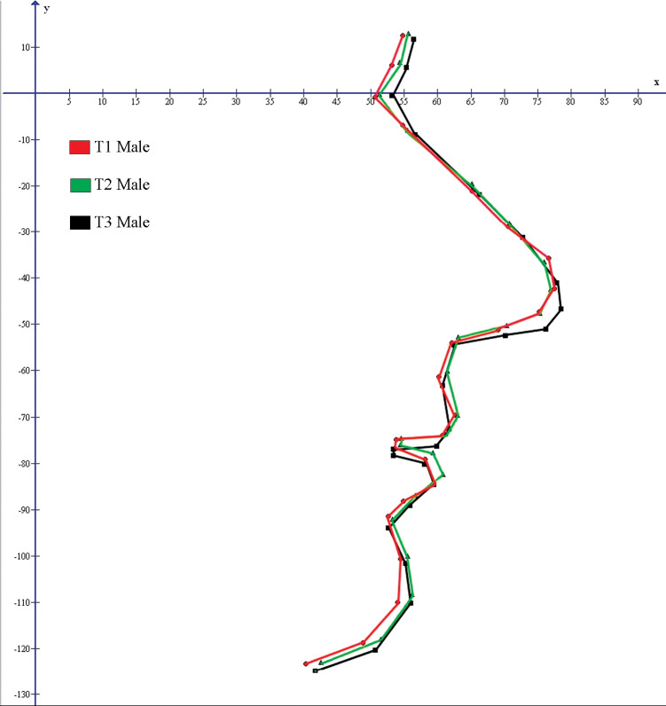
Figure 3 illustrates mean male profiles at T1, T2, and T3. In the upper facial profile, the area of frontal sinus grows evenly through T1 to T3 and is more pronounced than in women. The dorsum of the nose remains stable. The tip grows outward, as in women, but with less of a downward growth. As with women, the subnasal landmark remains fairly stable throughout the observation period. The lower facial profile shows the upper lip flattened out. The chin area grows in an outward and slightly downward direction.
Figure 3.
Mean male profile at T1, T2, and T3.
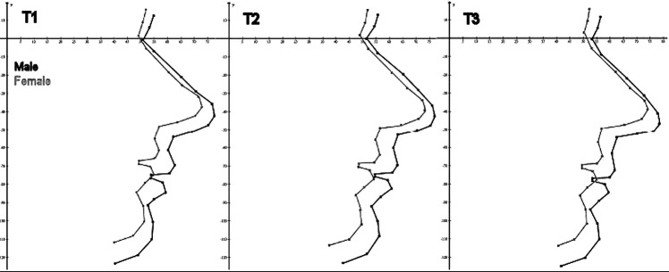
Figure 4 compares the male and female profiles at T1, T2, and T3. This view allows for a visual analysis of how the relation between the mean male and female profiles changes over time. It is, for example, apparent that the male profile is larger than the female profile.
Figure 4.
Male and female profile at T1, T2, and T3.
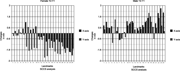
Bar graphs for men and women from the same time periods are side by side for easier comparison. Figure 5 shows all changes that occurred from T1 to T2. In women, the lower facial profile is displaced in a posterior and inferior direction. In men, the whole profile is displaced anteriorly and also slightly superiorly. Greater changes occur in the female profile than the male profile.
Figure 5.
All changes for the male and female profiles, period T2–T1.
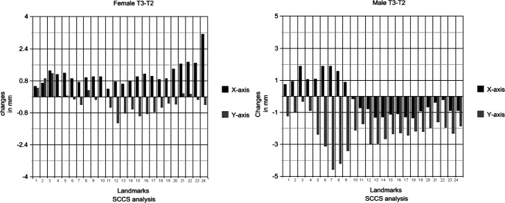
Figure 6 shows all the changes from T2 to T3. Major differences between the sexes are seen. The female profile experiences only minor changes during this period—almost no displacement in the inferior direction and only a minor displacement in the anterior direction (except for soft tissue menton). For men, the situation is different. The upper facial profile is displaced anteriorly, and the lower profile is displaced posteriorly. The whole profile is also displaced in the inferior direction. Greater changes occur in the male profile than the female profile.
Figure 6.
All changes for the male and female profiles, period T3–T2.
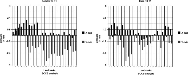
Figure 7 shows all changes that occurred during the whole observation period of 20 years. Even though the male and female profiles aged differently from the second to third and the third to fourth decades of life, Figure 7 shows that in the end the trends are the same. The upper facial profile is displaced in an anterior direction, and the whole profile is displaced inferiorly for both sexes. Bar graphs show that inferior displacement of landmarks along facial profile is greater than anterior displacement.
Figure 7.
All changes for the male and female profiles, period T3–T1.
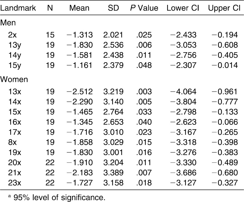
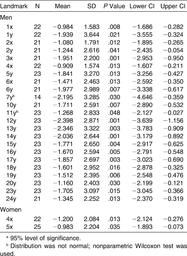
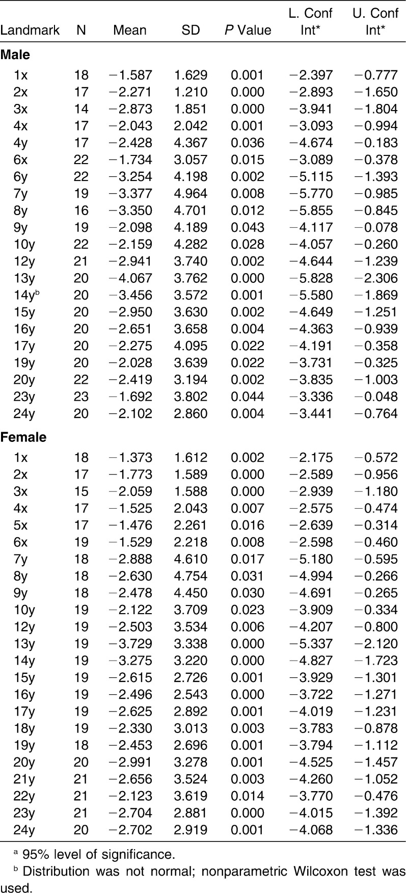
Tables 2 through 4 display all the landmarks for both sexes that experienced statistically significant changes during the respective time periods T2–T1, T3–T2, and T3–T1. The tables indicate the same thing as the bar graphs: female profile experiences greater changes from T1 to T2, and the male profile experiences greater changes from T2 to T3.
Table 2.
All Significant Changes T2–T1 (in millimeters) for the Horizontal (x) and Vertical (y) Directionsa
Table 3.
All Significant Changes T3–T2 (in millimeters) for the Horizontal (x) and Vertical (y) Directionsa
Table 4.
All Significant Changes T3–T1 (in millimeters) for the Horizontal (x) and Vertical (y) Directionsa
DISCUSSION
The results displayed in Tables 2 through 4 clearly indicate that significant changes are occurring in the soft tissue facial profile from the second to fourth decades of life. It can be also seen from the figures and tables that the soft tissue facial profile of women ages more from T1 to T2, whereas the facial profile of men ages more from T2 to T3. The data indicate that aging of the facial profile is not a gradual process; it occurs in spurts and at different periods of life for the two sexes. Even though it can be said that the aging of the facial profile begins 10 years later for men than for women, when the changes do occur, they are of greater magnitude.
Even though the male and female profiles age differently from second to third and from the third to fourth decades of life, Figure 7 shows that in the end the trends are the same. The upper facial profile is displaced in the anterior direction (the nose and frontal sinus area increase in size) and the whole profile is displaced inferiorly for both sexes. The greatest displacement along the facial profile was found to take place in the inferior direction for both men and women. The same fact was noted by Ferrario and colleagues.21
In women the average soft tissue profile of the chin moved in a posterior and inferior direction from the second to third decades of life. This could be partly due to a posterior rotation of mandible, observed by Bondevik.2 From the third to fourth decades, the average soft tissue profile of the chin is displaced anteriorly. This reversal may be due to many factors, including an increased length of the mandible.1 During the observation period the upper lip of the average male profile had visibly flattened. The flattening of lips has been noted in many other studies.2,22 The reason why the same characteristic was not observed in women could be because overjet and overbite decrease significantly only in men.23 The observation that the soft tissue of the chin in men continues to grow in an outward and slightly downward direction has been previously observed.3
Since this coordinate system analysis is based on stable reference points that are not displaced after early childhood, superimposition is not necessary as the coordinates for the landmarks can be compared directly from different time periods (Figures 2–4). This avoids the errors that may occur during superimpositioning. On the other hand, the [W–SE] line that makes up the horizontal axis is relatively short. A measurement error (especially in the vertical direction) on the landmarks W and SE may cause an undesirable rotational error of the coordinate system. The error can be minimized through operator experience and by simultaneously displaying onscreen the cephalograms being analyzed. Any differences then found between the reference landmarks can then be corrected for. It is clear that the error obtained using the standard method during superimposition has been replaced with another kind of error, and it would be interesting to compare the methods.
CONCLUSIONS
The results correspond well with the limited information available concerning the changes of the soft tissue profile from the second to fourth decades of life obtained from previous studies that used the standard method to obtain their results.
The soft tissue profiles of men and women age differently, however many similarities were also found. Even though it can be said that aging of the facial profile begins 10 years later for men than for women, when the changes do occur, they are of greater magnitude.
Aging of the soft tissue profile is not a gradual process; it occurs in spurts. Women experienced greater age-related changes from the second to third decades while men experienced greater age-related changes from the third to fourth decades. The greatest changes in the soft tissue profile for both sexes occurred in the inferior direction.
Acknowledgments
The authors thank Els-Marie Andersson, DDS, for her advice and support; Anita Åsheim, DDS, for her cooperation; Professor Emeritus Olav Bondevik, DDS, PhD, for use of his cephalometric material; Steffen Grønneberg, MS in mathematics, for his help with the statistical analysis, and Professor Ingar Olsen, DDS, PhD, for his advice.
REFERENCES
- 1.Björk A. Variations in the growth pattern of the human mandible: longitudinal radiographic study by the implant method. J Dent Res. 1963;42(pt 2):400–411. doi: 10.1177/00220345630420014701. [DOI] [PubMed] [Google Scholar]
- 2.Ghafari J, Engel F. E, Laster L. L. Cephalometric superimposition on the cranial base: a review and a comparison of four methods. Am J Orthod Dentofacial Orthop. 1987;91:403–413. doi: 10.1016/0889-5406(87)90393-3. [DOI] [PubMed] [Google Scholar]
- 3.Bondevik O. Growth changes in the cranial base and the face: a longitudinal cephalometric study of linear and angular changes in adult Norwegians. Eur J Orthod. 1995;17:525–532. doi: 10.1093/ejo/17.6.525. [DOI] [PubMed] [Google Scholar]
- 4.Sarhan O. A. Sella-nasion line revisited. J Oral Rehabil. 1995;22:905–908. doi: 10.1111/j.1365-2842.1995.tb00239.x. [DOI] [PubMed] [Google Scholar]
- 5.Satoh K, Wada T, Tachimura T, Fukuda J. Velar ascent and morphological factors affecting velopharyngeal function in patients with cleft palate and noncleft controls: a cephalometric study. Int J Oral Maxillofac Surg. 2005;34:122–126. doi: 10.1016/j.ijom.2004.05.002. [DOI] [PubMed] [Google Scholar]
- 6.Satoh K, Wada T, Tachimura T, Sakoda S, Shiba R. A cephalometric study of the relationship between the level of velopharyngeal closure and the palatal plane in patients with repaired cleft palate and controls without clefts. J Craniomaxillofac Surg. 1998;26:394–399. doi: 10.1054/bjom.1999.0187. [DOI] [PubMed] [Google Scholar]
- 7.Alves P. V, Mazucheli J, Vogel C. J, Bolognese A. M. A protocol for cranial base reference in cephalometric studies. J Craniofac Surg. 2008;19:211–215. doi: 10.1097/scs.0b013e31814fb80e. [DOI] [PubMed] [Google Scholar]
- 8.Wen-Ching K. E, Figueroa A. A, Polley J. W. Soft tissue profile changes after maxillary advancement with distraction osteogenesis by use of a rigid external distraction device: a 1-year follow-up. J Oral Maxillofac Surg. 2000;58:959–969; discussion 969–970. doi: 10.1053/joms.2000.8735. [DOI] [PubMed] [Google Scholar]
- 9.Björk A. Facial growth in man; x-ray studies with implanted metal indicators. Tandlaegebladet. 1955;59:55–66. [PubMed] [Google Scholar]
- 10.Björk A. Facial growth in man, studied with the aid of metallic implants. Acta Odontol Scand. 1955;13:9–34. doi: 10.3109/00016355509028170. [DOI] [PubMed] [Google Scholar]
- 11.Björk A. Sutural growth of the upper face studied by the implant method. Acta Odontol Scand. 1966;24:109–127. doi: 10.3109/00016356609026122. [DOI] [PubMed] [Google Scholar]
- 12.Björk A. Growth in width of the maxilla studied by the implant method. Scand J Plast Reconstr Surg. 1974;8:26–33. doi: 10.3109/02844317409084367. [DOI] [PubMed] [Google Scholar]
- 13.Björk A, Skieller V. Growth and development of the maxillary complex. Inf Orthod Kieferorthop. 1984;16:9–52. [PubMed] [Google Scholar]
- 14.Melsen B. The cranial base: the postnatal development of the cranial base studied histologically on human autopsy material. Acta Odont Scan. 1974 Vol 32, Suppl 62. [Google Scholar]
- 15.Arat M, Köklü A, Ozdiler E, Rübendüz M, Erdoğan B. Craniofacial growth and skeletal maturation: a mixed longitudinal study. Eur J Orthod. 2001;23:355–361. doi: 10.1093/ejo/23.4.355. [DOI] [PubMed] [Google Scholar]
- 16.Hahn von Dorsche S, Fanghänel J, Kubein-Meesenburg D, Nägerl H, Hanschke M. Interpretation of the vertical and longitudinal growth of the human skull. Ann Anat. 1999;181:99–103. doi: 10.1016/S0940-9602(99)80103-4. [DOI] [PubMed] [Google Scholar]
- 17.Nelson T. O. Analysis of the facial growth utilizing elements of the cranial base as registrations. Am J Orthod. 1960;46:379. [Google Scholar]
- 18.Athanasiou E. A. Orthodontic Cephalometry. Vol. 109 St Louis, MO: Mosby-Year Book; 1995. [Google Scholar]
- 19.Create a Graph. Available at: http://nces.ed.gov/nceskids/creategraph/default.aspx Accessed December 3, 2010. [Google Scholar]
- 20.Sayinsu K, Isik F, Trakyali G, Arun T. An evaluation of the errors in cephalometric measurements on scanned cephalometric images and conventional tracings. Eur J Orthod. 2007;29:105–108. doi: 10.1093/ejo/cjl065. [DOI] [PubMed] [Google Scholar]
- 21.Ferrario V. F, Sforza C, Serrao G, Ciusa V, Dellavia C. Growth and aging of facial soft tissues: a computerized three-dimensional mesh diagram analysis. Clin Anat. 2003;16:420–433. doi: 10.1002/ca.10154. [DOI] [PubMed] [Google Scholar]
- 22.Formby W. A, Nanda R. S, Currier G. F. Longitudinal changes in the adult facial profile. Am J Orthod Dentofacial Orthop. 1994;105:464–476. doi: 10.1016/S0889-5406(94)70007-9. [DOI] [PubMed] [Google Scholar]
- 23.Bondevik O. Changes in occlusion between 23 and 34 years. Angle Orthod. 1998;68:75–80. doi: 10.1043/0003-3219(1998)068<0075:CIOBAY>2.3.CO;2. [DOI] [PubMed] [Google Scholar]


