Abstract
Although large exciton binding energies of typically 0.6–1.0 eV are observed for monolayer transition metal dichalcogenides (TMDs) owing to strong Coulomb interaction, multilayered TMDs yield relatively low exciton binding energies owing to increased dielectric screening. Recently, the ideal carrier-multiplication threshold energy of twice the bandgap has been realized in multilayered semiconducting 2H-MoTe2 with a conversion efficiency of 99%, which suggests strong Coulomb interaction. However, the origin of strong Coulomb interaction in multilayered 2H-MoTe2, including the exciton binding energy, has not been elucidated to date. In this study, unusually large exciton binding energy is observed through optical spectroscopy conducted on CVD-grown 2H-MoTe2. To extract exciton binding energy, the optical conductivity is fitted using the Lorentz model to describe the exciton peaks and the Tauc–Lorentz model to describe the indirect and direct bandgaps. The exciton binding energy of 4 nm thick multilayered 2H-MoTe2 is approximately 300 meV, which is unusually large by one order of magnitude when compared with other multilayered TMD semiconductors such as 2H-MoS2 or 2H-MoSe2. This finding is interpreted in terms of small exciton radius based on the 2D Rydberg model. The exciton radius of multilayered 2H-MoTe2 resembles that of monolayer 2H-MoTe2, whereas those of multilayered 2H-MoS2 and 2H-MoSe2 are large when compared with monolayer 2H-MoS2 and 2H-MoSe2. From the large exciton binding energy in multilayered 2H-MoTe2, it is expected to realize the future applications such as room-temperature and high-temperature polariton lasing.
Subject terms: Materials science, Physics
Introduction
One of the key features of strong Coulomb interaction in two-dimensional (2D) van der Waals (vdW) layered materials is large exciton binding energy and spin–orbit coupling (SOC) in the monolayer TMDs1,2. Information on optical bandgap and exciton binding energy of a material is a primary concern for various applications in electronic, optical/optoelectronic, and photocatalytic devices3–5. Large exciton binding energy, particularly in a monolayer, can be attributed to neutral excitons as well as multi-excitons, such as trions and biexcitons, at room temperature6–9. Moreover, the large exciton binding energy can be used for room-temperature polariton lasing and heterostructure physics such as interlayer exciton10–12. The SOC in 2D transition metal dichalcogenide (TMD) materials originating from the d-orbitals of transition metals breaks inversion symmetry, splitting the valence band around K-point13,14. TMD materials with broken inversion symmetry are promising for applications applied to spin-valleytronics, in which their spin degree of freedom and valley polarization can be manipulated15. Despite the possibilities of a various applications, it is hard to apply in reality. Because material values such as bandgap, exciton binding energy, and SOC energy of TMD materials are stochastically scattered from material to material, let alone measurement tools.
In monolayer TMDs, the atoms are fully exposed to vacuum and involve strong Coulomb interaction and reduced dielectric screening effect, consequently exhibiting, in general, large exciton binding energies of typically 0.45–1.04 eV16–20, which are distinct by one order of magnitude from the lower exciton binding energies (2.7–40 meV) of 3D semiconducting materials such as Si, Ge, and GaAs21–23. The exciton binding energy strongly relies on materials. For example, monolayer 2H-MoS2 and 2H-WS2 exhibit relatively large exciton binding energies of 640–830 meV19,24, whereas some values are as small as ~ 160 meV in 2H-WS225. Such a large variance can be attributed to substrate effect, charge transfer, strain, and residual adsorbents under ambient conditions. Nevertheless, strong material dependence of exciton binding energy has been reported in the literatures.
The exciton binding energy in multilayered TMDs has not been sufficiently explored. In general, the exciton binding energies in multilayered TMDs vary with the thickness of the material as well as the atomic masses of the constituent atoms. The exciton binding energy decreases with increasing thickness of the material due to increased screening, whereas it decreases when the chalcogen atom is changed from the light S to the heavy Te16. Nevertheless, the exciton binding energies of bulk TMDs are still higher than those of 3D semiconducting materials. For example, the energies in the TMDs in 2H-MoS2 bulk26,27 are as high as 50–84 meV and approximately 150 meV in 2H-MoTe2 bulk28, and as low as 67 meV in 2H-MoSe2 bulk and 50 meV in 2H-WSe2 bulk26. It should be noted that the lower bound of binding energies is still higher than those (3–10 meV) of bulk materials21–23. This implies that the origin of large exciton binding energy in TMD materials does not simply rely on the thickness. The binding energy is further complicated by involving the SOC, dielectric constant and Coulomb interaction of the material.
The exciton binding energy and bandgap can be directly measured by combining optical absorption and/or photoluminescence (PL) with photocurrent spectroscopy or scanning tunneling spectroscopy (STS)29–31. Theoretical calculation of the quasi-particle bandgap may allow to estimate the exciton binding energy. The difficulty arises owing to the uncertainty caused by the two combined experimental systems or inaccurate estimation of bandgap from theory. For example, the measurements of exciton binding energy and bandgap in TMDs vary according to the different measurement systems used, such as PL combined with photocurrent, PL/STS, and PL/density functional theory (DFT)19,29–33. The reflectance/transmittance contrast spectrum is introduced to measure the exciton energy without explicitly measuring the bandgap28,34–37. Therefore, a robust measurement system is required to accurately measure the exciton energy and bandgap.
In this study, we report the thickness- and temperature-dependent optical properties of few-layered 2H-MoTe2 thin films grown by chemical vapor deposition (CVD). Fourier-transform infrared (FTIR) spectroscopy is used to measure the transmittance spectra of semiconducting 2H-MoTe2 thin films with various thicknesses 2 (3 layers), 4 (6 layers), and 10 (14 ~ 15 layers) nm, and their optical conductivities are obtained using a thin film analysis method, which is known as a transfer matrix method38,39. The optical conductivities are analyzed using the symmetric Lorentz and asymmetric Tauc–Lorentz models40–42 to extract information on direct/indirect bandgaps and exciton binding energy. We obtain significantly large exciton binding energy of around 300 meV at the K- and Γ-points in the Brillouin zone in multilayered 2H-MoTe2 compared to those of other TMDs with similar thicknesses. Our results provide detailed optical and electronic information on few-layered 2H-MoTe2 thin films, which can be applied to optoelectronic devices such as solar cells, detector, and polariton lasing.
Result and discussion
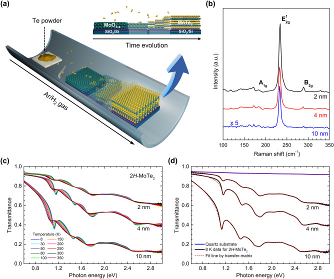
The 2H-MoTe2 films were synthesized using the CVD method with Mo thin films and Te pellets as Mo and Te sources, respectively. Figure 1a shows the schematic of a CVD system along with the growth process (the details are provided in Methods). Three different thicknesses of the samples (2, 4, and 10 nm; Figure S1, Supplementary Information) were characterized using Raman spectroscopy with a laser of wavelength 532 nm. The characteristic Raman active modes of A1g (~ 173 cm−1), E12g (~ 235 cm−1), and B12g (~ 290 cm−1) were clearly observed; these were independent of thickness beyond 2 nm. The B12g mode appeared for thin samples, which is absent in monolayer 2H-MoTe2 (Fig. 1b)43. The intensity of the B12g mode reduces with increasing thickness; it can be used as a measure for the thickness of the 2H-MoTe2 thin film43. In addition, 2H-MoTe2 films studied in this work have been characterized by using Raman mapping, X-ray diffraction (XRD), transmission electron microscopy (TEM) in previous report44. The samples were polycrystalline, and the crystallinity was 10 μm or more.
Figure 1.
Synthesis of 2H-MoTe2 film and measurement transmittance spectra. (a) Schematic of chemical vapor deposition system used to synthesize 2H-MoTe2 film on SiO2/Si substrate and illustration of growth process. The oxidized Mo thin film transformed gradually to the 2H-MoTe2 film as the processing time goes. (b) Thickness-dependent Raman spectra with three different thicknesses (2, 4, and 10 nm). (c) Measured transmittance spectra for 2H-MoTe2 films at various temperatures between 8 and 350 K. (d) Measured transmittance spectra of the three samples and their simulated spectra by using the transfer matrix method at 8 K. The transmittance and fit of quartz substrate are also displayed.
The 2H-MoTe2 films are prepared on a 6 × 6 mm2 quartz substrate for optical investigation, and the diameter of the optical probe beam is approximately 4 mm. The measured transmittance spectra of the three samples are shown in Fig. 1c. The transmittance increases as the 2H-MoTe2 film becomes thinner. The optical conductivities of the 2H-MoTe2 films were extracted from the measured transmittance spectra using a transfer matrix method, which is a rigorous method including multiple reflections in both film and substrate38,39. A more detailed description how we obtained the optical conductivity from the measured transmittance can be found in Supplementary Information. Representative data at 8 K and fits using the transfer matrix are shown in Fig. 1d.
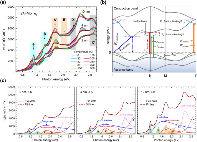
Figure 2a shows the optical conductivities of 2H-MoTe2 thin films at various temperatures and thicknesses 2, 4, and 10 nm. The well-known excitonic absorption peaks have shifted to the low-energy side and broadened with elevating temperature. Each exciton peak is clearly distinguished at low temperature. The optical conductivity in Fig. 2a is directly related to the electronic band structure of the material. The optical conductivity reflects all possible transitions from filled states below the Fermi level to empty states above it and is a function of only energy or frequency, i.e., the momentum averaged quantity over the Brillouin zone. Figure 2b shows the schematic of the band structure with conduction and valence bands in typical multilayered TMDs16,45–47. A direct bandgap appears at the K- (smaller Eg) and Г- (larger Eg) point, and the smallest indirect bandgap appears at the intermediate 0.52 Г-K point16. We note that the indirect transition can be possible by other processes which hold the momentum conservation law such as phonon-assisted process48. One prominent feature in TMD materials is the existence of the large SOC energy, giving rise to the splitting of energy bands, which is distinct from the case of small SOC in 3D materials16,26,46. As the SOC energy at the conduction band edge is relatively low compared to that at the valence band edge, the SOC energy at the conduction band edge is typically ignored in TMD materials. Exciton peaks are assigned as A and B near the K-point and Aʹ and Bʹ near the Г-point. It should be noted that the exciton absorption bands are symmetric, in contrast with asymmetric bandgap absorptions.
Figure 2.
Analysis of real part of optical conductivity and optical designation in band structure of TMDs. (a) Real part of optical conductivities for 2, 4, and 10 nm thick 2H-MoTe2 at various temperatures between 8 and 350 K. (b) Schematic of band structure of multilayered TMDs with optical designations. (c) Real part of optical conductivities fitted using Lorentz and TL models at 8 K.
In this study, we fully analyze symmetric and asymmetric absorption band structures in the optical conductivity using two models: Lorentz and Tauc–Lorentz (TL) models. The Lorentz model can be used to describe symmetric exciton absorptions (electron–hole pairs), whereas the TL model can be used to describe asymmetric direct and indirect bandgap absorptions40–42. The real part of optical conductivity can be described using the Lorentz and TL models as , where , , and are the oscillator strength, resonance frequency, and damping parameter of the th Lorentz component, respectively and , , and are the resonance frequency, bandgap (or absorption edge), oscillator strength, and damping parameter of the th Tauc–Lorentz component, respectively, and is the Heaviside step function. Here, the Drude component is not included since the systems are undoped semiconductors. For our multilayered TMD materials, four symmetric Lorentz components (A, B, A’, and B’) and three asymmetric TL components (indirect and direct bandgaps at the K- and Г-points) are included to fit the optical conductivity36,49,50. It is worth noting that the Г-point direct bandgap has been assigned as C mode in previous literature36,49.
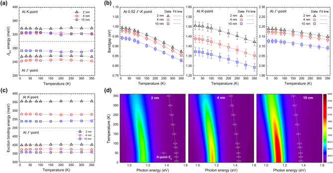
The observed conductivity with 2, 4, and 10 nm thick 2H-MoTe2 at 8 K is deconvoluted using the aforementioned Lorentz and TL components (Fig. 2c) (see Supplementary Information Figures S2, S3, and S4 for additional fittings at various temperatures between 8 and 350 K). The peak positions ( of four Lorentz components are extracted directly from the conductivity spectra with approximate spectral widths (). The optical conductivity is deconvoluted using additional asymmetric components (two direct bandgaps at the K- and Г-points and an indirect bandgap at 0.52 Г-K point); which is well fitted below 2 eV (see Supplementary Information Figure S5). These absorption modes including the prominent exciton modes are based on the calculated electronic band structure shown in Fig. 2b. Beyond 2 eV, the fitting is further improved by adding two unknown Lorentz components (X and X’). We note that X and X’ are not new; they were observed and denoted as ϕ and D in previous literature49. As far as we know, these two modes have not been clearly identified yet. It is also worth noting that we needed an additional weak Lorentz mode between A and B excitons. We think that this additional mode can be assigned as either the 2 s-like excited state of the exciton A or the ground state interlayer exciton, which were observed in a previous report28. The additional mode shows temperature-dependence; it becomes weaker as temperature increases (see Figures S2, S3 and S4 in the Supplementary Information). We show the temperature-and thickness-dependent width and position of A and B excitons of 2H-MoTe2 in Supplementary Information (see Supplementary Information Figure S6). The temperature-dependent positions of A and B excitons of MoTe2 are similar to reported results of WS2 and WSe224. The additional mode also shows thickness-dependence; it becomes stronger as the thickness increases (see Supplementary Information Figure S7). We also took the second derivative of the measured transmittance spectra to check any small features related to excitation states near the excitons and found a feature just above the peak energy of the exciton A, which might be related to the 2 s state. However, we could not see any higher excited states (see Supplementary Information Figure S7). The extracted spin–orbit splitting band energy (ΔSO), bandgap, and exciton binding energy are summarized in Fig. 3.
Figure 3.
Fitting parameters extracted using Lorentz and TL models. (a) ΔSO at K- and Γ-point for 2, 4, and 10 nm thick 2H-MoTe2 at various temperatures between 8 and 350 K. (b) Temperature- and thickness-dependent indirect and direct bandgaps of 2H-MoTe2 obtained using Lorentz and TL models. The fitting lines are calculated using temperature-dependent bandgap equation. (c) Exciton binding energy at K- and Γ-point for 2, 4, and 10 nm thick 2H-MoTe2 at various temperatures between 8 and 350 K. (d) Color maps () of the A exciton and the direct bandgap edge at K-point as functions of photon energy and temperature.
Figure 3a shows the thickness- and temperature-dependent ΔSO at the K- and Γ-point of 2H-MoTe2 with thicknesses of 2, 4, and 10 nm. The value of ΔSO at the K-point is 335.4 meV for the 2 nm sample at 8 K; it is mostly independent of the temperature range of 8–350 K and slightly lower for thicker samples. The value of ΔSO at the Γ-point for the 2 nm sample at 8 K is 262.9 meV, lower than that at the K-point, although its dependence on temperature and thickness are not evidently distinct. The bandgap decreases with temperature (Fig. 3b), similar to 3D bulk semiconductors. The indirect bandgap for the 2 nm 2H-MoTe2 at low temperature (8 K) is 0.96 eV; it reduces further to 0.87 eV at 10 nm, similar to that (0.88 eV) for bulk 2H-MoTe251. The dashed lines are fitting lines based on the temperature-dependent bandgap equation, , where is the bandgap at = 0, and are fitting parameters. It can be seen in Table 1 that the values of and are lower than those of indirect bulk materials such as Si and Ge. Fitting parameters of and decrease as increasing the thickness for the indirect bandgap. In contrast, the fitting parameter increases when the thickness increases for the direct (K-point) bandgap. Otherwise, fitting parameters do not follow the trends of the indirect and direct (K-point) bandgaps for the direct (Γ-point) bandgap. Furthermore, the extent of change is not proportional to the thickness. For the 2 nm sample, a relatively large direct bandgap of 1.5 eV appears at 8 K at the K-point, reducing to 1.39 eV for the 10 nm sample. The bandgaps are further reduced with temperature, similar to the indirect bandgaps. The variance in the direct bandgap at the Γ-point with thickness and similar fitting parameters is nearly negligible.
Table 1.
Parameters of temperature-dependent bandgap.
| Si | Ge | GaAs | 2H-MoTe2 | |||||||||
|---|---|---|---|---|---|---|---|---|---|---|---|---|
| Indirect | Direct (K-point) | Direct (Γ-point) | ||||||||||
| 2 nm | 4 nm | 10 nm | 2 nm | 4 nm | 10 nm | 2 nm | 4 nm | 10 nm | ||||
| Eg (0) [eV] | 1.16656 | 0.74456 | 1.51956 | 0.993 | 0.980 | 0.943 | 1.505 | 1.438 | 1.373 | 2.188 | 2.174 | 2.126 |
| α [10–4 eV/K] | 4.7356 | 4.7756 | 5.4156 | 3.70 | 3.50 | 2.70 | 3.40 | 3.50 | 3.60 | 3.80 | 3.90 | 4.50 |
| β [K] | 63656 | 23556 | 20456 | 145 | 125 | 90.5 | 110 | 220 | 290 | 740 | 540 | 400 |
The exciton binding energy at the K-point for the 2-nm-thick 2H-MoTe2 at 8 K is 355 meV; it reduces to 238 meV for the 10-nm-thick 2H-MoTe2 at the same temperature. The variance in temperature is nearly negligible. The exciton binding energy at the K-point is markedly large compared to previous reports28,52. Supplementary Information Tables S1 and S2 illustrate that the exciton binding energies of most TMD materials, such as 2H-MoS2, 2H-MoSe2, and 2H-WSe2, are in the range of 40–84 meV, one order of magnitude lower than that of the thin 2H-MoTe2 film. The exciton binding energy at the Γ-point is approximately 370 meV, almost independent of the sample thickness. Color maps () of the A exciton intensities as functions of photon energy and temperature for various thicknesses are represented in Fig. 3d. Here, we clearly observe almost temperature-independent exciton binding energies for all three 2H-MoTe2 film samples (see Fig. 3c) since both the peak position of exciton A and the direct bandgap edge at K- point show almost the same temperature-dependence (see Fig. 3d). The error bars in the bandgap edge curves are the confidence levels of the bandgap edge.
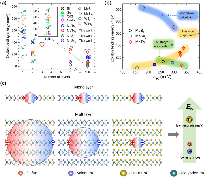
Thus far, we have learned from the absorption spectra that the exciton binding energy of multilayered 2H-MoTe2 is approximately 238–355 meV, which is unusually large, contrary to general belief, compared to the values of 24.8–79.4 meV obtained for multilayered 2H-MoS2 and 2H-MoSe2. Figure 4a summarizes the layer-dependent exciton binding energies of other TMDs, including 3D semiconductors, described in the literature. The reported experimental exciton binding energies of monolayer TMD materials are quite high and widely distributed, whereas those of thin 2D TMDs differ fundamentally from those of 3D semiconductors. The strong dielectric screening in 3D semiconductors allows the wave function to extend over a few to several nanometers, whereas the poor dielectric screening in 2D TMDs causes a significant increase in the exciton binding energy. The exciton binding energy is also closely related to SOC (Fig. 4b). The density-functional calculations for monolayer TMDs illustrate that the exciton binding energy reduces at high values of ΔSO—it reduces 710 meV for 2H-MoTe2, smaller than those for 2H-MoS2 and 2H-MoSe2; the value of ΔSO for 2H-MoTe2 is approximately 300 meV, which is larger than for others16. A similar trend is theoretically observed in multilayered TMDs. In contrast, experimentally, the situation for multilayered TMDs is different. We note that the large discrepancy between the experimental and calculated ΔSO dependent-exciton binding energies of 2H-MoTe2 is not known yet. The exciton binding energy of approximately 300 meV and ΔSO of approximately 325 meV in multilayered 2H-MoTe2 are unusually large, whereas relatively low values are observed in multilayered 2H-MoS2 and 2H-MoSe2. It is also worth noting that we applied the same approach for analyzing the measured spectra, which were obtained the same experimental technique. Therefore, the relative differences between TMD materials were quite reliable.
Figure 4.
Unusually large exciton binding energy in multilayered 2H-MoTe2. (a) Comparison of exciton binding energy with diverse semiconducting materials as function of thickness. Exciton binding energies were taken from previous reports 7,14,18,19,21–28,30,31,52,55. (b) Comparing exciton binding energy of MoX2 as a function of ΔSO. Calculated exciton binding energies of monolayer and multilayered MoX2 were taken from previous reports 16,52. (c) Schematics of possible scenarios for the exciton radii in screening effect and large exciton binding energy in multilayered 2H-MoTe2.
To rationalize the origin of the unusually large exciton binding energy in multilayered 2H-MoTe2, we introduce the Wannier–Mott exciton to examine the radii of excitons. For two-dimensional materials, the binding energy of a Wannier exciton can be written as , where is the Rydberg constant (13.6 eV), is the static relative dielectric constant, and is the reduced mass of the electron and hole; and are the effective masses of the electron and hole, respectively53. In terms of exciton radius, , where is the Bohr radius ()54, the exciton binding energy can also be written as .
The exciton radii of monolayer TMDs obtained from the hydrogenic Rydberg model are presented in Supplementary Information Table S3. These are smaller than 10 Å, indicating strong exciton confinement within the layer. Together with a relatively low dielectric constant, monolayer TMDs are expected to have high exciton binding energies owing to strong Coulomb interaction or reduced dielectric screening. The discrepancy in the exciton binding energies among different monolayers is reflected by the dielectric constant and exciton radius. The exciton radii of multilayered TMDs are summarized in Table 2. Further, we provide the real parts of optical conductivities and fits for 4 nm thick 2H-MoS2 and 2H-MoSe2 (Supplementary Information Figure S8) as well as the dielectric functions for multilayered 2H-MoX2 at 8 K (Supplementary Information Figure S9). We note that exciton radius of multilayered 2H-MoTe2 remains small with 7 Å, similar to that of monolayer 2H-MoTe2 (Supplementary Information Table S3). Therefore, multilayered 2H-MoTe2 is expected to have large exciton binding energy, which is slightly reduced comparing to that of monolayer 2H-MoTe2 owing to its high dielectric constant (Fig. 4c). In contrast, the exciton radii of multilayered 2H-MoS2 and 2H-MoSe2 are approximately 112.55 and 24.74 Å, respectively. In these cases, the Coulomb interaction is largely screened off by the adjacent layers, leading to smaller exciton binding energies by one order of magnitude when compared with 2H-MoTe2. Further, we expect that ΔSO should be also correlated with the exciton binding energy. As shown in Fig. 4b, ΔSO of multilayered 2H-MoTe2 is approximately 330 meV, larger than 220 and 150 meV for multi-layered 2H-MoSe2 and 2H-MoS2, respectively. This high spin–orbit splitting energy of 2H-MoTe2 is expected to affect the exciton binding energy. For TMDs, ΔSO strongly relies on the mass of the chalcogen atoms. Heavy atoms lead to large value of ΔSO.
Table 2.
Static dielectric constant, exciton binding energy, and exciton radius of multilayered 2H-MoX2 at 8 K.
| 4 nm MoS2 | 4 nm MoSe2 | 4 nm MoTe2 | 2 nm MoTe2 | 10 nm MoTe2 | |
|---|---|---|---|---|---|
| εr | 10.31 | 14.65 | 16.73 | 15.05 | 21.54 |
| Eb [meV] | 24.8 | 79.4 | 280 | 355 | 238 |
| re [Å] | 112.55 | 24.74 | 6.143 | 5.386 | 5.613 |
Conclusion
In conclusion, we measured the temperature- and thickness-dependent transmittance spectra of CVD-grown 2H-MoTe2. The transmittance spectra were analyzed using the transfer-matrix method to obtain the optical constants. To determine the exciton binding energy in multilayered 2H-MoTe2, the optical conductivity was fitted by using the Lorentz model to describe the exciton peaks and Tauc–Lorentz model to describe the indirect and direct bandgaps. The exciton binding energy of 4 nm-thick multilayered 2H-MoTe2 was approximately 280 meV, which varies slightly with thickness. This is unusually large, by one order of magnitude, compared to other multilayered TMD semiconductors such as 2H-MoS2 or 2H-MoSe2. This can be explained by the small exciton radius, based on the 2D Rydberg model. The exciton radius of multilayered 2H-MoTe2 is similar to that of monolayer 2H-MoTe2, whereas the exciton radii of multilayered 2H-MoS2 and 2H-MoSe2 are larger, by one order of magnitude, compared to those of monolayer 2H-MoS2 and 2H-MoSe2. Consequently, multilayered 2H-MoTe2 exhibits intralayer Coulomb interaction, in contrast with the strong interlayer screening exhibited by 2H-MoS2 or 2H-MoSe2. Therefore, we anticipate that multilayered 2H-MoTe2 can be used not only for fundamental studies on many-body effects and spin/valley Hall effects but also for exciton–polariton condensates and possible applications such as room-temperature or high-temperature polariton lasing due to large exciton binding energy which stabilizes the excitons at 300 K or above 300 K10–12.
Methods
Synthesis of 2H-MoTe2 films
To synthesize semiconducting 2H-MoTe2 thin films, 0.5, 1.3, and 4 nm Mo thin films were deposited on a 300-nm-thick SiO2/Si substrate using a sputter system with a high basal pressure of approximately 10–9 Torr. The prepared Mo substrate was mounted on a two-zone CVD system with Te source. A ceramic boat with a tellurium pellet of 0.5 g (Sigma-Aldrich) was placed in the first furnace zone (zone 1) and the Mo deposited on the SiO2/Si substrate was placed in the second furnace zone (zone 2) to separately control the temperatures of zones 1 and 2. After purging 1000 sccm of Ar gas for 30 min, zones 1 and 2 were heated up simultaneously with the ramping rate of zone 1 first reaching 495 °C in 11 min and then zone 2 reaching 650 °C in 15 min. During the growth process, the Ar and hydrogen (H2) gases were introduced at a flow rate of 50 and 4 sccm, respectively, to reduce the native oxide on the surface of Mo. When the temperature of zone 2 reached 650 °C, growth was allowed for 6 h. After growth, zone 1 was cooled down by opening the chamber. Thereafter, zone 2 was opened for 30 s, followed by cooling down zone 1. During the cooling process, 500 and 4 sccm of Ar and H2 gases, respectively, were flown to remove the reactants. Consequently, 2, 4, and 10 nm thick 2H-MoTe2 films were successfully synthesized from 0.5, 1.3, and 4 nm Mo thin films, respectively.
Transfer of 2H-MoTe2 onto the quartz substrate
To study the electronic properties of 2H-MoTe2 using optical spectroscopy, the 2H-MoTe2 films on SiO2/Si substrate were transferred onto a quartz substrate using the conventional polymethylmethacrylate (PMMA) method. Further details can be found in our previous report44.
Optical measurement (FTIR)
FTIR spectroscopy is an experimental technique used to obtain the absorbance spectra in a wide infrared range from far-infrared to visible. To measure the transmittance spectra of our samples, we used a commercial FTIR spectrometer (Bruker Vertex 80v). As Vertex 80v is a vacuum-type spectrometer, the measured power spectrum is free from moisture and air absorptions. The spectrometer consists of three major components: a tungsten lamp source, a CaF2 beam splitter, and an InGaAs detector for the near-infrared range (4000–12,000 cm−1 or 0.5–1.5 eV) and an Si-diode detector for the visible range (8000–25,000 cm−1 or 1.0–3.0 eV). We used a commercial cold finger-type Advanced Research Systems (ARS) optical cryostat and a commercial temperature controller (Lakeshore 325) to control the sample temperature, both below and above room temperature. We used liquid helium as a coolant and were able to control the sample temperature in the range 8–350 K.
Fitting strategy of optical conductivity with Lorentz and Tauc-Lorentz models
We first fitted the Lorentzian exciton modes and then fitted the indirect and direct bandgap absorptions with Tauc-Lorentzian modes by adjusting the intensities of the exciton modes. The peak positions and widths of the exciton modes are unambiguously determined. We constrained that the amplitudes of the exciton modes with the same origin were almost the same. We also constrained that the maximum of the indirect bandgap absorption was located in between two peaks of the A exciton and the direct transition at K-point. Additionally, the peak near 1.8 eV consists of two components: one is the peak of the exciton A’ and the other is associated with the maximum of the direct transition at K-point. Once we got a good fit for a spectrum at 8 K then we could get reliable fits for spectra at other temperatures by a systematic adjustment of the fitting parameters. For more in detailed description, refer to Supplementary Information. It is worth noting that the absorption modes including the exciton modes were based on the calculated electronic band structure. Particularly, near the bandgap, we used a minimal number of modes: The four exciton modes and the indirect and direct bandgaps.
Supplementary Information
Acknowledgements
This work was supported by the Institute for Basic Science of Korea (IBS-R011-D1). J.H. acknowledges financial support from the National Research Foundation of Korea (NRF-2017R1A2B4007387 and 2021R1A2C101109811).
Author contributions
These authors contributed equally. E.J. and J.C.P. are equally contributed and developed the work. J.-H.K., J. H. and Y.H.L. guided the manuscript. All the authors discussed the results and commented on the manuscript.
Competing interests
The authors declare no competing interests.
Footnotes
Publisher's note
Springer Nature remains neutral with regard to jurisdictional claims in published maps and institutional affiliations.
These authors contributed equally: Eilho Jung and Jin Cheol Park
Contributor Information
Ji-Hee Kim, Email: kimj@skku.edu.
Jungseek Hwang, Email: jungseek@skku.edu.
Young Hee Lee, Email: leeyoung@skku.edu.
Supplementary Information
The online version contains supplementary material available at 10.1038/s41598-022-08692-1.
References
- 1.Mueller T, Malic E. Exciton physics and device application of two-dimensional transition metal dichalcogenide semiconductors. NPJ 2D Mater. Appl. 2018;2:29. doi: 10.1038/s41699-018-0074-2. [DOI] [Google Scholar]
- 2.Qiu DY, da Jornada FH, Louie SG. Optical spectrum of MoS2: Many-body effects and diversity of exciton states. Phys. Rev. Lett. 2013;111:216805. doi: 10.1103/PhysRevLett.111.216805. [DOI] [PubMed] [Google Scholar]
- 3.Butler SZ, et al. Progress, challenges, and opportunities in two-dimensional materials beyond graphene. ACS Nano. 2013;7:2898–2926. doi: 10.1021/nn400280c. [DOI] [PubMed] [Google Scholar]
- 4.Duong DL, Yun SJ, van der Lee YH. Waals layered materials: Opportunities and challenges. ACS Nano. 2017;11:11803–11830. doi: 10.1021/acsnano.7b07436. [DOI] [PubMed] [Google Scholar]
- 5.Zong X, et al. Enhancement of photocatalytic H2 evolution on CdS by loading MoS2 as Cocatalyst under visible light irradiation. J. Am. Chem. Soc. 2008;130:7176–7177. doi: 10.1021/ja8007825. [DOI] [PubMed] [Google Scholar]
- 6.Jones AM, et al. Spin–layer locking effects in optical orientation of exciton spin in bilayer WSe2. Nat. Phys. 2014;10:130–134. doi: 10.1038/nphys2848. [DOI] [Google Scholar]
- 7.Yang J, et al. Robust excitons and trions in monolayer MoTe2. ACS Nano. 2015;9:6603–6609. doi: 10.1021/acsnano.5b02665. [DOI] [PubMed] [Google Scholar]
- 8.Szyniszewski M, Mostaani E, Drummond ND, Fal'ko VI. Binding energies of trions and biexcitons in two-dimensional semiconductors from diffusion quantum Monte Carlo calculations. Phys. Rev. B. 2017;95:081301. doi: 10.1103/PhysRevB.95.081301. [DOI] [Google Scholar]
- 9.Rigosi AF, Hill HM, Rim KT, Flynn GW, Heinz TF. Electronic band gaps and exciton binding energies in monolayer MoxW1−xS2 transition metal dichalcogenide alloys probed by scanning tunneling and optical spectroscopy. Phys. Rev. B. 2016 doi: 10.1103/PhysRevB.94.075440. [DOI] [Google Scholar]
- 10.Liu X, et al. Strong light–matter coupling in two-dimensional atomic crystals. Nat. Photonics. 2014;9:30–34. doi: 10.1038/nphoton.2014.304. [DOI] [Google Scholar]
- 11.Dufferwiel S, et al. Exciton-polaritons in van der Waals heterostructures embedded in tunable microcavities. Nat. Commun. 2015;6:8579. doi: 10.1038/ncomms9579. [DOI] [PMC free article] [PubMed] [Google Scholar]
- 12.Fraser MD, Hofling S, Yamamoto Y. Physics and applications of exciton-polariton lasers. Nat. Mater. 2016;15:1049–1052. doi: 10.1038/nmat4762. [DOI] [PubMed] [Google Scholar]
- 13.Zhu ZY, Cheng YC, Schwingenschlögl U. Giant spin-orbit-induced spin splitting in two-dimensional transition-metal dichalcogenide semiconductors. Phys. Rev. B. 2011;84:153402. doi: 10.1103/PhysRevB.84.153402. [DOI] [Google Scholar]
- 14.Mitioglu AA, et al. Optical investigation of monolayer and bulk tungsten diselenide (WSe2) in high magnetic fields. Nano Lett. 2015;15:4387–4392. doi: 10.1021/acs.nanolett.5b00626. [DOI] [PubMed] [Google Scholar]
- 15.Mai C, et al. Many-body effects in valleytronics: Direct measurement of valley lifetimes in single-layer MoS2. Nano Lett. 2014;14:202–206. doi: 10.1021/nl403742j. [DOI] [PubMed] [Google Scholar]
- 16.Ramasubramaniam A. Large excitonic effects in monolayers of molybdenum and tungsten dichalcogenides. Phys. Rev. B. 2012;86:115409. doi: 10.1103/PhysRevB.86.115409. [DOI] [Google Scholar]
- 17.Berkelbach TC, Hybertsen MS, Reichman DR. Theory of neutral and charged excitons in monolayer transition metal dichalcogenides. Phys. Rev. B. 2013;88:045318. doi: 10.1103/physrevb.88.045318. [DOI] [Google Scholar]
- 18.Wang G, et al. Exciton states in monolayer MoSe2: Impact on interband transitions. 2D Mater. 2015;2:045005. doi: 10.1088/2053-1583/2/4/045005. [DOI] [Google Scholar]
- 19.Hill HM, et al. Observation of excitonic rydberg states in monolayer MoS2 and WS2 by photoluminescence excitation spectroscopy. Nano Lett. 2015;15:2992–2997. doi: 10.1021/nl504868p. [DOI] [PubMed] [Google Scholar]
- 20.Bernardi M, Ataca C, Palummo M, Grossman JC. Optical and electronic properties of two-dimensional layered materials. Nanophotonics. 2017;6:479–493. doi: 10.1515/nanoph-2015-0030. [DOI] [Google Scholar]
- 21.Macfarlane GG, McLean TP, Quarrington JE, Roberts V. Exciton and phonon effects in the absorption spectra of germanium and silicon. J. Phys. Chem. Solids. 1959;8:388–392. doi: 10.1016/0022-3697(59)90372-5. [DOI] [Google Scholar]
- 22.Yu Y-M, et al. Band gap energy and exciton peak of cubic CdS/GaAs epilayers. J. Appl. Phys. 2002;92:1162–1164. doi: 10.1063/1.1486026. [DOI] [Google Scholar]
- 23.Sturge MD. Optical absorption of gallium arsenide between 0.6 and 2.75 eV. Phys. Rev. 1962;127:768–773. doi: 10.1103/PhysRev.127.768. [DOI] [Google Scholar]
- 24.Hanbicki AT, Currie M, Kioseoglou G, Friedman AL, Jonker BT. Measurement of high exciton binding energy in the monolayer transition-metal dichalcogenides WS2 and WSe2. Solid State Commun. 2015;203:16–20. doi: 10.1016/j.ssc.2014.11.005. [DOI] [Google Scholar]
- 25.Jo S, Ubrig N, Berger H, Kuzmenko AB, Morpurgo AF. Mono- and bilayer WS2 light-emitting transistors. Nano Lett. 2014;14:2019–2025. doi: 10.1021/nl500171v. [DOI] [PubMed] [Google Scholar]
- 26.Beal AR, Knights JC, Liang WY. Transmission spectra of some transition metal dichalcogenides, II: Group VIA: trigonal prismatic coordination. J. Phys. C Solid State Phys. 1972;5:3540–3551. doi: 10.1088/0022-3719/5/24/016. [DOI] [Google Scholar]
- 27.Saigal N, Sugunakar V, Ghosh S. Exciton binding energy in bulk MoS2: A reassessment. Appl. Phys. Lett. 2016;108:132105. doi: 10.1063/1.4945047. [DOI] [Google Scholar]
- 28.Arora A, et al. Interlayer excitons in a bulk van der Waals semiconductor. Nat. Commun. 2017;8:639. doi: 10.1038/s41467-017-00691-5. [DOI] [PMC free article] [PubMed] [Google Scholar]
- 29.Ugeda MM, et al. Giant bandgap renormalization and excitonic effects in a monolayer transition metal dichalcogenide semiconductor. Nat. Mater. 2014;13:1091–1095. doi: 10.1038/nmat4061. [DOI] [PubMed] [Google Scholar]
- 30.Klots AR, et al. Probing excitonic states in suspended two-dimensional semiconductors by photocurrent spectroscopy. Sci. Rep. 2014;4:6608. doi: 10.1038/srep06608. [DOI] [PMC free article] [PubMed] [Google Scholar]
- 31.Liu HJ, et al. Molecular-beam epitaxy of monolayer and bilayer WSe2: A scanning tunneling microscopy/spectroscopy study and deduction of exciton binding energy. 2D Mater. 2015;2:034004. doi: 10.1088/2053-1583/2/3/034004. [DOI] [Google Scholar]
- 32.Mak KF, Lee C, Hone J, Shan J, Heinz TF. Atomically thin MoS2: A new direct-gap semiconductor. Phys. Rev. Lett. 2010;105:136805. doi: 10.1103/PhysRevLett.105.136805. [DOI] [PubMed] [Google Scholar]
- 33.Tongay S, et al. Thermally driven crossover from indirect toward direct bandgap in 2D semiconductors: MoSe2 versus MoS2. Nano Lett. 2012;12:5576–5580. doi: 10.1021/nl302584w. [DOI] [PubMed] [Google Scholar]
- 34.Hsu C, et al. Thickness-dependent refractive index of 1L, 2L, and 3L MoS2, MoSe2, WS2, and WSe2. Adv. Op. Mater. 2019;7:1900239. doi: 10.1002/adom.201900239. [DOI] [Google Scholar]
- 35.Rigosi AF, Hill HM, Li Y, Chernikov A, Heinz TF. Probing interlayer interactions in transition metal dichalcogenide heterostructures by optical spectroscopy: MoS2/WS2 and MoSe2/WSe2. Nano Lett. 2015;15:5033–5038. doi: 10.1021/acs.nanolett.5b01055. [DOI] [PubMed] [Google Scholar]
- 36.Ruppert C, Aslan OB, Heinz TF. Optical properties and band gap of single- and few-layer MoTe2 crystals. Nano Lett. 2014;14:6231–6236. doi: 10.1021/nl502557g. [DOI] [PubMed] [Google Scholar]
- 37.Chernikov A, et al. Exciton binding energy and nonhydrogenic Rydberg series in monolayer WS2. Phys. Rev. Lett. 2014;113:076802. doi: 10.1103/PhysRevLett.113.076802. [DOI] [PubMed] [Google Scholar]
- 38.Hwang J, Tanner DB, Schwendeman I, Reynolds JR. Optical properties of nondegenerate ground-state polymers: Three dioxythiophene-based conjugated polymers. Phys. Rev. B. 2003 doi: 10.1103/PhysRevB.67.115205. [DOI] [Google Scholar]
- 39.Jo J, Jung E, Park JC, Hwang J. Comparative study of optical analysis methods for thin films. Curr. Appl. Phys. 2020;20:237–243. doi: 10.1016/j.cap.2019.11.010. [DOI] [Google Scholar]
- 40.Jellison GE, Modine FA. Parameterization of the optical functions of amorphous materials in the interband region. Appl. Phys. Lett. 1996;69:371–373. doi: 10.1063/1.118064. [DOI] [Google Scholar]
- 41.Tauc J, Grigorovici R, Vancu A. Optical properties and electronic structure of amorphous germanium. Phys. Status Solidi B. 1966;15:627–637. doi: 10.1002/pssb.19660150224. [DOI] [Google Scholar]
- 42.Galca AC, Socol G, Craciun V. Optical properties of amorphous-like indium zinc oxide and indium gallium zinc oxide thin films. Thin Solid Films. 2012;520:4722–4725. doi: 10.1016/j.tsf.2011.10.194. [DOI] [Google Scholar]
- 43.Yamamoto M, et al. Strong enhancement of Raman scattering from a bulk-inactive vibrational mode in few-layer MoTe2. ACS Nano. 2014;8:3895–3903. doi: 10.1021/nn5007607. [DOI] [PubMed] [Google Scholar]
- 44.Park JC, et al. Phase-engineered synthesis of centimeter-scale 1T'- and 2H-molybdenum ditelluride thin films. ACS Nano. 2015;9:6548–6554. doi: 10.1021/acsnano.5b02511. [DOI] [PubMed] [Google Scholar]
- 45.Kumar A, Ahluwalia PK. Electronic structure of transition metal dichalcogenides monolayers 1H-MX2 (M = Mo, W; X = S, Se, Te) from ab-initio theory: new direct band gap semiconductors. Eur. Phys. J. B. 2012;85:186. doi: 10.1140/epjb/e2012-30070-x. [DOI] [Google Scholar]
- 46.Böker T, et al. Band structure of MoS2, MoSe2, and α−MoTe2: Angle-resolved photoelectron spectroscopy and ab initiocalculations. Phys. Rev. B. 2001;64:235305. doi: 10.1103/PhysRevB.64.235305. [DOI] [Google Scholar]
- 47.Kumar A, Ahluwalia PK. Semiconductor to metal transition in bilayer transition metals dichalcogenides MX2(M= Mo, W; X= S, Se, Te) Modell. Simul. Mater. Sci. Eng. 2013;21:065015. doi: 10.1088/0965-0393/21/6/065015. [DOI] [Google Scholar]
- 48.Wooten F, Davis SP. Optical properties of solids. Am. J. Phys. 1973;41:939–940. doi: 10.1119/1.1987434. [DOI] [Google Scholar]
- 49.Wilson JA, Yoffe AD. The transition metal dichalcogenides discussion and interpretation of the observed optical, electrical and structural properties. Adv. Phys. 1969;18:193–335. doi: 10.1080/00018736900101307. [DOI] [Google Scholar]
- 50.Bromley RA, Murray RB, Yoffe AD. The band structures of some transition metal dichalcogenides, III: Group VIA—Trigonal prism materials. J. Phys. C Solid State Phys. 1972;5:759–778. doi: 10.1088/0022-3719/5/7/007. [DOI] [Google Scholar]
- 51.Lezama IG, et al. Indirect-to-direct band gap crossover in few-layer MoTe2. Nano Lett. 2015;15:2336–2342. doi: 10.1021/nl5045007. [DOI] [PubMed] [Google Scholar]
- 52.Komsa H-P, Krasheninnikov AV. Effects of confinement and environment on the electronic structure and exciton binding energy of MoS2 from first principles. Phys. Rev. B. 2012;86:241201(R). doi: 10.1103/PhysRevB.86.241201. [DOI] [Google Scholar]
- 53.Yang XL, Guo SH, Chan FT, Wong KW, Ching WY. Analytic solution of a two-dimensional hydrogen atom, I. Nonrelativistic theory. Phys. Rev. A. 1991;43:1186–1196. doi: 10.1103/physreva.43.1186. [DOI] [PubMed] [Google Scholar]
- 54.Fox, M. In Oxford Master Series in Physic (2nd Edition) 97 (Oxford University Press (Oxford, 2001).
- 55.Robert C, et al. Excitonic properties of semiconducting monolayer and bilayer MoTe2. Phys. Rev. B. 2016;94:155425. doi: 10.1103/PhysRevB.94.155425. [DOI] [Google Scholar]
- 56.Zeghbroeck, B. V. Principles of Electronic Deviceshttp://ecee.colorado.edu/~bart/book/book/chapter2/ch2_3.htm. Accessed June 2020 (2011).
Associated Data
This section collects any data citations, data availability statements, or supplementary materials included in this article.


