Abstract
Background:
This study aimed to compare the antibacterial effects of calcium hydroxide, curcumin, and Aloe vera as an intracanal medicament on 6-week-old Enterococcus faecalis biofilm.
Materials and Methods:
In this in vitro study, the solution containing E. faecalis ATCC® 29212™ was inserted into the canals of 72 single-rooted teeth to produce biofilm. The samples were divided into four groups, and the antibacterial agent as an intracanal drug was used for 1 week. Calcium hydroxide, curcumin, and A. vera were used as intracanal medicaments in three groups, respectively, and the fourth group was irrigated with normal saline. The collected debris was cultured by spread plate method for the bacterial count by colony count machine, and the number of bacteria in each sample per ml was reported in colony-forming unit per ml (CFU/ml). The data were analyzed using SPSS software. KruskalWallis and MannWhitney U-tests were used for comparison of CFU/ml between the study groups. P <0.05 was considered significant.
Results:
The mean CFU/ml in the groups of calcium hydroxide, curcumin, and A. vera were 749.44, 630.55, and 1529.16, respectively. Compared with the control group, curcumin, calcium hydroxide, and A. vera showed 99.5%, 99.41%, and 98.79% antimicrobial effects, respectively. All three groups were significantly effective than the control group (P = 0.023, P = 0.023, and P = 0.024, respectively) but were not significantly different from each other (P = 0.057).
Conclusion:
All three groups showed significant antibacterial activity compared to the control group, curcumin had the most significant effect, followed by calcium hydroxide and A. vera. Therefore, herbal materials can be considered safe alternatives to synthetic medicaments for intracanal usage.
Key Words: Aloe, biofilms, calcium hydroxide, curcumin, Enterococcus faecalis
INTRODUCTION
Bacteria and their by-products are the main reason for dental pulp necrosis and the formation of periapical lesions.[1] The main goal of root canal treatment is to eliminate these microorganisms and their products from the root canal system.[2] Due to the complexity of root canal systems, some microorganisms may not be eliminated completely even after thorough mechanical debridement. Therefore, chemical antibacterial agents are needed for enhancing the microorganism's elimination.[3]
Enterococcus faecalis is the most reported microorganism in teeth with failed endodontic treatment.[4] One of the most prominent features of E. faecalis is the ability to form a biofilm. As time passes, the biofilm structure matures by mineralization and calcifications and becomes more resistant to antibacterial agents.[5] In E. faecalis biofilm, this maturation occurs in the 6th week of formation.[6] Most previous studies were conducted on young biofilms, but the biofilms in root canals are completely matured in most clinical cases.[7]
One of the most common medications used as an intracanal antibacterial agent is calcium hydroxide.[8] Despite having many merits, calcium hydroxide can make the tooth structure susceptible to fractures[9] and also it is not effective against E. faecalis due to the proton pump of this bacterium.[10] Some herbal medications have been tested recently for their antimicrobial activities to overcome the disadvantage of calcium hydroxide and prevent antimicrobial resistance.[2,3]
Curcumin is a natural polyphenol derived from Curcuma longa.[11] In several studies, its antibacterial effects have been demonstrated.[12,13] A recent study has reported that photoactivated curcumin as an intracanal irrigator has more antibacterial activity than sodium hypochlorite.[14]
Aloe vera is a plant rich in vitamins, enzymes, minerals, and amino acids.[15] A. vera extract has been shown to have a valuable antibacterial effect. It also has been known to be effective against E. faecalis.[16] Among the ingredients of A. vera, Alloins and Barbodons are the ingredients responsible for its antibacterial effects.[15] Previous studies showed its effectiveness against E. faecalis,[17,18] but they studied the planktonic form which is more susceptible than the biofilm state.[19] Therefore, the purpose of this study is to evaluate the antibacterial effect of calcium hydroxide, curcumin, and A. vera on 6th-week-old E. faecalis biofilm as an intracanal medicament.
MATERIALS AND METHODS
In this in vitro study, 82 teeth were used (18 teeth in each group, five teeth for study under scanning electron microscopy [SEM], and five teeth as negative control). The teeth with single root and single root canals, fully closed apexes were included in the study. The teeth with cracked or fractured roots, curved roots, calcified canals, and the teeth which were previously received root canal treatment were excluded from the study.
Preparation of teeth
To prevent dehydration, the teeth were stored in 0.9% normal saline from the time they were extracted. The crowns were cut from the cementoenamel junction. After working, length determination, preparation, and shaping of the root canals were done with ProTaper Rotary Files (Denco, Shenzhen, China) and through the step-back technique to the apical size of 35 with 6% taper. To remove the smear layer from root canal walls, 5.25% sodium hypochlorite (NaOCl, Yekta, Paknam Co., Tehran, Iran) and 17% ethylenediaminetetraacetic acid (EDTA, Morvabon, Tehran, Iran) were, respectively, used for 3 min. Normal saline was also used as the final rinse. The teeth were autoclaved at 121°C for 20 min in 15 psi to eliminate all microorganisms from the teeth.
Formation of the biofilm
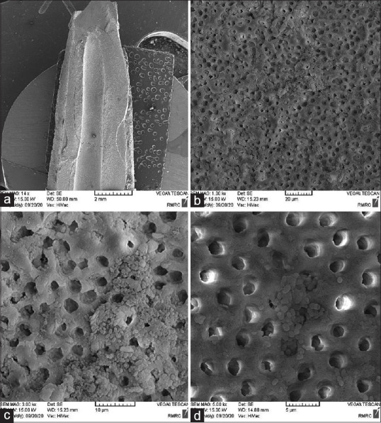
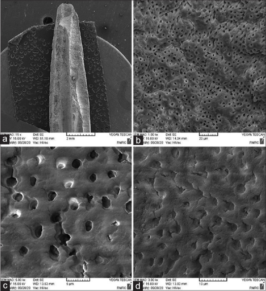
For biofilm formation, a pure culture of E. faecalis ATCC®29212™ was prepared in brain heart infusion (BHI) Broth in 37°C with the pressure of 10% CO2 for 24 h and bacterial suspension equal to 0.5 McFarland Standard. Sterilized teeth were separately placed in 1.5 ml sterile vials and 1 ml of the suspension was added to each vial. The vials were incubated for 6th week at 37°C, and the bacterial suspension was replaced daily. Then, five random samples and five negative control samples were examined to confirm biofilm formation under scanning electron microscopy (TESCAN VEGA, Kohoutovice, Czech Republic) [Figures 1 and 2].
Figure 1.

Scanning electron microscopy picture for the conformation of biofilm formation in study samples (a) with ×14 magnitude, (b) with ×1000 magnitude, (c) with ×3000 magnitude, (d) with ×5000 magnitude.
Figure 2.

Scanning electron microscopy picture for the conformation of lack of biofilm formation in control group (a) with ×14 magnitude, (b) with ×1000 magnitude, (c) with ×3000 magnitude, (d) with ×5000 magnitude.
Insertion of intracanal medicaments
The samples were divided into four groups (each containing 18 teeth), and the antibacterial agent as an intracanal drug was used for 1 week. In the first group, the intracanal drug was 1 ml of 2.24 g/cm3 calcium hydroxide (Morvabon, Tehran, Iran). In the second group, 1 ml of 6 g/ml curcumin paste (30 g natural curcumin was mixed with 15 ml sterile saline to obtain a paste with similar texture to calcium hydroxide) was used. In the third group, 1 ml natural A. vera gel extract (100% Aloe vera Gel, Sillaneh Co., Iran) was used, and in the fourth group, the control group, the teeth were only washed with normal saline. In all three groups, the medicament was injected with a 20 ml 18-gauge syringe, so to cover the entire length of the canals.
Preparation of specimens for microbiological analysis
After 1-week, antibacterial agents were irrigated from root canals using 10 ml sterile distilled water and after drying with sterile paper cones, number 4 and 5 Gates Glidden Drills (Mani, Tochigi, Japan) were used to collect debris from the entire root canal length. Then, the collected debris was transferred into sterile microtubes. To determine the number of E. faecalis colonies, the serial dilution method with a three-fold dilution was used. Finally, 100 μl of each diluted sample was inoculated into a BHI Agar plate by spread plate method and was incubated at 37°C for 24 h. Finally, the colony count machine (Funke-Gerber, Berlin, Germany) determined the number of bacteria in each sample and then CFU/ml was calculated by the following equation:
CFU/ml = (number of colonies × dilution factor)/volume of culture plate.
Statistical analyses
The data were analyzed using SPSS version 17 software (SPSS Inc., Chicago, IL, USA). The normality of data was evaluated by the KolmogorovSmirnov test. Then, the KruskalWallis analysis was used to compare colony-forming unit per ml (CFU/ml) between the study groups. The MannWhitney U-test was used to compare the mean CFU/ml of the study groups with the control group. P < 0.05 was considered significant. The regional ethics committee approved this study by the code of IR.TBZMED.REC.1398.1289.
RESULTS
The SEM study showed the formation of biofilm in study samples [Figure 1] and the lack of biofilm formation in the negative control group [Figure 2].
The mean CFU/ml in the groups of calcium hydroxide, curcumin, A. vera, and negative control is shown in Table 1. The CFU/ml levels in calcium hydroxide, curcumin, and A. vera groups were significantly higher than the control group [Table 2]. Compared with the control group, curcumin, calcium hydroxide, and A. vera showed 99.5%, 99.41%, and 98.79% antimicrobial activities, respectively. The KolmogorovSmirnov test evaluated the normality of data, and due to the normal distribution of data (P < 0.001), the mean CFU/ml of study groups was compared with the KruskalWallis test. Therefore, curcumin showed the highest antibacterial activity, and A. vera showed the least. All three groups were significantly effective than the control group but were not significantly different from each other (P = 0.057) [Table 3].
Table 1.
Mean colony-forming unit per ml (colony-forming unit/ml) in study groups
| Groups | Number | Mean±SD* | Minimum* | Maximum* |
|---|---|---|---|---|
| Control | 18 | 1.2×10⁵±8.1×10² | 8.6×10⁴ | 1.7×10⁵ |
| Ca (OH) 2 | 18 | 7.4×10²±5.2×10² | 10 | 1.4×10³ |
| Curcumin | 18 | 6.3×10²±4.6×10² | 12 | 1.2×10³ |
| Aleo vera | 18 | 1.5×10³±1.1×10³ | 37 | 4.8×10³ |
*CFU/ml, SD: Standard deviation, CFU: Colony-forming unit, Ca (OH)2: Calcium hydroxide
Table 2.
Comparison of mean colony-forming unit/ml of study groups with control group
| Study groups | Mean rank | Sum of ranks | P |
|---|---|---|---|
| Ca (OH)2 | 9.50 | 171.00 | 0.23 |
| Control | 19.50 | 39.00 | |
| Curcumin | 9.50 | 171.00 | 0.23 |
| Control | 19.50 | 39.00 | |
| Aloe vera | 9.00 | 153.00 | 0.24 |
| Control | 18.50 | 37.00 |
P value based on Mann-Whitney U-test. Ca (OH)2: Calcium hydroxide
Table 3.
Comparison of mean colony-forming unit/ml between study groups
| Study groups | Mean rank | P |
|---|---|---|
| Ca (OH)2 | 26.14 | 0.57 |
| Curcumin | 22.03 | |
| Aloe vera | 34.33 |
P value based on Kruskal-Wallis analysis. Ca (OH)2: Calcium hydroxide
DISCUSSION
For successful root canal treatment, the elimination of all microorganisms from the root canal is necessary. Microorganisms are found in planktonic and biofilm states in the root canal and the elimination of biofilm state is much more challenging.[7] E. faecalis has a high resistance to endodontic treatments,[20] which is due to its ability to penetrate to dentinal tubules,[21] tolerate high alkalinity,[22] and form biofilm.[23]
Most previous studies have used planktonic state of bacteria, but in the present study, the biofilm was used which is 1000 times more resistant to antibacterial agents and is more common in persistent infections.[6,7] Furthermore, in this study, to evaluate the antibacterial effect of medications, CFU/ml was calculated. However, most previous studies have used Agar disk diffusion which gives less reliable results.[24]
Calcium hydroxide is the gold standard for intracanal medicaments and its inhibitory effects on bacteria have been reported to be about 90%.[25] In the present study, calcium hydroxide showed 99.5% antimicrobial activity compared to the control group. Calcium hydroxide damages the bacterial cell wall and creates a high alkaline environment leading to protein denaturation and cell death but buffering features of dentin inhibits its effects to some degree.[26] Furthermore, E. faecalis can survive in the presence of calcium hydroxide due to its proton pump which balances the PH.[27] Due to some side effects and increased microbial resistance to calcium hydroxide, research on herbal drugs with less toxicity and fewer expenses is flourishing.[28]
As an anti-inflammatory and antibacterial agent, curcumin does not show any toxic effects on the human body, even in high doses.[29] The mechanism by which curcumin acts as an antibacterial agent has not been identified entirely, but it seems that curcumin inhibits the aggregation of protofilaments, inhibiting bacterial cell proliferation.[30]
Results of the present study indicate that curcumin, A. vera, and calcium hydroxide are all effective against E. faecalis, and the curcumin has the maximum antibacterial effect. Similar to the present study, a research conducted by Tyagi et al. showed that curcumin has a strong antibacterial effect against Pseudomonas aeruginosa, Escherichia coli, E. faecalis, and Staphylococcus aureus, and this effect escalates by an increase in time and dose, reaching 100% bacterial elimination.[31]
Curcumin's antibacterial effect as an intracanal medicament on planktonic bacteria has been shown to be significantly lower than chlorhexidine[28,32] but superior to A. vera and calcium hydroxide,[32] while in the present study conducted on biofilm state, the antibacterial activity of curcumin was equal to calcium hydroxide and superior to A. vera.
In a study by Swapnil et al. curcumin showed a lower antibacterial effect compared to Triphala and calcium hydroxide on planktonic state of E. faecalis. This difference in results from the present study may be due to the fact that in the Swapnil et al. study the Agar disk diffusion test was used and the bacteria were not in the biofilm state unlike the present study.[3]
A. vera is rich in anthraquinone, tannin, and myristic acid and has anti-inflammatory, antifungal, antiviral, antibacterial, and antioxidant characteristics. This substance is used in different medical settings such as treatment for recurrent aphthous stomatitis and lichen planus.[33] In a study conducted by Kurian et al. A. vera as an intracanal medicament on 3-week-old E. faecalis biofilm showed weaker results than calcium hydroxide in 3 days. However, the results were reversed on the 7th day, and A. vera had stronger antimicrobial activity. This finding is contrary to the results of the current study and the difference could be explained by different methods used to prepare pastes and different culturing techniques.[34]
In a study by Goud et al. A. vera as an intracanal irrigator against planktonic E. faecalis, measured in CFU showed to be less effective than 0.2% chlorhexidine but similar to 3% sodium hypochlorite.[35] In a confocal microscopic evaluation, A. vera had less antibacterial effects on 3-week-old E. faecalis biofilm compared to calcium hydroxide but the difference was not significant,[25] which is in accordance with the results of the present study.
It seems that variabilities in the findings of different studies is probably due to differences in the form of materials such as gel and paste or due to differences in the bacterial culture age, bacterial state, and evaluation methods.
CONCLUSION
Although none of the studied groups achieved 100% inhibitory effect, all three groups showed significant antibacterial activity compared to the control group. Curcumin had the most significant effect, followed by calcium hydroxide and A. vera. Therefore, considering the obtained results and easier access and biocompatibility, these herbal materials can be considered good alternatives to synthetic medicaments for intracanal usage.
Financial support and sponsorship
Tabriz University of Medical Sciences.
Conflicts of interest
The authors of this manuscript declare that they have no conflicts of interest, real or perceived, financial or nonfinancial in this article.
REFERENCES
- 1.Salem Milani A, Balaei Gajan E, Rahimi S, Moosavi Z, Abdollahi A, Zakeri-Milani P, et al. Antibacterial effect of diclofenac sodium on Enterococcus faecalis. J Dent (Tehran) 2013;10:16–22. [PMC free article] [PubMed] [Google Scholar]
- 2.Prabhakar A, Taur S, Hadakar S, Sugandhan S, Prabhakar A, Taur S, Hadakar S, Sugandhan S. Comparison of antibacterial efficacy of calcium hydroxide paste, 2% chlorhexidine gel and turmeric extract as an intracanal medicament and their effect on microhardness of root dentin: An in vitro study. Int J Clin Pediatr Dent. 2013;6:171–7. doi: 10.5005/jp-journals-10005-1213. [DOI] [PMC free article] [PubMed] [Google Scholar]
- 3.Swapnil SM, Sharma A, Shah N, Mandlik J, Ghogare AA. Antimicrobial efficacy of triphala and curcumin extract in comparison with calcium hydroxide against E. faecalis as an intracanal medicament an study. Paripex Indian J Res. 2017;6:876–9. [Google Scholar]
- 4.Kayaoglu G, Orstavik D. Virulence factors of Enterococcus faecalis: Relationship to endodontic disease. Crit Rev Oral Biol Med. 2004;15:308–20. doi: 10.1177/154411130401500506. [DOI] [PubMed] [Google Scholar]
- 5.Zand V, Milani AS, Amini M, Barhaghi MH, Lotfi M, Rikhtegaran S, et al. Antimicrobial efficacy of photodynamic therapy and sodium hypochlorite on monoculture biofilms of Enterococcus faecalis at different stages of development. Photomed Laser Surg. 2014;32:245–51. doi: 10.1089/pho.2013.3557. [DOI] [PubMed] [Google Scholar]
- 6.Stojicic S, Shen Y, Haapasalo M. Effect of the source of biofilm bacteria, level of biofilm maturation, and type of disinfecting agent on the susceptibility of biofilm bacteria to antibacterial agents. J Endod. 2013;39:473–7. doi: 10.1016/j.joen.2012.11.024. [DOI] [PubMed] [Google Scholar]
- 7.Wang Z, Shen Y, Haapasalo M. Effectiveness of endodontic disinfecting solutions against young and old Enterococcus faecalis biofilms in dentin canals. J Endod. 2012;38:1376–9. doi: 10.1016/j.joen.2012.06.035. [DOI] [PubMed] [Google Scholar]
- 8.Sathorn C, Parashos P, Messer H. Antibacterial efficacy of calcium hydroxide intracanal dressing: A systematic review and meta-analysis. Int Endod J. 2007;40:2–10. doi: 10.1111/j.1365-2591.2006.01197.x. [DOI] [PubMed] [Google Scholar]
- 9.Panchal V, Gurunathan D, Thangavelu L. Comparison of antibacterial efficacy of cinnamon extract and calcium hydroxide as intracanal medicament against E. faecalis: An in vitro study. Pharmacogn J. 2018;10:1165–8. [Google Scholar]
- 10.Krithikadatta J, Indira R, Dorothykalyani AL. Disinfection of dentinal tubules with 2% chlorhexidine, 2% metronidazole, bioactive glass when compared with calcium hydroxide as intracanal medicaments. J Endod. 2007;33:1473–6. doi: 10.1016/j.joen.2007.08.016. [DOI] [PubMed] [Google Scholar]
- 11.Esberard RM, Carnes DL, Jr, Del Rio CE. pH changes at the surface of root dentin when using root canal sealers containing calcium hydroxide. J Endod. 1996;22:399–401. doi: 10.1016/S0099-2399(96)80238-X. [DOI] [PubMed] [Google Scholar]
- 12.Saha S, Nair R, Asrani H. Comparative evaluation of propolis, metronidazole with chlorhexidine, calcium hydroxide and curcuma longa extract as intracanal medicament against E. faecalis – An in vitro study. J Clin Diagn Res. 2015;9:C19–21. doi: 10.7860/JCDR/2015/14093.6734. [DOI] [PMC free article] [PubMed] [Google Scholar]
- 13.Son HE, Kim EJ, Jang WG. Curcumin induces osteoblast differentiation through mild-endoplasmic reticulum stress-mediated such as BMP2 on osteoblast cells. Life Sci. 2018;193:34–9. doi: 10.1016/j.lfs.2017.12.008. [DOI] [PubMed] [Google Scholar]
- 14.Devaraj S, Jagannathan N, Neelakantan P. Antibiofilm efficacy of photoactivated curcumin, triple and double antibiotic paste, 2% chlorhexidine and calcium hydroxide against Enterococcus fecalis in vitro. Sci Rep. 2016;6:1–6. doi: 10.1038/srep24797. [DOI] [PMC free article] [PubMed] [Google Scholar]
- 15.Kusuma CS, Manjunath V, Gehlot PM. Comparative evaluation of neem, aloevera, chlorhexidine and calcium hydroxide as an intracanal medicament against E. faecalis – An in vitro study. J Clin Diagn Res. 2018;12:ZC21–5. [Google Scholar]
- 16.Ravishankar P, Lakshmi T, Kumar AS. Ethno-botanical approach for root canal treatment-an update. J Pharm Sci Res. 2011;3:1511–9. [Google Scholar]
- 17.Jain S, Rathod N, Nagi R, Sur J, Laheji A, Gupta N, et al. Antibacterial effect of Aloe vera gel against oral pathogens: An in vitro study. J Clin Diagn Res. 2016;10:C41–4. doi: 10.7860/JCDR/2016/21450.8890. [DOI] [PMC free article] [PubMed] [Google Scholar]
- 18.Batista VE, Olian DD, Mori GG. Diffusion of hydroxyl ions from calcium hydroxide and Aloe vera pastes. Braz Dent J. 2014;25:212–6. doi: 10.1590/0103-6440201300021. [DOI] [PubMed] [Google Scholar]
- 19.Jittapiromsak N, Sahawat D, Banlunara W, Sangvanich P, Thunyakitpisal P. Acemannan, an extracted product from Aloe vera, stimulates dental pulp cell proliferation, differentiation, mineralization, and dentin formation. Tissue Eng Part A. 2010;16:1997–2006. doi: 10.1089/ten.TEA.2009.0593. [DOI] [PubMed] [Google Scholar]
- 20.Hancock HH, 3rd, Sigurdsson A, Trope M, Moiseiwitsch J. Bacteria isolated after unsuccessful endodontic treatment in a North American population. Oral Surg Oral Med Oral Pathol Oral Radiol Endod. 2001;91:579–86. doi: 10.1067/moe.2001.113587. [DOI] [PubMed] [Google Scholar]
- 21.Sedgley CM, Lennan SL, Appelbe OK. Survival of Enterococcus faecalis in root canals ex vivo. Int Endod J. 2005;38:735–42. doi: 10.1111/j.1365-2591.2005.01009.x. [DOI] [PubMed] [Google Scholar]
- 22.Portenier I, Waltimo TM, Haapasalo M. Enterococcus faecalis–the root canal survivor and ‘star’ in post-treatment disease. Endod Topics. 2003;6:135–59. [Google Scholar]
- 23.Svensäter G, Bergenholtz G. Biofilms in endodontic infections. Endod Topics. 2004;9:27–36. [Google Scholar]
- 24.Fani M, Kohanteb J. Inhibitory activity of Aloe vera gel on some clinically isolated cariogenic and periodontopathic bacteria. J Oral Sci. 2012;54:15–21. doi: 10.2334/josnusd.54.15. [DOI] [PubMed] [Google Scholar]
- 25.Varshini R, Subha A, Prabhakar V, Mathini P, Narayanan S, Minu K. Antimicrobial efficacy of Aloe vera, Lemon, Ricinus communis, and calcium hydroxide as intracanal medicament against Enterococcus faecalis: A confocal microscopic study. J Pharm Bioallied Sci. 2019;11:S256–9. doi: 10.4103/JPBS.JPBS_5_19. [DOI] [PMC free article] [PubMed] [Google Scholar]
- 26.Jhamb S, Nikhil V, Singh V. An in vitro study of antibacterial effect of calcium hydroxide and chlorhexidine on Enterococcus faecalis. Indian J Dent Res. 2010;21:512–4. doi: 10.4103/0970-9290.74222. [DOI] [PubMed] [Google Scholar]
- 27.McHugh CP, Zhang P, Michalek S, Eleazer PD. pH required to kill Enterococcus faecalis in vitro. J Endod. 2004;30:218–9. doi: 10.1097/00004770-200404000-00008. [DOI] [PubMed] [Google Scholar]
- 28.Khetarpal S, Bansal A, Kukreja N. Comparison of anti-bacterial and anti-inflammatory properties of neem, curcumin and Aloe vera in conjunction with chlorhexidine as an intracanal medicament – An in vivo study. Dent J Adv Stud. 2014;2:130–7. [Google Scholar]
- 29.Gupta SC, Patchva S, Aggarwal BB. Therapeutic roles of curcumin: Lessons learned from clinical trials. AAPS J. 2013;15:195–218. doi: 10.1208/s12248-012-9432-8. [DOI] [PMC free article] [PubMed] [Google Scholar]
- 30.Kaur S, Modi NH, Panda D, Roy N. Probing the binding site of curcumin in Escherichia coli and Bacillus subtilis FtsZ--a structural insight to unveil antibacterial activity of curcumin. Eur J Med Chem. 2010;45:4209–14. doi: 10.1016/j.ejmech.2010.06.015. [DOI] [PubMed] [Google Scholar]
- 31.Tyagi P, Singh M, Kumari H, Kumari A, Mukhopadhyay K. Bactericidal activity of curcumin I is associated with damaging of bacterial membrane. PLoS One. 2015;10:e0121313. doi: 10.1371/journal.pone.0121313. [DOI] [PMC free article] [PubMed] [Google Scholar]
- 32.Yadav RK, Tikku AP, Chandra A, Verma P, Bains R, Bhoot H. A comparative evaluation of the antimicrobial efficacy of calcium hydroxide, chlorhexidine gel, and a curcumin-based formulation against Enterococcus faecalis. Natl J Maxillofac Surg. 2018;9:52–5. doi: 10.4103/njms.NJMS_47_17. [DOI] [PMC free article] [PubMed] [Google Scholar]
- 33.Monica B, Monisha R. Aloe vera in dentistry-a review. J Dent Med Sci. 2014;13:18–22. [Google Scholar]
- 34.Kurian B, Swapna D, Nadig RR, Ranjini M, Rashmi K, Bolar SR. Efficacy of calcium hydroxide, mushroom, and Aloe vera as an intracanal medicament against Enterococcus faecalis: An in vitro study. Endodontology. 2016;28:137–42. [Google Scholar]
- 35.Goud S, Aravelli S, Dronamraju S, Cherukuri G, Morishetty P. Comparative evaluation of the antibacterial efficacy of Aloe vera, 3% sodium hypochlorite, and 2% chlorhexidine gluconate against Enterococcus faecalis: An in vitro study. Cureus. 2018;10:e3480. doi: 10.7759/cureus.3480. [DOI] [PMC free article] [PubMed] [Google Scholar]


