Abstract
Objective:
To determine if a new unfilled experimental self-etching primer (SEP) adhesive system (SBP-40TX + C&B Metabond) that incorporates a methyl methacrylate–based 4-META/TBB (4-methacryloxyethyl trimellitate anhydride tri-n-butyl borane) resin can provide adequate shear bond strength (SBS) when used for bonding orthodontic brackets.
Methods and Materials:
Forty-eight human maxillary premolars were randomly divided into three groups of 16 specimens each. Brackets were bonded with three bonding systems. A filled Bis-GMA/TEGDM (triethylene glycol dimethacrylate)–based SEP adhesive system (Transbond Plus) and an unfilled conventional etch-and-rinse adhesive system (C&B Metabond) were used for comparison. The SBS for each sample was examined with a universal testing machine, and the Adhesive Remnant Index score was calculated. Enamel surfaces after conditioning were examined using a scanning electron microscope. Data were compared by one-way analysis of variance and a χ2 test.
Results:
The experimental SEP showed a milder etching pattern than Transbond Plus SEP. No statistically significant differences in the mean SBS were found between the specimens bonded with the unfilled experimental SEP adhesive system (10.0 MPa) and the filled SEP adhesive system (8.7 MPa). The unfilled experimental SEP adhesive system showed less residual adhesive than the filled SEP adhesive system.
Conclusions:
The unfilled experimental SEP adhesive system showed a clinically sufficient SBS that was equivalent to the filled SEP adhesive system.
Keywords: Self-etching primer, Adhesive, Resin, Shear bond strength, SEM
INTRODUCTION
Since the direct bonding of orthodontic appliances to enamel with composite resin was introduced by Newman in the mid 1960s,1 this technique has been widely accepted by most orthodontists.2 The major role of a composite resin adhesive in clinical orthodontics is to retain the brackets at precise locations during orthodontic treatment, which is essential for establishing ideal occlusion. Recently, Faltermeier and colleagues3 studied the effect of different filler contents of orthodontic composite resin adhesives on their bond strength and concluded that those with a higher filler content seem to provide greater bond strength than those with lower filler content or unfilled adhesives. On the other hand, bracket bonding with a self-etching primer (SEP) that simplifies bonding by combining etching and priming into a single step is now being used in clinical orthodontics.4–6 In addition to saving time and reducing procedural errors with such a primer, their lower etching ability, due to their higher pH compared to phosphoric acid, might minimize enamel loss.7–10 Previous studies have reported the effects of the acidity,11,12 application time,13–16 and agitation14–16 on etching efficacy.
A new SEP (experimental code name SBP-40TX, Sun Medical, Shiga, Japan) that can be used for bracket bonding in combination with C&B Metabond (Sun Medical), a methyl methacrylate (MMA)–based adhesive resin that contains 4-META/TBB (4-methacryloxyethyl trimellitate anhydride tri-n-butyl borane) and no inorganic fillers in the polymer powder, has been developed. Because no information is available regarding the bracket bond strength of this experimental SEP and C&B Metabond, the purpose of this study was to determine if this new unfilled SEP adhesive system can provide adequate shear bond strength (SBS) for bonding orthodontic brackets. A Bis-GMA/TEGDMA (triethylene glycol dimethacrylate)–based SEP adhesive system, Transbond Plus (3M Unitek, Monrovia, Calif), and a conventional etch-and-rinse MMA-based 4-META/TBB adhesive system, C&B Metabond, were used for comparison.
MATERIALS AND METHODS
Materials
Forty-eight non-caries human maxillary premolars were used in this study. The teeth had been extracted for orthodontic reasons with the patients' informed consent. The teeth were randomly divided into three groups of 16 specimens each for measurements of SBS. Selection criteria included the absence of any visible decalcification or cracking of the enamel surface under a stereoscopic microscope (SMZ 1500, Nikon, Tokyo, Japan) at a magnification of ×10. The extracted teeth were stored in a 0.5% chloramine solution at approximately 4°C. The buccal surfaces of all teeth were cleaned using nonfluoridated pumice. The teeth were also polished using a rubber cup, thoroughly washed, and dried using a moisture-free air source.
Group 1: Unfilled experimental SEP adhesive system (SBP-40TX + C&B Metabond)
Experimental SEP, SBP-40TX, was applied to the enamel surfaces for 20 seconds using a sponge pledget. An air jet was lightly applied to the enamel. After appropriate amounts of the monomer (Super-Bond C&B Quick Monomer, Sun Medical) and the catalyst were mixed in a well and some amount of polymer powder was added to the well, metal brackets for upper premolars (Victory Series, 3M Unitek, Monrovia, Calif) with a base area of 10.0 mm2 were bonded using the brush-dip technique.
Group 2: Unfilled etch-and-rinse adhesive system (C&B Metabond)
The enamel surfaces were treated with 20% phosphoric acid etching gel for 20 seconds, washed for 20 seconds, and dried with an oil-free air stream. Metal brackets for upper premolars (Victory Series, 3M Unitek) were bonded with the monomer, the catalyst and polymer powder by the brush-dip technique.
Group 3: Filled adhesive system (Transbond Plus SEP system)
Transbond Plus SEP was applied and rubbed on the enamel surfaces for 3 seconds. An air jet was lightly applied to the enamel, and the brackets were bonded with Transbond XT composite.
The excess bonding material was removed with a small scaler. Samples bonded with a Transbond Plus self-etching adhesive system were light-cured for 20 seconds (10 seconds from each proximal side). After the bonding procedures, the specimens were stored in artificial saliva at 37°C for 24 hours. The SBS was then measured. The specimens were fixed to a custom-fabricated acryl resin block using Model Repair II (Densply-Sankin, Tokyo, Japan), and the block was fixed to a universal testing machine (EZ Test, Shimadzu, Kyoto, Japan). A knife-edged shearing blade was secured to the crosshead with the direction of force parallel to the buccal surface and the bracket base. Force was applied directly to the bracket-tooth interface. The brackets were debonded at a crosshead speed of 0.5 mm/min.
After bond failure, the bracket bases and enamel surfaces were examined with a stereoscopic microscope at a magnification of ×10. Adhesive Remnant Index (ARI) scores were used to assess the amount of adhesive left on the enamel surface. ARI scores ranged from 0 to 3: 0 = no adhesive left on the tooth surface, that is, the failure site was between the adhesive and enamel; 1 = less than half of the adhesive was left on the tooth surface; 2 = half or more of the adhesive was left on the tooth; 3 = all of the adhesive was left on the tooth surface, that is, the failure site was between the adhesive and bracket base.
To assess the etching efficacy of intact enamel using a scanning electron microscope (SEM; SSX-550, Shimadzu, Kyoto, Japan), buccal enamel surfaces were conditioned with two different SEPs (SBP-40TX for 20 seconds and Transbond Plus for 3 seconds), and the primers were then rinsed off. As a control, an enamel surface was etched with 20% phosphoric acid for 20 seconds and then washed for 20 seconds. After conditioning or etching, the specimens were dehydrated in increasing concentrations of ethanol and water up to 100% ethanol. The specimens were sputter-coated with gold (SC-701 AT, Sanyu Electron, Tokyo, Japan) and examined by an SEM operating at 15 kV.
Statistical Analysis
Statistical analysis was performed using the Statistical Package for the Social Sciences software (version 16.0J for Windows, SPSS, Chicago, Ill). The bond strength data were tested for normality with the Kolmogorov-Smirnov test. The mean SBS, along with the standard deviation (n = 16), for the groups of bonding materials were compared by one-way analysis of variance (ANOVA), followed by the Tukey Kramer honestly significant difference test. The χ2 test was used to evaluate the significance of differences in the ARI scores among the different groups. For the statistical analysis, ARI scores of 0 and 1 were combined, as were ARI scores of 2 and 3. For all statistical tests, significance was predetermined at P < .05.
RESULTS
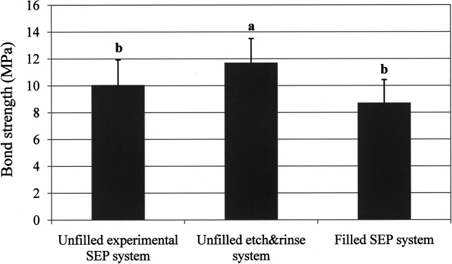
The results regarding SBS are shown in Figure 1. In one-way ANOVA, specimen bonded with the unfilled experimental SEP adhesive system showed significantly lower mean SBS (10.0 MPa) than that bonded with the unfilled etch-and-rinse adhesive system (11.8 MPa). However, no statistically significant differences were found between the specimens bonded with the unfilled experimental SEP adhesive system and with the filled SEP adhesive system (8.7 MPa).
Figure 1.
Mean and standard deviation of the bond strength (MPa) in the three specimen groups. For bars with identical letters, the average values are not significantly different (P < .05, Tukey test).
A χ2 analysis of the ARI scores for the three adhesives revealed a significant difference in the distribution of frequencies among the ARI categories for the three adhesive groups. The unfilled experimental SEP adhesive system had a greater frequency of ARI scores of 0 and 1 (Table 1).
Table 1.
Frequency Distribution of ARI Scores of Tested Groups
The enamel surface etched with 20% phosphoric acid for 20 seconds showed a very porous surface, and numerous enamel prisms could be observed (Figure 2a), which reflected a typical honeycomb pattern. For the Transbond Plus SEP, enamel prisms could be observed in some areas, but were less prominent (Figure 2c). On the other hand, SBP-40TX produced less surface roughness than Transbond Plus SEP; many scratches and fossae were seen (Figure 2b).
Figure 2.
SEM photomicrographs of etching efficacy. (a) Superbond etching. (b) Superbond self-etching primer. (c) Transbond Plus self-etching primer.
DISCUSSION
To study the effect of acidity on etching, a previous study used three SEPs with different pH values and found that the etching patterns on aprismatic enamel depended on the acidity of the primer. However, there was no correlation between the acidity and the strength of the bond to intact enamel.11 The effects of application time on the etching pattern12,13 and bond strength14,15 have also been investigated. Perdigao and colleagues12 found that doubling the enamel conditioning time might increase the bond strength for specific SEP adhesive systems. Two recent studies13,16 reported that an increase in the application time of Transbond Plus SEP showed slightly increased etching efficacy but did not significantly increase the SBS. In the present study, SBP-40TX (experimental SEP) with an application time of 20 seconds gave a milder etching pattern than Transbond Plus SEP with an etching time of 3 seconds. This result indicates that SBP-40TX does not etch as strongly as Transbond Plus SEP.
Because a previous study3 showed that adhesives with a higher filler content seem to provide greater bond strength than those with a lower filler content or unfilled adhesives, the addition of fillers to a resin adhesive might increase the bracket bond strength. However, this is not consistent with the present finding that the experimental unfilled SEP adhesive system showed equivalent bracket bond strength with the filled SEP adhesive system (Transbond Plus SEP system). A previous study17 used X-ray photoelectron spectroscopy and atomic absorption spectrophotometry to compare the chemical bonding efficacy of three functional monomers (10-MDP, 4-MET, phenyl-P) and found that the bonding potential of 10-MDP to hydroxyapatite is significantly greater than that of 4-MET and phenyl-P. The results suggest that, in addition to the influence of micro-mechanical interlocking between the resin adhesive and enamel on bracket bond strength, the performance of monomer (4-META/TBB and Bis-GMA/TEGDMA) used in this study might also influence the bond strength with the resin adhesive. However, further research is necessary to verify these hypotheses.
When a bracket bonded with resin adhesive is removed from the enamel, failure can occur at one of three interfaces: between the adhesive and the bracket, within the adhesive itself, or between the adhesive and the enamel surface. The failure site might be influenced by the strength of the bond between the enamel and the adhesive resin, the strength of the bond between the adhesive resin and the bracket base, and the mechanical properties of the adhesive resin. In this study, the unfilled experimental SEP adhesive system had a greater frequency of ARI scores of 0 and 1, which means less residual adhesive resin was seen on the enamel surface after debonding. This result is intriguing because this system showed similar bracket bond strength with the filled SEP adhesive system. There are few possible explanations for this result; for example, (1) the composite resin paste of the filled adhesive system may not have penetrated sufficiently into the bracket mesh, and (2) very short resin tags that formed a hybrid layer remained on the surface of enamel of a specimen bonded with the unfilled experimental SEP adhesive system, and it may not be possible to observe this layer by a stereomicroscope at low magnification. To verify these hypotheses, further research is required to investigate debonded enamel by the SEM with energy-dispersive spectrometry.
Because unfilled adhesive systems do not contain fillers, it is easy to remove any adhesive after debonding, and this may be a clinical advantage. However, the brush-dip method for C&B Metabond, which was used in the unfilled adhesive systems in this study, may require more time than the light-cured method with Transbond SEP system. Although the use of fillers helps to strengthen the adhesive itself, the addition of fillers may not be necessary if the chemical bond strength between the enamel and resin is sufficient.
CONCLUSIONS
The experimental SEP (SBP-40TX) produced a milder etching pattern than Transbond Plus SEP.
The unfilled experimental SEP system (SBP-40TX + C&B Metabond) showed equivalent bracket bond strength with the filled SEP adhesive system (Transbond Plus SEP system).
The unfilled experimental SEP system might be durable for a normal orthodontic treatment period and easier to remove the residual adhesive after bracket debonding.
Acknowledgments
The authors thank Sun Medical Company for supplying the experimental self-etching primer.
REFERENCES
- 1.Newman G. V. Epoxy adhesives for orthodontic attachments: progress report. Am J Orthod. 1965;51:901–912. doi: 10.1016/0002-9416(65)90203-4. [DOI] [PubMed] [Google Scholar]
- 2.Eliades T, Eliades G. Orthodontic adhesive resins. In: Brantley W. A, Eliades T, editors. Orthodontic Materials Scientific and Clinical Aspects. Stuttgart, Germany: Thieme; 2001. pp. 201–219. [Google Scholar]
- 3.Faltermeier A, Rosentritt M, Faltermeier R, Reicheneder C, Müßig D. Influence of filler level on the bond strength of orthodontic adhesives. Angle Orthod. 2007;77:494–498. doi: 10.2319/0003-3219(2007)077[0494:IOFLOT]2.0.CO;2. [DOI] [PubMed] [Google Scholar]
- 4.Bishara S. E, Ajlouni R, Laffoon J. F, Warren J. J. Effect of a fluoride-releasing self-etch acidic primer on the shear bond strength of orthodontic brackets. Angle Orthod. 2002;72:199–202. doi: 10.1043/0003-3219(2002)072<0199:EOAFRS>2.0.CO;2. [DOI] [PubMed] [Google Scholar]
- 5.Arhun N, Arman A, Sesen C, Karabulut E, Korkmaz Y, Gokalp S. Shear bond strength of orthodontic brackets with 3 self-etch adhesives. Am J Orthod Dentofacial Orthop. 2006;129:547–550. doi: 10.1016/j.ajodo.2005.12.006. [DOI] [PubMed] [Google Scholar]
- 6.Scougall Vilchis R. J, Yamamoto S, Kitai N, Hotta M, Yamamoto K. Shear bond strength of a new fluoride-releasing adhesive. Dent Mater J. 2007;26:45–51. doi: 10.4012/dmj.26.45. [DOI] [PubMed] [Google Scholar]
- 7.Fjeld M, Øgaard B. Scanning electron microscopic evaluation of enamel surfaces exposed to 3 orthodontic bonding systems. Am J Orthod Dentofacial Orthop. 2006;130:575–581. doi: 10.1016/j.ajodo.2006.07.002. [DOI] [PubMed] [Google Scholar]
- 8.Scougall Vilchis R. J, Hotta Y, Yamamoto K. Examination of enamel-adhesive interface with focused ion beam and scanning electron microscopy. Am J Orthod Dentofacial Orthop. 2007;131:646–650. doi: 10.1016/j.ajodo.2006.11.017. [DOI] [PubMed] [Google Scholar]
- 9.Iijima M, Ito S, Yuasa T, Muguruma T, Saito T, Mizoguchi I. Bond strength comparison and scanning microscopic evaluation of three orthodontic bonding systems. Dent Mater J. 2008;27:392–399. doi: 10.4012/dmj.27.392. [DOI] [PubMed] [Google Scholar]
- 10.Iijima M, Muguruma T, Brantley W. A, Ito S, Yuasa T, Saito T, Mizoguchi I. Effect of bracket boding on nanomechanical properties of enamel. Am J Orthod Dentofacial Orthop. doi: 10.1016/j.ajodo.2009.01.028. (In press) [DOI] [PubMed] [Google Scholar]
- 11.Pashley D. H, Tay F. R. Aggressiveness of contemporary self-etching adhesives. Part II: Etching effects on unground enamel. Dent Mater. 2001;17:430–444. doi: 10.1016/s0109-5641(00)00104-4. [DOI] [PubMed] [Google Scholar]
- 12.Perdigao J, Gomes G, Lopes M. M. Influence of conditioning time on enamel adhesion. Quintessence Int. 2006;37:35–41. [PubMed] [Google Scholar]
- 13.Ostby A. W, Bishara S. E, Laffoon J, Warren J. J. Influence of self-etching application time on bracket shear bond strength. Angle Orthod. 2007;77:885–889. doi: 10.2319/092006-383. [DOI] [PubMed] [Google Scholar]
- 14.Cehreli S. B, Eminkahyagil N. Effect of active pretreatment of self-etching primers on the ultramorphology of intact primary and permanent tooth enamel. J Dent Child. 2006;73:86–90. [PubMed] [Google Scholar]
- 15.Velasquez L. M, Sergent R. S, Burgess J. O, Mercante D. E. Effect of placement agitation and placement time on the shear bond strength of 3 self-etching adhesives. Oper Dent. 2006;31:426–430. doi: 10.2341/05-52. [DOI] [PubMed] [Google Scholar]
- 16.Iijima M, Ito S, Yuasa T, Muguruma T, Saito T, Mizoguchi I. Effects of application time and agitation for bonding orthodontic brackets with two self-etching primer systems. Dent Mater J. 2009;28:89–95. doi: 10.4012/dmj.28.89. [DOI] [PubMed] [Google Scholar]
- 17.Yoshida Y, Nagakane K, Fukuda R, et al. Comparative study on adhesive performance of functional monomers. J Dent Res. 2004;83:454–458. doi: 10.1177/154405910408300604. [DOI] [PubMed] [Google Scholar]


