Abstract
Objectives:
To determine root lengths of fully developed permanent teeth of cleft lip and palate (CLP) patients and to define their crown-root (C/R) ratios.
Method:
Crown height and root length of permanent teeth were measured from panoramic radiographs of 44 CLP patients and 37 controls. A total of 1397 teeth were measured, and C/R ratios were calculated.
Results:
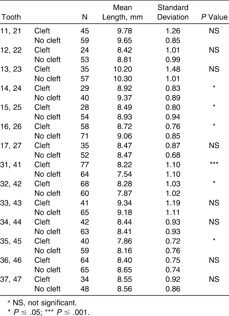
Higher C/R ratios were found in CLP patients; this was statistically significant for both maxillary and mandibular incisors and canines. Bilateral CLP subjects showed higher C/R ratios in general than unilateral CLP subjects. Roots of maxillary incisors, canines, and some other teeth were significantly shorter in CLP patients than in controls.
Conclusions:
CLP patients should be considered to have unfavorable C/R ratios, which could be the result of short root lengths for some teeth.
Keywords: Crown height, Crown-root ratio, Root length, Panoramic, Cleft lip and palate
INTRODUCTION
Cleft lip and palate (CLP) account for a large fraction of all human birth defects and are notable for their significant life-long morbidity and complex etiology. CLP is not just a localized, transient disruption in development, in that patients with CLP have considerably more dental anomalies than do individuals without clefts.1–3 Common findings include reduced size of crowns and roots (altered crown-root ratio), aberrant root forms, simplified crown morphology, and malformed teeth.4,5 Systemic, compromised growth potential in these patients is expressed as decreased tooth size and amplified asymmetry, both of which affect all the teeth in both arches.5 On the other hand, the cleft itself is at least partially responsible for the observed reduction in growth potential.4
An increased incidence of morphologic dental crown abnormalities associated with various expressions of CLP has been reported by several investigators; these abnormalities have affected both upper and lower arches and both anterior and posterior teeth.1,5–8 Similarly, several studies have been carried out to assess root development in cleft patients; however, most of these focused on root development of the lateral incisor in the vicinity of the cleft.9–11 Unlike bony structures, teeth do not remodel, so transient insults will be recorded in those teeth undergoing morphogenesis at that time, and chronic debilitations will affect multiple teeth.
The morphologic events associated with tooth root formation in a variety of animals have been thoroughly described; however, the mechanisms involved in human tooth root formation are not well understood.12,13 Some types of environmental insults during tooth development were found to result in short-rooted teeth; these include chemotherapy14 and radiation therapy.15 Short roots also have been observed in some disorders such as scleroderma,16 Stevens-Johnson syndrome,17 Down syndrome,18 and Turner syndrome.19,20 Short roots, resulting in high unfavorable crown-root (C/R) ratios, may affect the prognosis of teeth, especially in patients with chronic periodontitis, and may complicate orthodontic or prosthodontic treatment planning. The main reasons for short dental roots are disturbances during dental root development and resorption of originally well-developed roots.21
A more extreme condition known as short root anomaly has been described by some authors.22,23 In this condition, the short roots are not due to resorption, nor are they due to any systemic disturbance associated with generalized shortness of the roots. The roots in this condition have been described as developmentally very short, blunt roots of the maxillary incisor teeth.23
Underexplored topics regarding CLP subjects include tooth root length and C/R ratio. We could find no reports that compare the C/R ratios in CLP patients with those in healthy patients with fully developed dentitions. Therefore the aims of this study are (1) to define C/R ratios, and (2) to determine the root lengths of fully developed permanent teeth of CLP patients and healthy Jordanian controls. This information could be valuable for clinical application during orthodontic or prosthodontic treatment.
MATERIALS AND METHODS
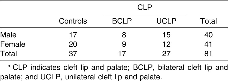
Information for the present investigation was obtained from cleft palate patients' records at the Oral and Maxillofacial Surgery Outpatient Clinic at King Abdullah University Hospital, and from orthodontic patients' records at the Dental Teaching Center at Jordan University of Science and Technology. Subjects were included if they met the following criteria: (1) had a diagnosis of unilateral CLP (UCLP) or bilateral CLP (BCLP) with no other recognizable syndromes, (2) were older than 12 years of age (when most permanent tooth roots are completed, excluding third molars), and (3) had a clear panoramic radiograph. The study sample consisted of 44 CLP patients ranging in age from 12 to 31 years (mean, 18.5 ± 3.6 years) and 37 age- and sex-matched controls who were selected randomly from patient records in the orthodontic department. Ages of controls ranged from 12 to 30 years (mean, 19.3 ± 2.2 years). The distribution of the sample is summarized in Table 1.
Table 1.
Total Samplea
Measurements
Under ideal conditions, including the use of subdued lights, film masking, a magnifying lens, and a conventional viewing box, the outlines of the permanent maxillary and mandibular teeth were apparent. The outlines of these teeth were marked with a special pencil. Crown heights and root lengths were measured using the method of Lind, which was adapted for use in posterior teeth,23 and measurements were made with a sliding digital caliber on acetate sheets (Mitutoyo, Tokyo, Japan). All measurements were rounded to the nearest tenth decimal.
For the purposes of tooth length measurements, three parallel reference lines were drawn. An incisal/occlusal reference line formed a tangent to an incisal tip or a buccal cusp and was visually placed perpendicular to the long axis of the tooth. The cervical reference line was the line joining the mesial and distal cervical margins of enamel. The apical reference line touched the outermost part of the root, and in teeth with two buccal roots, the longer root was measured; this line was visually placed perpendicular to the long axis of the tooth.23 The palatal roots of the maxillary molars were omitted. Crown height was the perpendicular line from the midpoint of the cervical reference line to the incisal/occlusal reference line. Root length was the perpendicular line from the midpoint of the cervical reference line to the apical reference line. The C/R ratio of an individual tooth was calculated by dividing crown height by root length.
Individual teeth were excluded if (1) teeth showed obvious distortion, (2) the apex was not closed, (3) root resorption was evident, (4) the teeth were impacted, or (5) marked attrition or abrasion of the crown was noted.
To assess intraexaminer reproducibility and the reliability of measurements, 300 teeth on 13 panoramic radiographs were remeasured at a minimum interval of 2 months.
Statistical Analysis
The Statistical Package for the Social Sciences (SPSS), version 15.0 (SPSS Inc, Chicago, Ill), was used for statistical analysis. Mean values for C/R ratios of CLP and control subjects were calculated. Differences in mean C/R ratios between CLP subjects and controls, BCLP and UCLP, CLP males and females, control males and females, and cleft side and noncleft side of UCLP patients, in addition to differences in mean crown and root lengths in CLP subjects, were studied using independent sample t-tests.
The precision of the measurement was calculated by means of the method of error (ME), according to the following formula:
where d is the difference between duplicate measurements, and n is the number of duplicate measurements.24 Intraexaminer reproducibility was examined by means of paired Student's t-tests. A statistical significance level of P = .05 was selected.
RESULTS
Crown heights and root lengths were measured and C/R ratios calculated for a total of 1397 teeth (600 for CLP, 797 for controls). Many teeth were not traced, especially from the CLP sample, mainly because they were missing, distorted, dilacerated, impacted, or incompletely developed. No teeth had marked attrition evident on panoramic radiographs.
Reproducibility testing of the two sets of data were correlated (r > 0.97), and no statistically significant difference was found (P > .05). The method of the error was 0.02 mm.
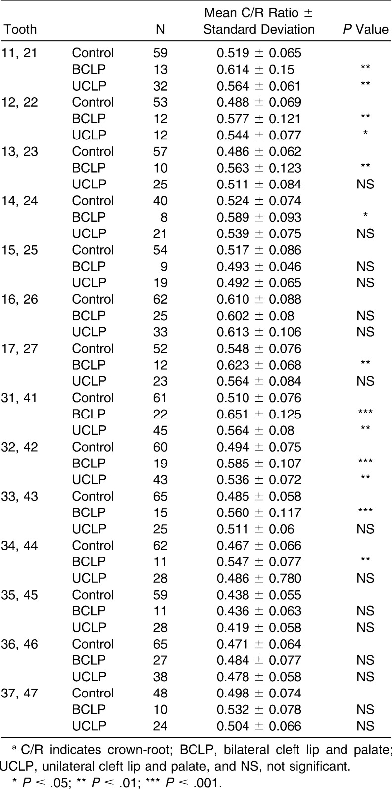
C/R ratios were higher for both BCLP and UCLP subjects than for controls, and this difference was statistically significant for all incisors. It was noted that C/R ratios of canines, premolars, and molars in both jaws were not affected in the UCLP group (Table 2). When data from the two CLP groups were pooled together, the C/R ratios of maxillary and mandibular incisors and of canines were significantly higher than those of controls. This was also true for the maxillary second molar and the mandibular first premolar.
Table 2.
Differences in Mean C/R Ratios Between UCLP and Control, BCLP and Controla
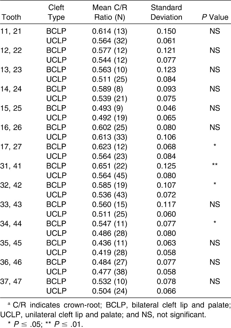
Within the CLP group, BCLP subjects showed higher C/R ratios in general than did UCLP subjects, and for some teeth, this was significant (P < .05) (Table 3). When cleft-side and non–cleft-side maxillary teeth C/R ratios in UCLP subjects were compared, no statistically significant difference was found (P > .05).
Table 3.
Differences in Mean C/R Ratios According to Cleft Typea
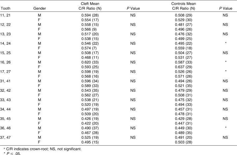
C/R ratios did not show a consistent relation with gender in control subjects, for instance, they were higher in females for some teeth, but for the rest males had higher values. No differences between female and male C/R ratios reached statistical significance (Table 4).
Table 4.
Differences in Mean C/R Ratios According to Gender in Cleft and Control Samplesa
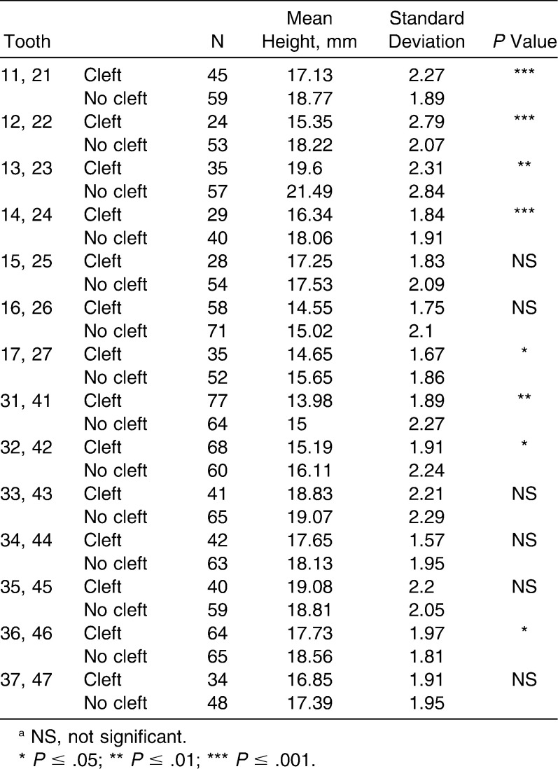
When crown heights and root lengths were compared separately in CLP subjects and their controls, it was noted that some roots were significantly shorter in CLP patients than in controls (Table 5). With regard to crown height, results showed that the crowns of some teeth were significantly shorter, but those of other teeth were significantly longer, in CLP subjects than in control subjects (Table 6).
Table 5.
Differences in Mean Root Lengths Between Cleft and Control Subjectsa
Table 6.
Differences in Mean Crown Heights of All Teeth Between Cleft and Control Subjectsa
DISCUSSION
Several studies have demonstrated that accurate reproducibility of panoramic radiographs and their diagnostic quality are heavily dependent on careful attention to positioning and processing.25–27 The problem of vertical distortion, which is usually encountered when absolute heights and lengths are reported from panoramic radiographs, would be overcome by ratio calculations, as proportions of crown and root parts of the tooth would remain unchanged.28 When C/R ratio measurements from panoramic radiographs were tested in one study, the authors concluded that tooth lengths and C/R ratios could be measured accurately from panoramic radiographs.29
A problem in studying unusual clinical conditions is use of a small study sample, which makes it difficult for investigators to reach relevant conclusions. This is why we had to add many data within the cleft group to have good numbers for comparisons. Within this limitation, C/R ratios were higher in the CLP group than in the control group, and were even higher in the BCLP group than in the UCLP group. This unfavorable ratio could be the result of shorter roots or longer crowns in CLP patients. No studies that have investigated C/R ratios and root lengths in these patients are available for comparison. Delayed root development, which is one type of disturbance in root development,21 would result in short roots; this in turn may produce increased and unfavorable C/R ratios. This possibility is valid for causing short roots in CLP subjects, as several studies had found root development to be delayed in these patients when compared with normal reference populations.30,31 In a recent study investigating differences in dental development between UCLP and BCLP patients, a significantly greater delay was noted for BCLP subjects than for UCLP subjects.31
Teeth that showed less favorable C/R ratios in BCLP than in UCLP would be expected to be the least likely affected by the cleft (maxillary second molar, mandibular incisors, and first premolar). Findings that BCLP subjects had comparable C/R ratios to those in UCLP for the maxillary anterior teeth, and that UCLP cleft-side and non–cleft-side teeth C/R ratios were also comparable, lead to the suggestion of a shared genetic basis. This shared genetic basis may have a greater impact on C/R ratios than the direct effect of the cleft itself has on primordial tissues because effects are more pronounced when the patient has a bilateral rather than a unilateral cleft.
Males and females exhibited no differences in C/R ratios within the CLP group, but females had greater C/R ratios than males in the control group. This finding could be the result of longer roots in males or shorter roots in females. Many studies that investigated the effects of X and Y chromosomes on root growth in sex chromosome abnormalities have concluded that the promoting effect of the Y chromosome on growth of root length is greater than that of the X chromosome19,32,33; this could have been the cause of longer roots in males, thus reducing their C/R ratios. In a study investigating root-crown ratios in a healthy Finnish population, it was found that males had more favorable ratios—a fact that was concluded to be the result of their longer roots.21 The fact that the CLP group was not affected could be the result of delay or could be the root-shortening effect of the etiologic factor of the cleft dominating over the sex gene effect.
It is interesting to note that maxillary incisors and canines had significantly shorter roots than those in the control group, but at the same time, this was not consistently so. In one study, root development rather than root length was compared for maxillary lateral incisors using cleft and noncleft sides.9 It was found that root development was delayed for the cleft side, which was in agreement with the findings of previous studies.11,34 Similarly, Demirjian's study35 concluded that mechanisms controlling dental development are independent of somatic and sexual maturity and are highly influenced by the same etiologic factor as the cleft. Because some types of environmental insults during tooth development and genetic factors may result in short-rooted teeth,14–20 CLP patients should be considered as potentially having short roots, resulting in unfavorable C/R ratios.
With regard to crown height comparisons, the results were somehow inconsistent in that tooth crowns were shorter in some cleft patients, and in others they were longer (Table 6). In CLP subjects, enamel defects and abnormalities in shape and size of both deciduous and permanent teeth are far more common than in normal subjects1,8,36; this explains the deviation in crown height from normal in the current CLP sample. A recent study showed that occlusogingival measurements of tooth crowns in the casts of CLP patients were smaller than those of controls—not only in the affected maxillary dental arch, but also in the mandibular dental arch.37 It is important, however, to be aware that study methods varied in that investigators used casts rather than radiographic assessments.
Early diagnosis of short roots in CLP patients may influence their orthodontic treatment strategy. In addition, these patients may require fixed prostheses to close edentulous spaces because missing teeth are commonly prevalent.
CONCLUSIONS
CLP patients should be considered to have short roots and unfavorable C/R ratios.
BCLP patients had significantly higher C/R ratios than did UCLP patients for some teeth.
UCLP cleft-side and non–cleft-side C/R ratios were affected similarly, with higher C/R ratios.
No differences were noted between male and female C/R ratios within the CLP group.
REFERENCES
- 1.Aljamal G, Hazza'a A, Rawashdeh M. Prevalence of dental anomalies in a population ofcleft lip and palate patients. Cleft Palate Craniofac J. 2010:22. doi: 10.1597/08-275.1. [Epub ahead of print.] [DOI] [PubMed] [Google Scholar]
- 2.Dixon D. A. Defects of structure and formation of the teeth in persons with cleft palate and the effect of reparative surgery on the dental tissues. Oral Surg Oral Med Oral Pathol. 1968;25:435–446. doi: 10.1016/0030-4220(68)90019-4. [DOI] [PubMed] [Google Scholar]
- 3.Ranta R, Rintala A. Tooth anomalies associated with congenital sinuses of the lower lip and cleft lip/palate. Angle Orthod. 1982;52:212–221. doi: 10.1043/0003-3219(1982)052<0212:TAAWCS>2.0.CO;2. [DOI] [PubMed] [Google Scholar]
- 4.Harris E. F, Hullings J. G. Delayed dental development in children with isolated cleft lip and palate. Arch Oral Biol. 1990;35:469–473. doi: 10.1016/0003-9969(90)90210-2. [DOI] [PubMed] [Google Scholar]
- 5.Werner S. P, Harris E. F. Odontometrics of the permanent teeth in cleft lip and palate: systemic size reduction and amplified asymmetry. Cleft Palate J. 1989;26:36–41. [PubMed] [Google Scholar]
- 6.Jordan R. E, Kraus B. S, Neptune C. M. Dental abnormalities associated with cleft lip and/or palate. Cleft Palate J. 1966;3:22–55. [PubMed] [Google Scholar]
- 7.Kraus B. S, Jordan R. E, Pruzansky S. Dental abnormalities in the deciduous and permanent dentitions of individuals with cleft lip and palate. J Dent Res. 1966;45:1736–1746. doi: 10.1177/00220345660450062601. [DOI] [PubMed] [Google Scholar]
- 8.Rawashdeh M. A, Bakir I. F. The crown size and sexual dimorphism of permanent teeth in Jordanian cleft lip and palate patients. Cleft Palate Craniofac J. 2007;44:155–162. doi: 10.1597/05-197.1. [DOI] [PubMed] [Google Scholar]
- 9.Deepti A, Muthu M. S, Kumar N. S. Root development of permanent lateral incisor in cleft lip and palate children: a radiographic study. Indian J Dent Res. 2007;18:82–86. doi: 10.4103/0970-9290.32426. [DOI] [PubMed] [Google Scholar]
- 10.Pioto N. R, Costa B, Gomide M. R. Dental development of the permanent lateral incisor in patients with incomplete and complete unilateral cleft lip. Cleft Palate Craniofac J. 2005;42:517–520. doi: 10.1597/04-045r.1. [DOI] [PubMed] [Google Scholar]
- 11.Ribeiro L. L, das Neves L. T, Costa B, Gomide M. R. Dental development of permanent lateral incisor in complete unilateral cleft lip and palate. Cleft Palate Craniofac J. 2002;39:193–196. doi: 10.1597/1545-1569_2002_039_0193_ddopli_2.0.co_2. [DOI] [PubMed] [Google Scholar]
- 12.Thomas H. F. Root formation. Int J Dev Biol. 1995;39:231–237. [PubMed] [Google Scholar]
- 13.Wright T. The molecular control of and clinical variations in root formation. Cells Tissues Organs. 2007;186:86–93. doi: 10.1159/000102684. [DOI] [PubMed] [Google Scholar]
- 14.Jaffe N, Toth B. B, Hoar R. E, Ried H. L, Sullivan M. P, McNeese M. D. Dental and maxillofacial abnormalities in long-term survivors of childhood cancer: effects of treatment with chemotherapy and radiation to the head and neck. Pediatrics. 1984;73:816–823. [PubMed] [Google Scholar]
- 15.Holtta P, Hovi L, Saarinen-Pihkala U. M, Peltola J, Alaluusua S. Disturbed root development of permanent teeth after pediatric stem cell transplantation: dental root development after SCT. Cancer. 2005;103:1484–1493. doi: 10.1002/cncr.20967. [DOI] [PubMed] [Google Scholar]
- 16.Foster T. D, Fairburn E. A. Dental involvement in scleroderma. Br Dent J. 1968;16;124:353–356. [PubMed] [Google Scholar]
- 17.Thornton J. B, Worley S. L. Short root anomaly in a patient with a history of Stevens-Johnson syndrome: report of case. ASDC J Dent Child. 1991;58:256–259. [PubMed] [Google Scholar]
- 18.Prahl-Andersen B, Oerlemans J. Characteristics of permanent teeth in persons with trisomy G. J Dent Res. 1976;55:633–638. doi: 10.1177/00220345760550041501. [DOI] [PubMed] [Google Scholar]
- 19.Lahdesmaki R, Alvesalo L. Root growth in the permanent teeth of 45,X/46,XX females. Eur J Orthod. 2006;28:339–344. doi: 10.1093/ejo/cji121. [DOI] [PubMed] [Google Scholar]
- 20.Midtbo M, Halse A. Root length, crown height, and root morphology in Turner syndrome. Acta Odontol Scand. 1994;52:303–314. doi: 10.3109/00016359409029043. [DOI] [PubMed] [Google Scholar]
- 21.Holtta P, Nystrom M, Evalahti M, Alaluusua S. Root-crown ratios of permanent teeth in a healthy Finnish population assessed from panoramic radiographs. Eur J Orthod. 2004;26:491–497. doi: 10.1093/ejo/26.5.491. [DOI] [PubMed] [Google Scholar]
- 22.Apajalahti S, Holtta P, Turtola L, Pirinen S. Prevalence of short-root anomaly in healthy young adults. Acta Odontol Scand. 2002;60:56–59. doi: 10.1080/000163502753472014. [DOI] [PubMed] [Google Scholar]
- 23.Lind V. Short root anomaly. Scand J Dent Res. 1972;80:85–93. doi: 10.1111/j.1600-0722.1972.tb00268.x. [DOI] [PubMed] [Google Scholar]
- 24.Dahlberg G. Statistical Methods for Medical and Biological Students. London, UK: G. Allen & Unwin Ltd; 1940. [Google Scholar]
- 25.Larheim T. A, Svanaes D. B, Johannessen S. Reproducibility of radiographs with the orthopantomograph 5: tooth-length assessment. Oral Surg Oral Med Oral Pathol. 1984;58:736–741. doi: 10.1016/0030-4220(84)90045-8. [DOI] [PubMed] [Google Scholar]
- 26.Stramotas S, Geenty J. P, Darendeliler M. A, Byloff F, Berger J, Petocz P. The reliability of crown-root ratio, linear and angular measurements on panoramic radiographs. Clin Orthod Res. 2000;3:182–191. doi: 10.1034/j.1600-0544.2000.030404.x. [DOI] [PubMed] [Google Scholar]
- 27.Thanyakarn C, Hansen K, Rohlin M. Measurements of tooth length in panoramic radiographs. 2: Observer performance. Dentomaxillofac Radiol. 1992;21:31–35. doi: 10.1259/dmfr.21.1.1397449. [DOI] [PubMed] [Google Scholar]
- 28.Brook A. H, Holt R. D. The relationship of crown length to root length in permanent maxillary central incisors. Proc Br Paedod Soc. 1978;8:17–20. [PubMed] [Google Scholar]
- 29.Stramotas S, Geenty J. P, Petocz P, Darendeliler M. A. Accuracy of linear and angular measurements on panoramic radiographs taken at various positions in vitro. Eur J Orthod. 2002;24:43–52. doi: 10.1093/ejo/24.1.43. [DOI] [PubMed] [Google Scholar]
- 30.Brouwers H. J, Kuijpers-Jagtman A. M. Development of permanent tooth length in patients with unilateral cleft lip and palate. Am J Orthod Dentofacial Orthop. 1991;99:543–549. doi: 10.1016/S0889-5406(05)81631-2. [DOI] [PubMed] [Google Scholar]
- 31.Hazza'a A. M, Rawashdeh M. A, Al-Jamal G, Al-Nimri K. S. Dental development in children with cleft lip and palate: a comparison between unilateral and bilateral clefts. Eur J Paediatr Dent. 2009;10:90–94. [PubMed] [Google Scholar]
- 32.Lahdesmaki R, Alvesalo L. Root growth in the teeth of 46,XY females. Arch Oral Biol. 2005;50:947–952. doi: 10.1016/j.archoralbio.2005.03.002. [DOI] [PubMed] [Google Scholar]
- 33.Lahdesmaki R, Alvesalo L. Root lengths in the permanent teeth of Klinefelter (47,XXY) men. Arch Oral Biol. 2007;52:822–827. doi: 10.1016/j.archoralbio.2007.02.002. [DOI] [PubMed] [Google Scholar]
- 34.Solis A, Figueroa A. A, Cohen M, Polley J. W, Evans C. A. Maxillary dental development in complete unilateral alveolar clefts. Cleft Palate Craniofac J. 1998;35:320–328. doi: 10.1597/1545-1569_1998_035_0320_mddicu_2.3.co_2. [DOI] [PubMed] [Google Scholar]
- 35.Demirjian A, Goldstein H, Tanner J. M. A new system of dental age assessment. Hum Biol. 1973;45:211–227. [PubMed] [Google Scholar]
- 36.Ranta R. A review of tooth formation in children with cleft lip/palate. Am J Orthod Dentofacial Orthop. 1986;90:11–18. doi: 10.1016/0889-5406(86)90022-3. [DOI] [PubMed] [Google Scholar]
- 37.Akcam M. O, Toygar T. U, Ozer L, Ozdemir B. Evaluation of 3-dimensional tooth crown size in cleft lip and palate patients. Am J Orthod Dentofacial Orthop. 2008;134:85–92. doi: 10.1016/j.ajodo.2006.05.048. [DOI] [PubMed] [Google Scholar]


