



Abstract
In 2016, two adult male sperm whales beached off of Yangkou Port in Nantong City, Jiangsu Province, China. The local government planned to preserve them as specimens, one was entrusted to Dalian Hoffen Biological Co., Ltd., and thus became the first sperm whale to be preserved by plastination. The other sperm whale was preserved in Nantong by the traditional stripping method (The skin was preserved, and then the prosthesis was filled into the skin to preserve the specimens. The material of the prosthesis was polyurethane. The outline of the animal was sculpted by suturing the skin like a bag and filling it with polyurethane). Plastination of such a large marine mammal allowed us to view the mutual adaptations of its internal structure to its specific living environment and daily habits. This sperm whale is the largest specimen in the world and this is the first time a sperm whale has been preserved using the plastination method. The plastination process also provides a method for studying the anatomy of large marine mammals for humans to understand deep‐sea organisms at close contact and visual level. The plastination of this sperm whale promises to be a world class resource holding tremendous scientific, educational, and artistic value.
We recorded the detailed process of the world's first plastination sperm whale. The plastination of a large marine mammal shows the mutual adaptability of its internal structure, living environment, and living habits. The plastination sperm whale promise to be an enduring asset of tremendous scientific, educational, and artistic value.

1. INTRODUCTION
Sperm whales are the largest of the toothed whales (Watwood et al., 2006). A previous study has shown that sperm whales frequently dive between 400 and 1200 m (Watwood et al., 2006), it is also reported that they can submerge to depths below 2 km. In some cases, whales may end up in shallow waters, which increases the probability that the sperm whale will beach on land (Steiner et al., 2012). In the era of commercial whaling, overfishing of most large whale species has resulted in a drastic reduction in the number of sperm whales (Xu, 2020). Whales are iconic biological organisms known for their extraordinary anatomical and physiological adaptation to specialized marine survival (Hirasaki et al., 2018; Miller et al., 2017). The challenges and limitations of this aquatic lifestyle may affect organ development and physiology (Miller et al., 2017). Sperm whales inhabit the pelagic zone and people focus mainly on how they hunt (Fais et al., 2016), how the environment affects them (Farmer et al., 2018; Janc et al., 2018), and how to better protect them from overfishing (Foskolos et al., 2020; Schnitzler et al., 2017). There is only one pure anatomical study on sperm whales (Alam et al., 2016), especially considering their internal anatomical structures and specific environmental and general adaptability to their pelagic lifestyle. Understanding the sperm whales' internal anatomical structure and the topographic relations between each structure will promote the study of the sperm whales themselves and further comparison of homologous animals and the overall understanding of biological evolution (Clement et al., 2018). The sheer size of a sperm whale makes it a huge challenge to restore the overall structure relative to its correct anatomical position. Anatomical knowledge in cetacean research is often obtained by dissection of stranded individuals followed by morphological or histological examinations (Liu et al., 2018; Mass et al., 2013; Moore et al., 2004). Traditionally peeled specimens can only show the anatomical information of a certain part (Alstrup et al., 2017; Soldevilla et al., 2005). They are good at showing the characteristics of biodiversity but show disadvantages in the causes of diversity.
Plastination preserves macroscopic anatomical structures, without being limited by the size of the specimen (von Horst et al., 2019). Studying the anatomical structures of an animal in situ has considerably enriched the knowledge of its internal anatomy (Okoye & Sui, 2019). By replacing tissue fluid with a curable polymer, the artificial longevity of biological tissues is increased allowing the preservation of specimens (von Horst et al., 2019). Plastination has become a common method for the preservation of rare or unique specimens with inherent scientific significance, such as land animals (horses or elephants, von Horst et al., 2019), marine animals, for example the heart of a whale shark (Hirasaki et al., 2018), a ringed seal (Smodlaka et al., 2008), and a blue whale (Miller et al., 2017). This study reports the whole plastination process of an adult male sperm whale stranded on the beach of Yangkou Port in Nantong City, Jiangsu Province, China. This sperm whale is the largest plastinated specimen in the world and the first time a sperm whale has been preserved using this method. We show that plastination provides an important anatomical method for the scientific community to understand the anatomical characteristics of large marine mammals in the future and to further understand deep‐sea mammals.
2. MATERIALS AND METHODS
2.1. Materials
In 2016, two adult male sperm whales beached in Yangkou Port in Nantong City, Jiangsu Province, China (Sui et al., 2020). After discussions between the Nantong municipal government and experts, an adult male sperm whale with a length of 14.88 m and a weight of about 35 tons had been gifted to Hoffen Biotechnology Co., Ltd., Dalian City, Liaoning Province for preservation by plastination. The other sperm whale was to be preserved in Nantong as a traditionally stripped specimen, involving the steps of skinning the whale, tanning the skin, filling with polyurethane prosthetic, and suturing and repairing the skin (Sui et al., 2020).
2.2. Methods
2.2.1. Exhaust and ex‐explosion
By percussion of the body of the sperm whale, it was determined where the putrefaction gas accumulated, and then a sharpened steel pipe was inserted into the body to vent the gas.
2.2.2. Formalin perfusion
Five tons of 10% formaldehyde solution was injected into the abdominal cavity of the sperm whale using hydrostatic pressure to prevent further decay.
2.2.3. Preparation for motorized transport
The innermost layer of the outer packaging of the sperm whale was a cotton cloth soaked in formaldehyde to prevent the sperm whale from further decay. Next, it was wrapped in plastic to prevent the formaldehyde from evaporating and finally cooled with ice and sealed with a canvas for a fast and safe transportation (Figure 1).
FIGURE 1.

Preservation techniques applied for transportation of the sperm whale. (a) cotton cloth soaked in formaldehyde. (b) plastic sheeting to prevent formaldehyde from evaporating. (c) the canvas was airtight
2.2.4. Segmentation
Upon arrival in Dalian (Figure 2), the sperm whale was skinned (Figure 3). The peeling principle was as follows: The skin of the head and trunk was stripped along the median sagittal axis and each fin retained the rest of its 15‐ to 20‐cm marginal skin to peel off later, the skin under peeling was measured and recorded for correct reinsertion (Figure 4). After the abdominal cavity and thoracic cavity were opened, the internal organs of the sperm whale were removed from caudal to rostral according to the accessibility of the individual viscera. Then the sperm whale was divided into four segments; the skull and cervical vertebrae were the first segment (Figure 5), the thoracic vertebrae were the bony frame of the second segment (Figure 6). The third segment consisted of the lumbar and upper caudal vertebrae (Figure 7). The fourth segment consisted of the tail and measured 4.5 m in the cranial plane (Figure 8). The sperm whale's caudal fin and forelimbs were cut and preserved separately. Each part has been individually marked with digitized labels made of high‐density polyethylene. The numbers have been engraved with an iron pen. High‐density polyethylene is a plastic that is insoluble in organic solvents, thus resisting all steps of plastination.
FIGURE 2.
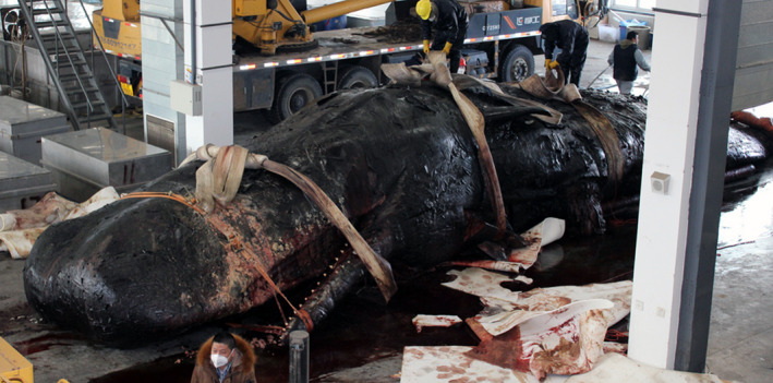
Sperm whale before skinning
FIGURE 3.

The sperm whale while being skinned
FIGURE 4.

Length, widths, and thickness of a skin segment derived from the dorsal side of the neck of the sperm whale measuring 86, 52, and 17 cm, respectively
FIGURE 5.

Head and neck segment of the sperm whale
FIGURE 6.
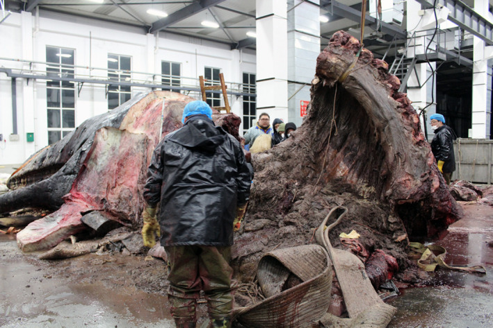
Chest segment of the sperm whale
FIGURE 7.
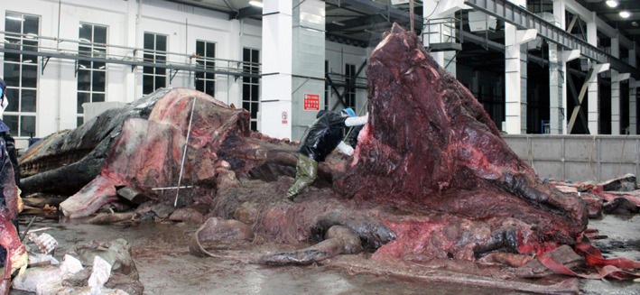
Waist segment of the sperm whale
FIGURE 8.

Lumbar segment of the sperm whale including the caudal fins
2.2.5. Fixation
he individual parts of the body (visceral organs and four body segments) and the separated skin layers underwent immersion fixation using 10% formaldehyde solution for 8 months.
2.2.6. Dissection
The general approach was: on the left side of the sperm whale, the muscles were removed to expose the bones such as the skull, vertebrae, ribs, forelimb bones, etc., as well as the internal organs such as heart, lungs, liver, stomach, intestines, and so on (Figure 9). All organs were extracted and measured (Table 1). On the right, the fat layer was removed to reveal the superficial muscles. The skin of the caudal fin was removed to expose the veinous distribution. The penis was kept intact and in situ. The process of dissection was as follows:
FIGURE 9.
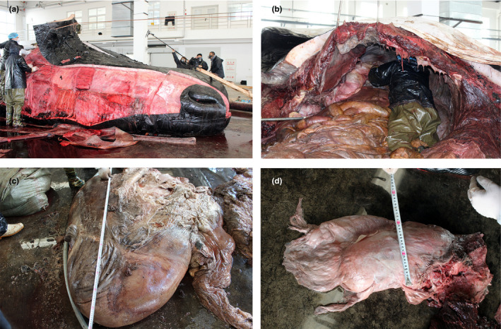
Sperm whale being dissected. (a) sperm whale head muscles were separated. (b) sperm whale organs were separated. (c) sperm whale's heart, maximal diameter in the long axis = 115 cm. (d) sperm whale's stomach, maximal upper, and lower diameter = 23 cm
TABLE 1.
The dimensions of viscera after the dissection
| Visceral organs | Diameter in the long axis (cm) | Antero posterior diameter (cm) | Transverse diameter (cm) |
|---|---|---|---|
| Heart | 115 | 60 | 115 |
| Liver | 190 | 120 | 170 |
| Left lung a | 150 | 115 | 50 |
| Left kidney a | 95 | 46 | 20 |
| Stomach | 170 | 62 | 23 |
Measurement was subject to a maximal diameter of the organs in an upright position (the same as the upright posture of the organ in Figure 12).
Sperm whale had already undergone autolysis which led to the deformation of lungs and kidneys. It forced us to ignore the size differences between the paired organs during plastination.
The muscles, nerves, and blood vessels of the sperm whale were dissected and separated from the skeletal ligaments in the craniocaudal direction. The muscles were removed from their origins in the direction towards their insertions paying special attention to the interconnections found to be present within the layer; special attention was also paid to the deep small muscles.The ongoing dissection was accompanied by photography and verbal records.
2.2.7. Specimen preparation
Following dissection, each part was dehydrated, degreased, and impregnated with polymer using Hoffen‐S10 technology. Dehydration and degreasing were carried out according to the standard procedure described by Okoye and Sui (Okoye & Sui, 2019). After selecting a suitable container for the samples, the silicone rubber that would cover the upper surface of the specimen was mixed with the Hoffen S3 reagent (developed by Hoffen independently) and poured into the blender. The weight of the S3 reagent is 2% of the weight of the silicone rubber. The stirring revolutions was set at 2200 rpm and after stirring for 2 h the polymer was stored in a low‐temperature environment. Then atmospheric pressure impregnation and negative pressure impregnation were carried out until no bubbles were left in the polymer. Each step lasted for about 4 months.
2.2.8. Internal support frame
In order to restore the original structure of the sperm whale, we designed an internal support frame for this large mammal to be plastinated (Figure 10). It was manufactured by the Biomechanics Laboratory of Dalian University of Technology.
FIGURE 10.

The internal support frame of the sperm whale. (a) holistic view. (b) right lateral view. (c) anterior view
2.2.9. Assembly
Using the support frame, the individual parts of the whale body were restored from tail to head. We started to assemble the bones and joints then inserted the viscera and finally attached the muscles, skin, and the fins in the correct anatomical order. During assembly, it was necessary to position the individual parts of the sperm whale in place by means of a pulley. Therefore, a large range of fixating frames was placed above the support frame to allow fixation of the pulley where necessary (Figure 11a). The structural damage caused during the process of segmentation was repaired by professional personnel (Figure 11b). The recovered sperm whale was placed in a sealed environment for hardening. The curing agent (Hoffen S6, developed by Hoffen independently) was steamed to fumigate the samples for one month.
FIGURE 11.

Sperm whale in assembly. (a) pulley and fixation frames during assembly. (b) sperm whale was rearranged by professionals
3. RESULTS
The sperm whale was partitioned into about 600 cubes, each sizing about 0.8 m3. Among them, there were more than 120 bones and more than 300 muscles, which were cleaned and fixed, respectively, and then reassembled and returned to the correct anatomical location. The 15‐m adult male sperm whale was successfully preserved by plastination technology (Figure 12). To show the structure of the sperm whale in its natural condition, on the left side the viscera and on the right‐side part of its muscles are displayed (trapezius, and lumbar and caudal muscles). The dimensions of the viscera were recorded and are presented in Table 1. Macroscopically, both the external and internal structures of the plastinated sperm whale have been well preserved, thus offering a great visual impression. The plastinated specimen is dry, non‐toxic, and does not require any additional preservation medium, which adds to its significance in the museum's scientific collection.
FIGURE 12.

The completely assembled sperm whale. Internal organs such as the heart, stomach, and kidney were assembled on the left side of the sperm whale, and the muscles of the sperm whale, such as the trapezius, were shown on the right side of the sperm whale
4. DISCUSSION
The whale is an iconic emblem of conservation (Hirasaki et al., 2018). Therefore, there are few opportunities to study the anatomical structure of this endangered species and every effort should be made to preserve important anatomical materials via a long‐lasting method. In this paper, the preservation of a whole sperm whale by plastination is reported. It was performed to ensure that this rare specimen is available for educational purposes for future generations. The major hurdle during preservation was the sperm whales’ large size, along with its wet and slippery nature. Hence, a crane was needed to move the sperm whale through each stage of the plastination process and to segment the animal.
In the past, traditional anatomical methods were commonly used to elucidate the external and internal structures of whales (Liu et al., 2018; Mass et al., 2013; Moore et al., 2004), but these methods have some limitations in exploring the causes of biodiversity, especially the mutual adaptability between the internal structure of animals and their living habits and living environment (Marselle et al., 2021; Naeem et al., 2016). Plastination is a proven method for fully displaying the internal structure of specimens, making it a useful tool for studying morphological sciences and anatomical and pathological sciences at the macro‐ and micro‐scopic levels (Ernesto et al., 2018). Therefore, the relationship between in situ anatomical structure and living environment in animals can be analyzed by plastination.
Due to natural selection, advantageous traits and variations are preserved within future generations of each species and passed down to offspring (Darwin et al., 1859). For example, the kidney (Sui et al., 2021) of the sperm whale has undergone great changes in biological evolution. Its outer surface resembles the grape pod, indicating the existence of a compound kidney composed of many anterior lobes, which individually can maintain the function of the kidney and all are able to efficiently eliminate salt. The kidney of cattle presents with a grooved and multiple nippled surface, thus representing a form of a single kidney and the grooves border the renal lobes, hence resembling the outer shape of the whale kidney. The whale stomach and the stomach of ruminants on land, such as cattle, sheep, and horses, are also very similar in morphology and also consists of four chambers. By comparing these homologous organs, we can better explain the evolution of whales and deduce their presumable ancestors. The present anatomical dissection has also revealed the presence of regressed hind limb bones (remnants) in the body of the sperm whale (Figure 12). Thus, the ancestors of ancient whales potentially possessed functional active hind limbs. Sperm whales have two nostrils, but only the left one is open for breathing, and the right one is inherently obstructed (Xu, 2020). Plastination of the sperm whale restored the left tilted nasal passage, which was decided to act as a teaching aid to highlight the characteristic differences between toothed and baleen whales. In the tail of the sperm whale, the muscles are grouped along the longitudinal axis of its body. This arrangement is ideal for propulsion in the ocean.
The Royal Ontario Museum in Canada has a special specimen‐ the heart of a blue whale (Miller et al., 2017). The blue whale is the largest of cetacean species and is the largest animal in history. In 2014, when a well‐preserved blue whale beached on the coast of Newfoundland, Canada, the Royal Ontario Museum of Canada removed a 440‐pound (about 200 kg) heart from the blue whale carcass and used plastination technology for preservation (Miller et al., 2017). That processed heart specimen is on display in the museum (as of 2017). The display of this huge heart specimen has greatly increased the museum's appeal to audiences around the world.
It is worth noting that the successful plastination of the sperm whale provides an opportunity for the study of existing oversized animals. The plastination allows detailed anatomical information of a certain part to be studied in situ, without being limited by the size of the specimen. The plastination technique can preserve the integrity of rare specimens. It is important to note that this project provides unique insights into the challenge of plastination of oversized animals by requiring us to adapt the method to the extraordinary size of this large animal. In this study, the general principles of plastination: specimen preparation, dehydration/defatting, impregnation, and curing, were similar to those of previous smaller more conventional specimens (Ekim et al., 2017; Henry & Gary, 2007; Nader et al., 2019). The main difference is the time span of impregnation, which is probably one of the moast critical aspects of the plastination procedure, as it has to be carried out at the same pressure for both large and small specimens. The time of impregnation was twice as long as for most smaller specimens hitherto reported (Miller et al., 2017). Importantly, the world's first successful plastination of a sperm whale has increased the possibility of preserving similar rare biomaterials in the future, providing direction for exhibitions in museums around the world. Close contact with deep‐sea creatures has also greatly promoted the popularization of marine education which will encourage conservation.
5. LIMITATIONS
Since this plastination of the sperm whale is the first in the world, we lacked a large amount of data for this preservation, which ensured the groundbreaking nature of this study and there were a lot of unknowns which we had to develop methods to deal with. Although we have made great efforts to do this work, there is no guarantee that this work was carried out perfectly as this was the first time this has ever been done.
6. CONCLUSION
Using the specifically adapted plastination technique, we successfully preserved the unique specimen of a sperm whale, thus providing a highly valuable contribution to the understanding of certain processes of mammalian adaptation.
AUTHORS' CONTRIBUTIONS
S‐B. Y and H‐J. S, conceived and designed the experiments. W‐B. J, J H, X‐W M, and H L involved in data acquisition. W‐B. J drafted the manuscript. H‐J. S is a plastination instructor and also carried out critical revision of the manuscript. All authors were involved in data interpretation.
ACKNOWLEDGMENTS
We thank Professor Xin Zhao of Dalian Medical University for assistance with photography and Dr. Campbell Gilmore of St. George's University of London for proofreading the manuscript. The authors declare no conflicts of interest associated with this study.
Jiang, W.‐B. , Han, J. , Ma, X.‐W. , Liu, H. , Yu, S.‐B. & Sui, H.‐J. (2022) Plastination of a sperm whale. Journal of Anatomy, 240, 669–677. Available from: 10.1111/joa.13581
The first authors: Wen‐Bin Jiang.
Funding information
This study was supported by the Dalian Key Science and Technology Project (No.2019YF22SN045).
Contributor Information
Jian Han, Email: hanjian@hoffen.com.cn.
Xue‐Wei Ma, Email: maxuewei@hoffen.com.cn.
Hu Liu, Email: 8233901@qq.com.
Sheng‐Bo Yu, Email: ysbdmu@126.com.
Hong‐Jin Sui, Email: suihj@hotmail.com.
REFERENCES
- Alam, P. , Amini, S. , Tadayon, M. , Miserez, A. & Chinsamy, A. (2016) Properties and architecture of the sperm whale skull amphitheatre. Zoology, 119, 42–51. [DOI] [PubMed] [Google Scholar]
- Alstrup, A.K.O. , Munk, O.L. , Jensen, T.H. , Jensen, L.F. , Hedaya, A. & Hansen, B. (2017) Magnetic resonance imaging and computed tomography as tools for the investigation of sperm whale (Physeter macrocephalus) teeth and eye. Acta Veterinaria Scandinavica, 59, 38. [DOI] [PMC free article] [PubMed] [Google Scholar]
- Clement, A.M. , King, B. , Giles, S. , Choo, B. , Ahlberg, P.E. , Young, G.C. et al. (2018) Neurocranial anatomy of an enigmatic Early Devonian fish sheds light on early osteichthyan evolution. Elife, 7.e34349. [DOI] [PMC free article] [PubMed] [Google Scholar]
- Darwin, C.R. (1859) On the origin of species. wikisource. [online] Available at: <https://en.wikisource.org/wiki/On_the_Origin_of_Species_(1859)> [accessed 25 May 2021].
- Ekim, O. , Haziroglu, R.M. , Insal, B. , Bakici, C. , Akgun, R.O. & Tunali, S. (2017) A modified S10B silicone plastination method for preparation and preservation of scaled reptile specimens. Ankara Universitesi Veteriner Fakultesi Dergisi, 64, 155–160. [Google Scholar]
- Ernesto, O.N. , Baptista, C.A.C. , Latorre, R. , Felipe, B.H. , Del, S.M. & Fuentes, R. (2018) E12 sheet plastination: techniques and applications. Clinical Anatomy, 31, 742–756. [DOI] [PubMed] [Google Scholar]
- Fais, A. , Johnson, M. , Wilson, M. , Soto, N.A. & Madsen, P.T. (2016) Sperm whale predator‐prey interactions involve chasing and buzzing, but no acoustic stunning. Scientific Reports, 6, 28562. [DOI] [PMC free article] [PubMed] [Google Scholar]
- Farmer, N.A. , Baker, K. , Zeddies, D.G. , Denes, S.L. , Noren, D.P. , Garrison, L.P. et al. (2018) Population consequences of disturbance by offshore oil and gas activity for endangered sperm whales (Physeter macrocephalus). Biological Conservation, 227, 189–204. [Google Scholar]
- Foskolos, I. , Koutouzi, N. , Polychronidis, L. , Alexiadou, P. & Frantzis, A. (2020) A taste for squid: the diet of sperm whales stranded in Greece, Eastern Mediterranean. Deep Sea Research Part I: Oceanographic Research Papers, 155, 103164. [Google Scholar]
- Henry, GvH & Gary, S. (2007) Silicone plastination of biological tissue: cold‐temperature technique ‐ biodur S10/S15 technique and products. Journal of the International Society for Plastination, 22, 2–14. [Google Scholar]
- Hirasaki, Y. , Tomita, T. , Yanagisawa, M. , Ueda, K. , Sato, K. & Okabe, M. (2018) Heart anatomy of rhincodon typus: three‐dimensional x‐ray computed tomography of plastinated specimens. Anatomical Record‐Advances in Integrative Anatomy and Evolutionary Biology, 301, 1801–1808. [DOI] [PubMed] [Google Scholar]
- Janc, A. , Richard, G. , Guinet, C. , Arnould, J.P.Y. , Villanueva, M.C. , Duhamel, G. et al. (2018) How do fishing practices influence sperm whale (Physeter macrocephalus) depredation on demersal longline fisheries? Fisheries Research, 206, 14–26. [Google Scholar]
- Liu, P. , Li, C. , Zheng, N. , Yuan, X. , Zhou, Y. , Chun, P.U. et al. (2018) The myodural bridges' existence in the sperm whale. PLoS One, 13, e0200260. [DOI] [PMC free article] [PubMed] [Google Scholar]
- Marselle, M.R. , Hartig, T. , Cox, D.T.C. et al. (2021) Pathways linking biodiversity to human health: a conceptual framework. Environment International, 150, 106420. [DOI] [PubMed] [Google Scholar]
- Mass, A.M. , Supin, A.Y. , Abramov, A.V. , Mukhametov, L.M. & Rozanova, E.I. (2013) Ocular anatomy, ganglion cell distribution and retinal resolution of a killer whale (Orcinus orca). Brain, Behavior and Evolution, 81, 1–11. [DOI] [PubMed] [Google Scholar]
- Miller, J.R. , Henry, R.W. , Nader, P. et al. (2017) The challenges of plastinating a Blue Whale (Balaenoptera musculus) heart. The Journal of Plastination, 29, 22–29. [Google Scholar]
- Moore, M.J. , Kraus, S.D. , McLellan, W.A. & Bonde, R.K. (2004) Morphometry, gross morphology and available histopathology in North Atlantic right whale (Eubalaene glacialis) mortalities (1970–2002). Journal of Cetacean Research and Management, 6, 199–214. [Google Scholar]
- Nader, P.B. , Henry, R.W. & Sui, H.J. (2019) Plastination of larger and massive specimens‐With Silicone. Anatomia Histologia Embryologia, 48, 547–551. [DOI] [PubMed] [Google Scholar]
- Naeem, S. , Chazdon, R. , Duffy, J.E. , Prager, C. & Worm, B. (2016) Biodiversity and human well‐being: an essential link for sustainable development. Proceedings of the Royal Society B: Biological Sciences, 283(1844), 20162091. [DOI] [PMC free article] [PubMed] [Google Scholar]
- Okoye, C.S. & Sui, H.J. (2019) Updated protocol for the hoffen P45 sheet plastination technique. The Journal of Plastination, 31, 22–25. [Google Scholar]
- Schnitzler, J.G. , Frédérich, B. , Früchtnicht, S. , Schaffeld, T. , Baltzer, J. , Ruser, A. et al. (2017) Size and shape variations of the bony components of sperm whale cochleae. Scientific Reports, 7, 46734. [DOI] [PMC free article] [PubMed] [Google Scholar]
- Smodlaka, H. , Henry, R.W. , Schumacher, J. & Reed, R.B. (2008) Macroscopic anatomy of the heart of the ringed seal (Phoca hispida). Anatomia Histologia Embryologia, 37, 30–35. [DOI] [PubMed] [Google Scholar]
- Soldevilla, M.S. , McKenna, M.F. , Wiggins, S.M. , Shadwick, R.E. , Cranford, T.W. & Hildebrand, J.A. (2005) Cuvier's beaked whale (Ziphius cavirostris) head tissues: physical properties and CT imaging. Journal of Experimental Biology, 208, 2319–2332. [DOI] [PubMed] [Google Scholar]
- Steiner, L. , Lamoni, L. , Plata, M.A. , Jensen, S.K. , Lettevall, E. & Gordon, J. (2012) A link between male sperm whales, Physeter macrocephalus, of the Azores and Norway. Journal of the Marine Biological Association of the United Kingdom, 92, 1751–1756. [Google Scholar]
- Sui, H.J. (2020) The sperm whale casted away. Science Press. [online] Available at: <http://cms.sciencepress.cn/2020550027004/472140.jhtml> [accessed 18 May 2021]. [Google Scholar]
- Sui, H.J. , Fan, Y.X. , Li, J. (2021) Legend of the great whale. Beijing, China: Science Press. [Google Scholar]
- von Horst, C. , von Hagens, R. , Sora, C.‐M. & Henry, R.W. (2019) History and development of plastination techniques. Anatomia Histologia Embryologia, 48, 512–517. [DOI] [PubMed] [Google Scholar]
- Watwood, S.L. , Miller, P.J.O. , Johnson, M. , Madsen, P.T. & Tyack, P.L. (2006) Deep‐diving foraging behaviour of sperm whales (Physeter macrocephalus). Journal of Animal Ecology, 75, 814–825. [DOI] [PubMed] [Google Scholar]
- Xu, W.T. (2020) Introduction of the sperm whales. Popular science china. [online] Available at: <https://baike.baidu.com/item/%E6%8A%B9%E9%A6%99%E9%B2%B8/709965#6> [accessed 24 May 2021]. [Google Scholar]