Abstract
Lipomas are slow-growing, benign mesenchymal masses. Most lipomas are small, weighing only a few grams; however, if their size becomes exceptionally large, they are called giant lipomas. Giant lipoma of the breast is infrequently observed due to the rarity of size and location, with very few case reports available in the literature. Here, we report the case of a 48-year-old female patient who presented with a painless, huge lump in her right breast. The patient underwent surgical removal of the mass with a histologic examination confirming the diagnosis of a giant breast lipoma.
Keywords: excision surgery, lipoma, breast mass, giant lipoma, benign breast condition
Introduction
Lipoma is one of the most common benign neoplasms, derived from fatty tissue, and represents 16% of all mesenchymal tumors [1]. Lipomas usually occur in the age group of 40-60 years. They are slow-growing, without pain or functional impairment [2]. Lipomas are usually freely mobile underneath the skin and develops as well-circumscribed, encapsulated masses [3]. They are mostly small (less than 5 cm in diameter) with a weight of a few grams only [4]. They could occur in any part of the body and 20% are located in the chest wall [5]. Breast lipomas are usually small and may be difficult to diagnose due to the normal adipose tissue of the breast [6]. When the diameter of a lipoma is at least 10 cm or its weight exceeds 1,000 grams, it is considered as giant lipoma [7]. A giant breast lipoma is a rare manifestation so making a correct diagnosis is crucial in the prevention of the overtreatment of this mass, which otherwise may be managed as a malignant tumor. Here, we report a rare case of a 48-year-old female patient with a giant lipoma in the right breast measuring 30 x 18 x 3 cm causing breast asymmetry.
Case presentation
A 48-year-old female presented to the surgery clinic with a painless lump in her right breast for three years. The lump grew gradually in size. The patient is medically free with no previous history of breast trauma or disease. She has a history of tongue papilloma that was excised five years ago. She did not have a family history of breast carcinoma. Physical examination of the right breast revealed a well-defined, homogeneous, firm, non-tender, mobile mass, measuring approximately 30 × 18 × 3 cm in diameter, with dilated veins on the surface. The overlying skin of the mass and the nipple-areola complex appeared to be normal. There was no nipple discharge. Axillary lymphadenopathy was not present. The left breast examination was unremarkable.
Mammogram images revealed a right breast replacement by a huge homogeneously isoechoic encapsulated mass with an internal tiny cyst with peripheral calcification measuring about 16 × 14 cm causing medial compression of the breast tissue. No skin thickening or nipple retraction was observed. There was no intralesional vascularity or enlarged axillary lymph nodes detected (Figure 1).
Figure 1. Mammographic view of the right breast shows several round, well-circumscribed, encapsulated, radiolucent masses.
In correlation to the mammogram, it represented a large lipoma with central fat necrosis, consistent with breast imaging-reporting and data system (BI-RADS) assessment category 2, showing benign findings. Fine needle aspiration cytology (FNAC) was not performed as it was not needed.
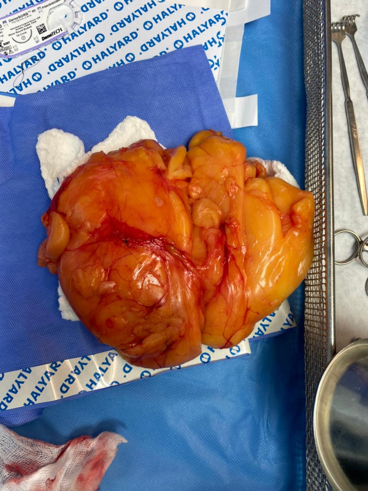
An elective surgical excision was scheduled and performed under general anesthesia. The patient was placed in the supine position on the table; aseptic technique was followed, and draping was provided. The right breast revealed an intracapsular, lobular, solitary mass. A skin incision was made around the areola followed by dissection around the lipoma to excise it. The mass weighed 1000 g (Figure 2).
Figure 2. Gross appearance of the mass.
Hemostasis was achieved with electrocautery. Washing and drain insertion were applied under negative pressure. Closure was done intradermally with a subcuticular suture, and a pressure dressing was applied. Histopathologic examination of the specimen revealed one huge soft consistency, irregular surface, and yellow-colored tissue measuring 30 × 18 × 3 cm. Cut sections showed adipose tissue without evidence of atypia or malignant change. The findings were consistent with lipoma of the breast. The patient had an uneventful postoperative course. She was discharged the next day after drain removal, to be followed up in the outpatient clinic.
Discussion
Lipomas are one of the most common adipose tumors. They are usually benign, well-circumscribed, and covered by a thin capsule [8]. The etiology of lipomas remains unidentified. However, it has been shown to be sporadic or linked to an inherited condition [9,10]. They can arise in any part of the body, with a prevalence rate of 2.1 per 1000 people [9]. Of lipomas, 20% are found in the chest wall, and the breast is a common site for this pathology [5,11]. They vary in size with the vast majority being small in size weighing only a few grams [11]. Hawary et al. defined a giant breast lipoma by the presence of a lesion with a dimension of at least 5 cm and a weight more than 500 g [12]. However, according to Sanchez et al., a giant lipoma has a dimension of 10 cm [7]. A mass that weighed 5700 g and measured 35 x23 x20 cm, reported by Ribeiro et al., appears to be the largest in literature [13]. Lipomas are usually solitary but might be hard to detect in large or postmenopausal breasts [14].
Patients typically present when lipomas cause anatomical changes that may lead to cosmetic concerns or interfere with function [15]. The presence of size disparity and architectural distortion mandates a comprehensive history, clinical examination, and investigations [16]. Because of the post-traumatic fat necrosis, lipomas may also appear as firm, fixed masses that may be clinically misinterpreted as malignancies [17]. Imaging techniques such as ultrasonography and mammography prove to be beneficial and useful for a more accurate diagnosis, especially in tailoring a specific management plan for every patient [6]. On mammography, a radiolucent nodule surrounded by a thin radiopaque capsule that may include ring-like calcifications owing to fat necrosis can establish the diagnosis of a lipoma. A correct diagnosis is critical since breast lipomas can be misdiagnosed as malignancies, phyllodes tumors, fibroadenomas, and duct papillomas [18]. Achieving the correct diagnosis might be challenging, necessitating an invasive approach such as FNAC or punch biopsy [17]. According to Lanng et al, the diagnosis of breast lipoma can be a diagnostic dilemma with difficulties in establishing the diagnosis through clinical examination, imaging studies, and FNAC [1]. In our case, FNAC was not done as the diagnosis was established according to the mammogram findings and confirmed through the histopathological examination that was done after the excision of the mass. Santi et al. reported a similar case of a 27-year-old female who presented with significant breast asymmetry. Preoperatively, she was diagnosed with breast liposarcoma. After excision and histopathology study was done, the result revealed the presence of a giant breast lipoma measuring 15.5 ×9.3 ×4.5 cm and weighing 257 grams; on histopathology, a uniform fatty composition with benign adipocytes was discovered [19].
Surgical excision has been the mainstay of treatment for lipoma historically. However, advanced medical treatment might cause a reduction in lipoma size. Two to three injections of deoxycholate have shown a decrease in approximately 75% of the lesion size in a case series [20]. Liposuction-assisted excision could be also considered as another definitive management of lipoma [7]. This technique is preferred in areas where larger scars should be avoided because it allows the incision to be placed in an inconspicuous location [21]. Although it's associated with better cosmetic results, it has been shown to increase the risk of recurrence and hematoma formation due to incomplete capsule removal [7]. Furthermore, skin irregularities such as dimpling, numbness, paresthesia, and pigmentation changes are all common complications after the liposuction technique [22-23]. The main surgical indications for breast lipoma removal include cosmetic deformity, patient discomfort, and the rapid growth of the lump [15]. For many uncomplicated cases, the excision can be performed in the minor operating room under local anesthesia [24]. However, since our patient presented with a huge mass that may have invaded the deep fascia or muscles, we decided to manage the patient with an advanced procedure under general anesthesia. The skin incisions should follow the langer's lines and be elliptical in order to obtain the best cosmetic result [24]. Hematomas and seromas are considered the most common postoperative complications [25]. Giant breast lipomas always cause discomfort and heaviness in patients. So, surgical intervention is often required for cosmetic reasons or to alleviate the patient's symptoms [15].
Conclusions
Breast lipomas are usually observed when they reach considerable dimensions and produce breast asymmetries and cosmetic problems that determine the patient's need to proceed with surgery. Whenever breast lipomas are diagnosed, they should be entirely excised and sent for histopathological studies in order to confirm a positive diagnosis of benignity. Long-term follow-up is necessary since lipomas can relapse even after several years.
The content published in Cureus is the result of clinical experience and/or research by independent individuals or organizations. Cureus is not responsible for the scientific accuracy or reliability of data or conclusions published herein. All content published within Cureus is intended only for educational, research and reference purposes. Additionally, articles published within Cureus should not be deemed a suitable substitute for the advice of a qualified health care professional. Do not disregard or avoid professional medical advice due to content published within Cureus.
The authors have declared that no competing interests exist.
Human Ethics
Consent was obtained or waived by all participants in this study
References
- 1.Lipoma of the breast: a diagnostic dilemma. Lanng C, Eriksen BØ, Hoffmann J. Breast. 2004;13:408–411. doi: 10.1016/j.breast.2004.04.011. [DOI] [PubMed] [Google Scholar]
- 2.Benign skin lesions: lipomas, epidermal inclusion cysts, muscle and nerve biopsies. Pandya KA, Radke F. Surg Clin North Am. 2009;89:677–687. doi: 10.1016/j.suc.2009.03.002. [DOI] [PubMed] [Google Scholar]
- 3.Giant lipoma of the breast. Ramírez-Montaño L, Vargas-Tellez E, Dajer-Fadel WL, Espinosa Maceda S. https://www.ncbi.nlm.nih.gov/pmc/articles/PMC3665869/ Arch Plast Surg. 2013;40:244–246. doi: 10.5999/aps.2013.40.3.244. [DOI] [PMC free article] [PubMed] [Google Scholar]
- 4.Use of the oncoplastic reduction pattern technique following removal of a giant breast lipoma. Grossman J, Menes T, Lahat G, Gur E, Weiss J, Barnea Y. Ann Plast Surg. 2011;67:106–108. doi: 10.1097/SAP.0b013e3181f3e3cc. [DOI] [PubMed] [Google Scholar]
- 5.Giant infiltrating lipoma of the face: CT and MR imaging findings. Cappabianca Salvatore, Barberi Antonio, Walter Del Vecchio, et al. http://www.ajnr.org/content/24/2/283.short. Am J Neuroradiol. 2003;24:283–286. [PMC free article] [PubMed] [Google Scholar]
- 6.Giant lipoma of the breast: a case report and review of the literature. Li YF, Lv MH, Chen LF, Wu YF. Clin Breast Cancer. 2011;11:420–422. doi: 10.1016/j.clbc.2011.04.005. [DOI] [PubMed] [Google Scholar]
- 7.Giant lipoma: case report and review of the literature. Sanchez MR, Golomb FM, Moy JA, et al. J Am Acad Dermatol. 1993;28:266–268. doi: 10.1016/s0190-9622(08)81151-6. [DOI] [PubMed] [Google Scholar]
- 8.Size, site and clinical incidence of lipoma. Factors in the differential diagnosis of lipoma and sarcoma. Rydholm A, Berg NO. Acta Orthop Scand. 1983;54:929–934. doi: 10.3109/17453678308992936. [DOI] [PubMed] [Google Scholar]
- 9.What should be the treatment modality in giant cutaneous lipomas? Review of the literature and report of 4 cases. Silistreli OK, Durmuş EU, Ulusal BG, Oztan Y, Görgü M. Br J Plast Surg. 2005;58:394–398. doi: 10.1016/j.bjps.2004.09.005. [DOI] [PubMed] [Google Scholar]
- 10.Familial multiple lipomatosis: report of a case and a review of the literature. Leffell DJ, Braverman IM. https://www.jaad.org/article/S0190-9622(86)70166-7/pdf. J Am Acad Dermatol. 1986;15:275–279. [PubMed] [Google Scholar]
- 11.Gigantic lipomas. Hakim E, Kolander Y, Meller Y, Moses M, Sagi A. Plast Reconstr Surg. 1994;94:369–371. doi: 10.1097/00006534-199408000-00025. [DOI] [PubMed] [Google Scholar]
- 12.Giant breast tumors. Hawary MB, Cardoso E, Mahmud S, Hassanain J. Ann Saudi Med. 1999;19:174–176. doi: 10.5144/0256-4947.1999.174. [DOI] [PubMed] [Google Scholar]
- 13.Breast reconstruction with parenchymal cross after giant lipoma removal. Ribeiro RC, Saltz R, España Quintera LF. Aesthetic Plast Surg. 2008;32:695–697. doi: 10.1007/s00266-008-9164-4. [DOI] [PubMed] [Google Scholar]
- 14.Giant lipoma of the breast. Schmidt J, Schelling M, Lerf B, Vogt M. Breast J. 2009;15:107–108. doi: 10.1111/j.1524-4741.2008.00681.x. [DOI] [PubMed] [Google Scholar]
- 15.Case report: giant lipoma of the breast. Çitgez B, Akgün I, Ferhatoğlu F, Kartal A, Akçakaya A. http://citeseerx.ist.psu.edu/viewdoc/download?doi=10.1.1.821.7874&rep=rep1&type=pdf Breast Case. 2012;1:20–22. [Google Scholar]
- 16.Lipomatous tumors and tumors with fatty component: MR imaging potential and comparison of MR and CT results. Dooms GC, Hricak H, Sollitto RA, Higgins CB. Radiology. 1985;157:479–483. doi: 10.1148/radiology.157.2.4048459. [DOI] [PubMed] [Google Scholar]
- 17.Giant lipoma of the breast. Rodriguez LF, Shuster BA, Milliken RG. Br J Plast Surg. 1997;50:263–265. doi: 10.1016/s0007-1226(97)91157-7. [DOI] [PubMed] [Google Scholar]
- 18.Imaging of giant breast masses with pathological correlation. Muttarak M, Chaiwun B. http://www.smj.org.sg/sites/default/files/4503/4503pe1.pdf. Singapore Med J. 2004;45:132–139. [PubMed] [Google Scholar]
- 19.Giant breast lipoma: a rare cause of breast asymmetry. delli Santi G, Bellioni M, Loreti A, Stagnitto D, La Pinta M, Dell'Osso A. Plast Reconstr Surg. 2006;117:1068–1069. doi: 10.1097/01.prs.0000201322.91566.42. [DOI] [PubMed] [Google Scholar]
- 20.Lipomas treated with subcutaneous deoxycholate injections. Rotunda AM, Ablon G, Kolodney MS. J Am Acad Dermatol. 2005;53:973–978. doi: 10.1016/j.jaad.2005.07.068. [DOI] [PubMed] [Google Scholar]
- 21.Liposuction of giant lipoma. Nichter LS, Gupta BR. Ann Plast Surg. 1990;24:362–365. doi: 10.1097/00000637-199004000-00011. [DOI] [PubMed] [Google Scholar]
- 22.Liposuction-assisted excision of cervicofacial lipomas. Calhoun KH, Bradfield JJ, Thompson C. https://pubmed.ncbi.nlm.nih.gov/7567011/ Otolaryngol Head Neck Surg. 1995;113:401–403. doi: 10.1016/S0194-59989570075-7. [DOI] [PubMed] [Google Scholar]
- 23.Another indication for liposuction: small facial lipomas. Wilhelmi BJ, Blackwell SJ, Mancoll JS, Phillips LG. Plast Reconstr Surg. 1999;103:1864–1867. doi: 10.1097/00006534-199906000-00008. [DOI] [PubMed] [Google Scholar]
- 24.Comparison of the surgical outcomes of punch incision and elliptical excision in treating epidermal inclusion cysts: a prospective, randomized study. Lee HE, Yang CH, Chen CH, Hong HS, Kuan YZ. Dermatol Surg. 2006;32:520–525. doi: 10.1111/j.1524-4725.2006.32105.x. [DOI] [PubMed] [Google Scholar]
- 25.Minimal-scar segmental extraction of lipomas: study of 122 consecutive procedures. Chandawarkar RY, Rodriguez P, Roussalis J, Tantri MD. Dermatol Surg. 2005;31:59–4. doi: 10.1111/j.1524-4725.2005.31005. [DOI] [PubMed] [Google Scholar]


